中文English
ISSN 1001-5256 (Print)
ISSN 2097-3497 (Online)
CN 22-1108/R

Correlation of the serum levels of adiponectin, omentin, and visfatin with the severity of acute pancreatitis

DOI: 10.12449/JCH240920
Research funding:

Xiamen City Health Guidance Project (3502Z20199063)

More Information
  • Corresponding author: XU Xin, xuxing19891031@126.com (ORCID: 0000-0002-2966-9459)
  • Received Date: 2024-01-19
  • Accepted Date: 2024-04-07
  • Published Date: 2024-09-25
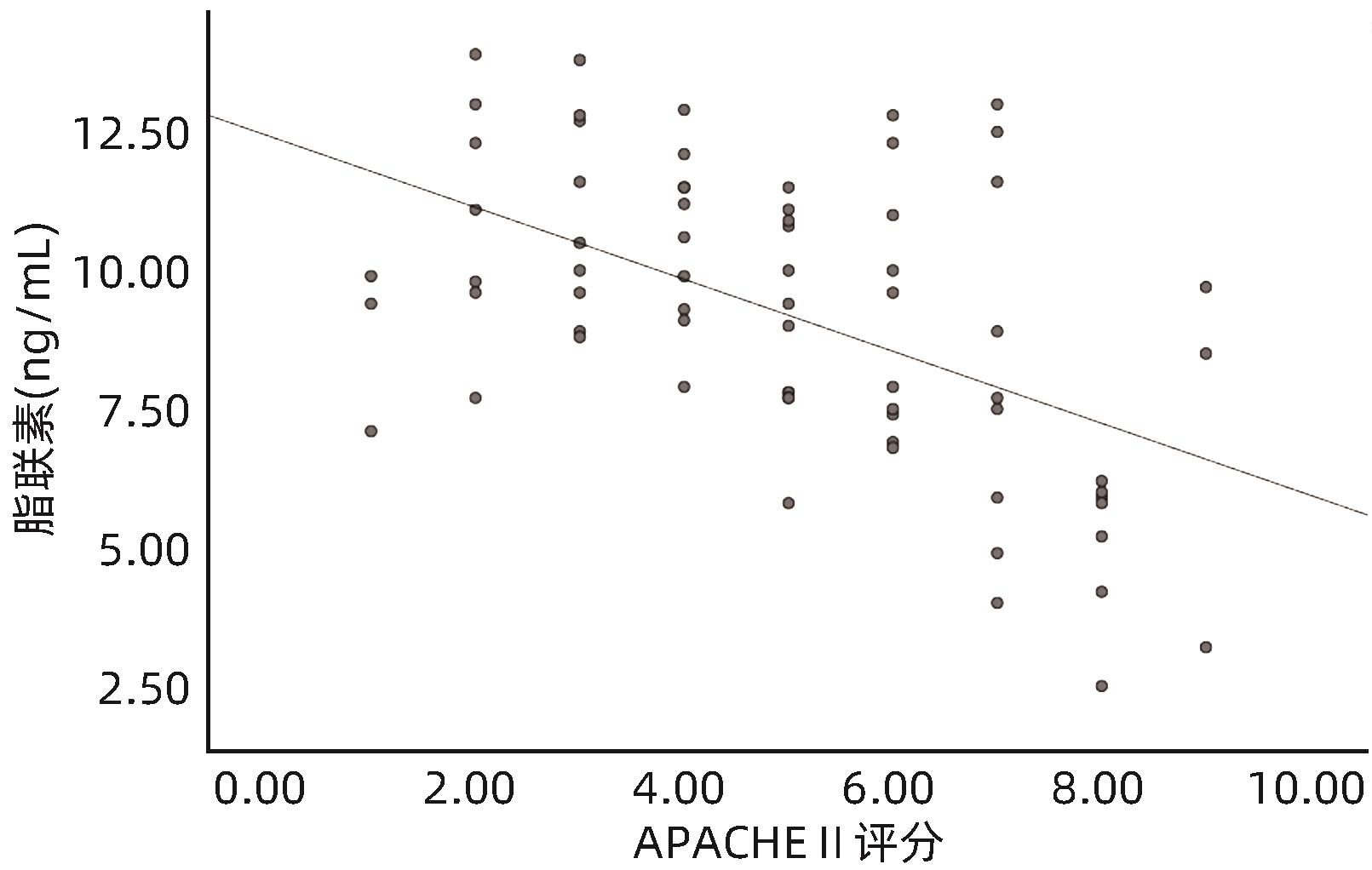
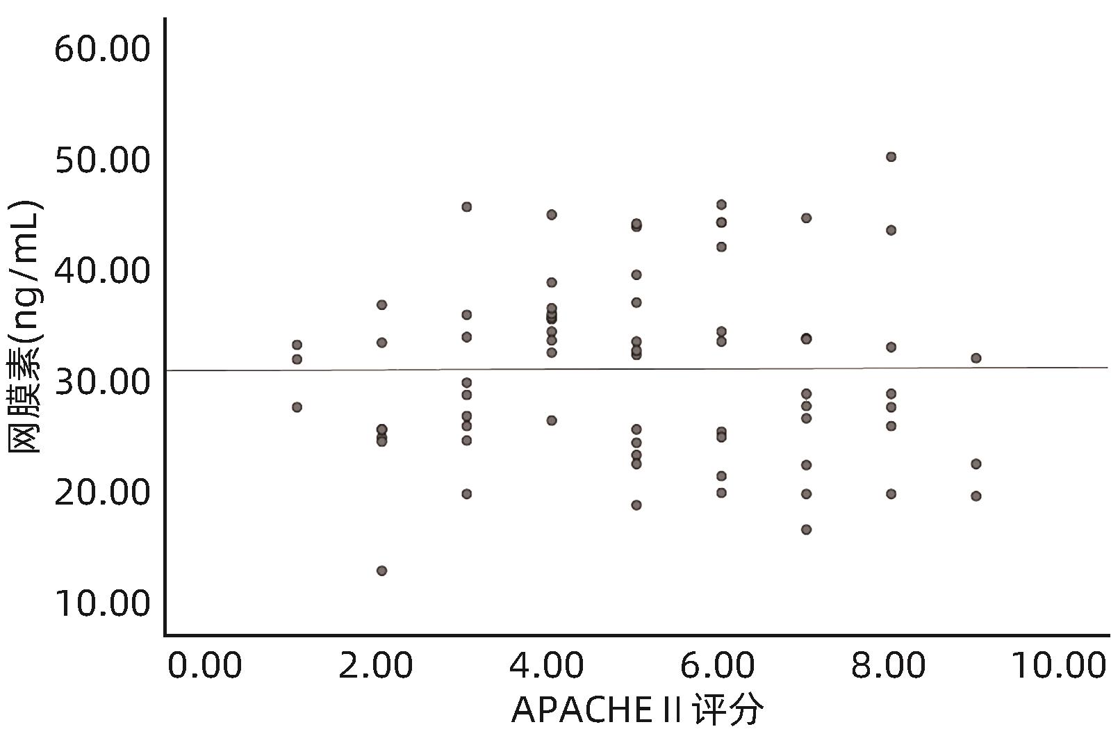
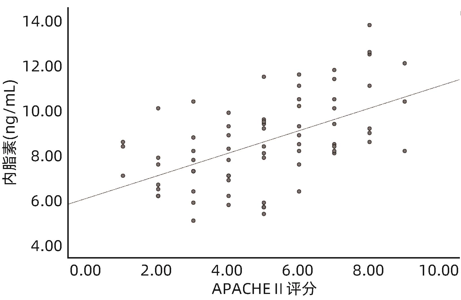
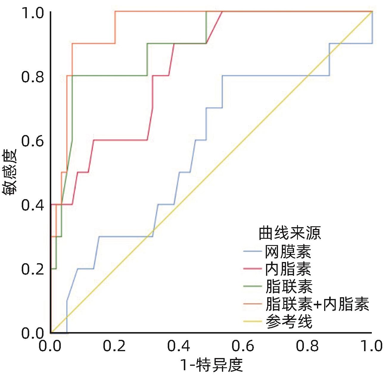
  •   Objective  To investigate the correlation of the serum levels of adiponectin, omentin, and visfatin with the severity of acute pancreatitis (AP).  Methods  Blood samples were collected from 35 healthy individuals in the control group and 70 patients with AP who were admitted to Chenggong Hospital Affiliated to Xiamen University from March 2022 to October 2023, and enzyme-linked immunosorbent assay was used to measure the serum levels of adiponectin, omentin, and visfatin in each group within 24 hours after admission. The analysis of variance was used for comparison of continuous between groups, and the LSD-t test was used for further comparison between two groups; the chi-square test was used for comparison of categorical data between groups. The Pearson correlation analysis was used to investigate the correlation between indicators, and the receiver operating characteristic (ROC) curve was used to investigate the value of the three indicators in predicting severe acute pancreatitis (SAP).  Results  Compared with the control group, the AP group had significantly higher serum levels of omentin and visfatin (t=5.51 and 9.41, both P<0.01), and the level of visfatin gradually increased with the aggravation of the disease (F=43.32, P<0.01), while there was no significant change in omentin with the aggravation of the disease (F=0.47, P>0.05). The AP group had a significantly lower level of adiponectin than the control group (t=-14.47, P <0.01), and the level of adiponectin gradually decreased with the aggravation of the disease (F=35.61, P<0.01). The serum level of visfatin was positively correlated with Acute Physiology and Chronic Health Evaluation II (APACHE-II) score (r=0.547, P<0.01), and the level of adiponectin was negatively correlated with APACHE-II score (r=-0.520, P<0.01), while there was no significant correlation between omentin APACHE-II score (r=0.007, P>0.05). The three indicators had an area under the ROC curve (AUC) of 0.893, 0.570, and 0.829, respectively, in predicting SAP, and combined measurement of adiponectin and visfatin with an AUC of >0.7 showed an AUC of 0.953 in predicting SAP, with a sensitivity of 0.900 and a specificity of 0.933.  Conclusion  The serum levels of adiponectin and visfatin are correlated with the severity of AP and have an important clinical significance in predicting SAP, and combined measurement of the two indicators has a higher value, while further studies are needed to investigate the correlation of omentin level with the severity of AP.


  • 成年人体内约有37.2万亿个细胞,每天死亡的细胞约占0.4%1。凋亡细胞的清除效率很高,因此即使在新陈代谢很高的组织中,凋亡细胞数量也很少。吞噬细胞通过其胞葬作用参与凋亡细胞的清除,从而在维持组织稳态方面发挥重要作用。吞噬细胞胞葬功能障碍使凋亡细胞无法清除,可发生继发性坏死,导致病理性炎症和引起自身免疫反应2。另有研究3表明,吞噬细胞的胞葬作用可抑制炎症细胞因子和效应T淋巴细胞的产生,而与肿瘤的进展相关。肝脏疾病是影响全球的重要公共健康问题。据估计,我国有超过1/5的人口患有某种形式的肝病,如病毒性肝炎、非酒精性脂肪性肝病(NAFLD)、酒精性肝病、药物性肝损伤、肝硬化和肝细胞癌(HCC)等,是我国人群肝病发病率和死亡率较高的主要原因之一4。目前针对不同类型的肝病虽有一些相应的治疗手段,但尚不能完全满足对各种肝病治疗的需要。肝巨噬细胞是肝脏中具有胞葬功能的专职吞噬细胞5,近年来,越来越多的证据表明肝巨噬细胞的胞葬作用与急性肝损伤、酒精性肝病、自身免疫性肝病、肝纤维化和HCC等多种急慢性肝病相关6,是当前肝病领域研究的热点之一。本文主要从巨噬细胞与凋亡细胞胞葬相关分子的表达、胞葬过程及巨噬细胞胞葬功能在不同肝病中的作用和相关治疗等方面开展综述。

    巨噬细胞的胞葬作用与经典的吞噬作用不同,其中凋亡细胞和巨噬细胞胞葬相关分子(包括胞葬配体、受体和桥接分子)的表达对巨噬细胞清除凋亡细胞至关重要(表1)。凋亡配体与巨噬细胞胞葬受体结合诱导巨噬细胞骨架重排并促进对凋亡细胞的吞噬,随后吞噬体与溶酶体融合降解凋亡细胞中的物质7

    表  1  凋亡细胞和巨噬细胞胞葬相关分子
    Table  1.  Funeral-related molecules in apoptotic cells and macrophages
    凋亡细胞胞葬配体 巨噬细胞胞葬受体 桥接分子
    LPC、S1P、ATP、UTP、PS 直接受体:G2A、S1PR、P2Y2、LRP1、SRB1、BAI1、TIM-4、RAGE、TREM2 间接受体:TAM、Integrins Gas6、ProS、MFGE8
    下载: 导出CSV 
    | 显示表格

    凋亡细胞胞葬配体的表达是诱导巨噬细胞对其吞噬清除的首要步骤,其主要的配体分子包括溶血磷脂酰胆碱(lysophosphatidylcholine,LPC)、鞘氨醇-1-磷酸(sphingosine-1-phosphate,S1P)、腺苷三磷酸(adenosine triphosphate,ATP)、尿苷三磷酸(uridine triphosphate,UTP)等。上述信号分子被吞噬细胞上的受体感知,诱导吞噬细胞向凋亡细胞迁移。磷脂酰丝氨酸(phosphatidylserine,PS)是凋亡细胞表达的另一种胞葬相关配体,其通过与巨噬细胞中相应的受体结合,介导巨噬细胞对凋亡细胞的摄取和吞噬8

    巨噬细胞胞葬受体根据与其配体的结合方式不同可分为直接受体和间接受体。其中G蛋白偶联受体(G-protein-coupled receptor,G2A)、嘌呤能受体(purinergic receptors,P2Y2)、鞘氨醇-1-磷酸受体(sphingosine-1-phosphate receptor,S1PR)可与凋亡细胞表达的LPC、ATP和UTP、S1P直接结合,从而使巨噬细胞迁移至凋亡细胞周围。而低密度脂蛋白受体相关蛋白1(low-density lipoprotein receptor related proteins 1,LRP1)、清道夫受体B1(scavenger receptor1,SRB1)、脑特异性血管生成抑制剂1(brain-specific angiogenesis inhibitor 1,BAI1)、T淋巴细胞免疫球蛋白黏蛋白受体4(T cell immunoglobulin mucin receptor4,TIM-4)、晚期糖基化终产物受体(advanced glycation end-product receptor,RAGE)和骨髓源性细胞表达的触发受体2(triggering receptor expressed in myeloid cells 2,TREM2)等则可与凋亡细胞的PS直接结合,继而诱导巨噬细胞对凋亡细胞的吞噬。间接受体是指通过桥接分子与凋亡细胞中的PS相结合,主要包括TYRO3/Axl/MER酪氨酸激酶受体(TYRO3/Axl/MER tyrosine kinase receptor,TAM)和整合素(Integrins)。巨噬细胞胞葬受体分子被激活后可触发不同的信号通路促进细胞骨架重排,完成对凋亡细胞的吞噬及随后的降解68-9

    桥接分子有2个受体结合域,在巨噬细胞和凋亡细胞之间起桥梁作用。主要的桥接分子包括生长阻滞特异性蛋白6 (growth arrest specific 6,Gas6)、蛋白S(protein S,ProS)、乳脂球表皮生长因子8(milk fat globule epidermal growth factor 8,MFGE8)等,这些桥接分子的分泌可促进巨噬细胞对凋亡细胞的吞噬和清除10

    巨噬细胞的胞葬过程主要分为4个阶段:(1)招募阶段。凋亡细胞表达和释放的LPC、S1P、ATP、UTP、趋化因子C-X3-C-基序配体1等信号被巨噬细胞相应的受体分子感知,招募巨噬细胞至凋亡细胞的周围。(2)识别阶段。巨噬细胞的胞葬受体如趋化因子C-X3-C-基序受体1、LRP1、TREM2等识别并结合凋亡细胞表达的PS等配体分子。(3)吞噬阶段。胞葬配体与受体结合后激活巨噬细胞Rho GTP酶(Rho GTPases)介导对凋亡细胞的吞噬。(4)降解阶段。胞葬细胞通过线粒体裂解增加吞噬小体封闭所需的胞质钙水平,并通过合成磷脂酰肌醇3,4,5-三磷酸和产生活性氧,促进LC3相关的吞噬,随后吞噬溶酶体融合、酸化,最终导致凋亡细胞的降解7-9

    急性肝损伤是由多种肝毒性因素如肝炎病毒、药物和免疫损伤等因素导致短时间内大量肝细胞死亡11。研究12表明,急性肝损伤时,包括TAM家族在内的多种胞葬相关受体在巨噬细胞上表达上调,巨噬细胞胞葬功能增强,在清除死亡细胞的同时,分泌一些细胞因子如IL-10、IL-4、IL-6和TNF-α等,可抑制炎症和刺激组织再生,从而有利于急性肝损伤后肝组织的修复。

    IRI是肝外伤、肝部分切除术和肝移植过程中发生的一种不可避免病理结果,可损害患者的肝功能13。巨噬细胞的胞葬作用通过促进对死亡细胞的清除、抑制炎症反应,在IRI中发挥积极作用。研究12表明,MER酪氨酸激酶(mer tyrosine kinase receptor,MerTK)和Gas6信号被抑制的小鼠,其促炎细胞因子如TNF和IL-1β的mRNA水平升高,而激活巨噬细胞中的Gas6/MerTK信号通路则抑制这些细胞因子的产生,显著降低了IRI后暴发性肝衰竭发生的风险。巨噬细胞中TIM-4和TREM2受体被激活后可促进IL-10产生和Ras-related C3肉毒毒素底物1(Ras-related C3 botulinum toxin substrate 1,Rac1)的激活,促进巨噬细胞对凋亡细胞的吞噬和抑制IRI后炎症反应14-15

    ALF是由各种因素导致的肝细胞短期内发生广泛死亡,从而使肝脏发生无法控制的炎症反应16。研究1217-18表明,巨噬细胞TAM受体在ALF中具有保护作用。与健康人相比,ALF患者肝脏中MerTK+巨噬细胞显著增多,通过胞葬作用清除凋亡的中性粒细胞,促进ALF后的炎症消退。其机制与肝X受体(liver X receptor,LXR)和5-脂氧合酶密切相关。巨噬细胞激活的MerTK信号除可诱导炎症抑制因子LXR丰度的恢复,还可通过细胞外调节蛋白激酶/肌质网钙泵2/钙/钙调素依赖性蛋白激酶2通路抑制5-脂氧合酶的磷酸化,促进脂素A4的合成,最终促进炎症消退。Axl是TAM受体家族中的另一个成员,Axl与Gas6结合后其细胞质内的酪氨酸残基发生磷酸化,诱导巨噬细胞自噬,抑制NOD样受体蛋白3(oligomerization domain-like receptors protein 3,NLRP3)炎性小体的激活,进而改善肝损伤。

    巨噬细胞通过胞葬作用促进对死亡细胞的清除,抑制炎症反应可促进急性肝损伤的修复,但在不同类型的慢性肝病中巨噬细胞的胞葬功能可改善或推动疾病的进展,这可能与多种不同因素及复杂的分子机制有关。

    酒精性肝病是由于长期饮酒导致的慢性肝脏疾病,未经有效干预,可从酒精性脂肪肝发展为酒精性脂肪性肝炎、肝纤维化和HCC19。巨噬细胞胞葬功能障碍在酒精性肝病的发展及相关并发症如败血症的发生中具有重要作用。(1)酒精可抑制巨噬细胞MFGE8基因的表达,影响巨噬细胞的胞葬功能,使凋亡细胞不能清除而发生继发性坏死,促炎因子释放增加,加重肝损伤。(2)酒精性肝病患者血清中高迁移率族蛋白B1表达上调,其可作为损伤相关分子模式蛋白激活促炎Toll样受体(Toll like receptor,TLR)和NLRP3信号,同时与MFGE8竞争性结合PS抑制巨噬细胞对凋亡细胞的吞噬。(3)酒精性肝病患者肠道屏障功能遭到破坏,可合并细菌感染引起的败血症。中性粒细胞浸润增加并通过形成中性粒细胞胞外陷阱(neutrophil extracellular trap,NET)使微生物死亡是控制感染的有效手段。为了抑制NET后的炎症反应,NET通过巨噬细胞的胞葬作用而清除。酒精可显著抑制肝脏NET的形成和巨噬细胞对中性粒细胞的清除,从而延长肝脏炎症和损伤。研究20-22表明,重组人MFG-E8通过增强巨噬细胞的胞葬功能可降低酒精相关败血症患者血清中的TNF-α、IL-6和内毒素水平,提示重组人MFG-E8在酒精性肝病及其相关败血症的治疗方面可能具有较好的疗效。

    NAFLD是常见的慢性肝脏疾病之一,脂肪积累和炎症是NAFLD进展的主要原因,根据病变的严重程度可分为非酒精性脂肪肝(NAFL)和非酒精性脂肪性肝炎(NASH)23-24。超重及肥胖是NAFLD发生重要因素,而巨噬细胞的胞葬功能障碍可促进NASH的发展,胞葬受体TREM2的表达对巨噬细胞吞噬清除凋亡细胞具有重要作用。长期高营养状态可导致肝脏促炎因子如TNF、IL6和IL-1β的产生,这些炎症细胞因子通过解聚素金属蛋白酶17(a disintegrin and metalloprotease 17,ADAM17)依赖的方式诱导TREM2脱落,TREM2的表达下调导致巨噬细胞的胞葬功能减弱,凋亡的肝细胞异常积累,炎症细胞因子产生增加,促进NAFL向NASH方向进展。因此,靶向ADAM17的抑制剂或TREM2的激动剂可能是NAFLD的潜在治疗靶点25

    自身免疫性肝病是以自身抗体产生为主要特征的慢性肝病,主要包括自身免疫性肝炎、原发性胆汁性胆管炎及原发性硬化性胆管炎26。在自身免疫性肝病中,细胞凋亡是肝细胞和胆管上皮细胞死亡的主要方式,而巨噬细胞对凋亡细胞的清除障碍与自身免疫性肝病的发生相关627。巨噬细胞的胞葬相关受体如TIM、Axl和MerTK等表达下调导致其胞葬功能减弱,凋亡细胞不能被清除而发生继发性坏死,导致自身抗原成分和炎症细胞因子释放,继而吸引免疫攻击,促进疾病进展。此外,巨噬细胞胞葬功能障碍除了可促进炎症细胞因子释放激活肝星状细胞外,还可同抑制多种基质金属蛋白酶的产生而促进肝纤维化的发生,使自身免疫性肝病朝不可逆的方向发展28-29。靶向巨噬细胞TIM、Axl、MerTK的激动剂的增强巨噬细胞胞葬作用可能为自身免疫性肝病开辟新的治疗前景。

    肝纤维化是各种慢性肝病的晚期阶段,可进展为肝硬化、HCC,严重影响患者的生存质量30。巨噬细胞的胞葬功能在肝纤维化中的作用是具有争议的。巨噬细胞表达的stabilin-1与PS及丙二醛低密度脂蛋白结合可促进对凋亡细胞的清除,抑制促纤维化因子产生,从而对肝纤维化的发生起到积极的保护作用5。TIM-4是巨噬细胞表达的另一个胞葬相关受体,也可抑制肝纤维化的发生,其机制主要包括:(1)TIM-4与TAM受体协同作用促进巨噬细胞对凋亡肝细胞的清除,抑制肝细胞线粒体损伤相关分子模式蛋白的释放和肝星状细胞的激活;(2)TIM-4与PS结合激活LKB1/AMPKα信号通路抑制NLRP3炎症小体,减少促炎细胞因子的释放,从而起到抑制肝纤维化的作用12。与此相反的是,巨噬细胞中MerTK的表达可通过细胞外调节蛋白激酶1/2途径使TGF-β产生增加,继而激活肝星状细胞而对肝纤维化发生具有促进作用。研究31表明,全反式维甲酸通过激活ADAM17裂解MerTK分子可抑制TGF-β的产生,故其可能是肝纤维化的潜在治疗靶点。

    HCC是肝恶性肿瘤之一,也是多种慢性肝病的终末期形式。HCC患者的预后较差,长期生存率较低32。巨噬细胞胞葬分子的表达与HCC的进展密切相关。巨噬细胞PS受体激活将干扰NF-κB信号传导和Ⅰ型干扰素的产生,进而限制抗肿瘤免疫反应。在HCC中,MerTK+巨噬细胞在清除凋亡的肿瘤细胞时可产生免疫抑制因子如IL-13和TGF-β等,使免疫系统沉默,促进肿瘤细胞逃脱免疫监视和攻击33-35。由在肿瘤生长、血管生成和转移中的作用,针对PS及其受体TIM、TAM阻断性抗体正成为抗肿瘤治疗的研究热点。例如,新型抗TIM3抗体与抗PD1抗体的联合可用于HCC的治疗36。研究5证实,HCC细胞通过上调“非吃我信号”CD47分子的表达,可阻止巨噬细胞对其识别,通过阻断CD47与巨噬细胞信号调节蛋白α1的相互作用,增强巨噬细胞对肿瘤细胞的吞噬,可抑制HCC的进展。综上所述,巨噬细胞和HCC细胞胞葬分子的表达可通过抑制免疫反应、阻碍巨噬细胞对肿瘤细胞的识别而促进HCC的进展。

    近年来,越来越多的证据表明巨噬细胞的胞葬功能与多种肝病密切相关,但其机制尚未完全明确。巨噬细胞的胞葬功能在不同类型肝病中的作用不同。一方面,巨噬细胞的胞葬功能障碍可能促进酒精性肝病、非酒精性肝病及自身免疫性肝病的进展;另一方面,巨噬细胞的胞葬作用可能在肝纤维化、HCC的进展中具有”双刃剑”效应,这可能与巨噬细胞胞葬受体的冗余性有关。凋亡配体与不同受体的结合可能通过不同的信号通路而产生不同的效果。深入研究巨噬细胞胞葬分子在不同肝病的表达情况及分子机制,开发靶向多种胞葬分子的药物,有望使更多肝病患者受益。

  • [1]
    CHEN SC, ZHU JF, SUN LQ, et al. LincRNA-EPS alleviates severe acute pancreatitis by suppressing HMGB1-triggered inflammation in pancreatic macrophages[J]. Immunology, 2021, 163( 2): 201- 219. DOI: 10.1111/imm.13313.
    [2]
    BARRETO SG, HABTEZION A, GUKOVSKAYA A, et al. Critical thresholds: Key to unlocking the door to the prevention and specific treatments for acute pancreatitis[J]. Gut, 2021, 70( 1): 194- 203. DOI: 10.1136/gutjnl-2020-322163.
    [3]
    HU XY, YANG ZY, ZHAO CJ, et al. Research progress in acute pancreatitis scoring systems in predicting the severity of disease[J/OL]. Chin J Hepatic Surg(Electronic Edition), 2024, 13( 2): 239- 243. DOI: 10.3877/cma.j.issn.2095-3232.2024.02.021.

    胡欣芫, 杨智義, 赵成俊, 等. 急性胰腺炎评分系统预测病情严重程度的研究进展[J/OL]. 中华肝脏外科手术学电子杂志, 2024, 13( 2): 239- 243. DOI: 10.3877/cma.j.issn.2095-3232.2024.02.021.
    [4]
    HARSHIT KUMAR A, SINGH GRIWAN M. A comparison of APACHE II, BISAP, Ranson’s score and modified CTSI in predicting the severity of acute pancreatitis based on the 2012 revised Atlanta Classification[J]. Gastroenterol Rep, 2018, 6( 2): 127- 131. DOI: 10.1093/gastro/gox029.
    [5]
    DI MY, LIU H, YANG ZY, et al. Prediction models of mortality in acute pancreatitis in adults: A systematic review[J]. Ann Intern Med, 2016, 165( 7): 482- 490. DOI: 10.7326/M16-0650.
    [6]
    SILVA-VAZ P, ABRANTES AM, CASTELO-BRANCO M, et al. Multifactorial scores and biomarkers of prognosis of acute pancreatitis: Applications to research and practice[J]. Int J Mol Sci, 2020, 21( 1): 338. DOI: 10.3390/ijms21010338.
    [7]
    KUMARI R, KUMAR S, KANT R. An update on metabolic syndrome: Metabolic risk markers and adipokines in the development of metabolic syndrome[J]. Diabetes Metab Syndr, 2019, 13( 4): 2409- 2417. DOI: 10.1016/j.dsx.2019.06.005.
    [8]
    ZHAO CL, SHANG DF, ZHOU C, et al. Mechanism of lipid metabolism mediated by hepatokines and adipokines in nonalcoholic fatty liver disease[J]. J Clin Hepatol, 2023, 39( 1): 168- 174. DOI: 10.3969/j.issn.1001-5256.2023.01.026.

    赵晨露, 尚东方, 周铖, 等. 肝因子和脂肪因子介导的脂代谢在非酒精性脂肪性肝病中的作用机制[J]. 临床肝胆病杂志, 2023, 39( 1): 168- 174. DOI: 10.3969/j.issn.1001-5256.2023.01.026.
    [9]
    YIN JY, WANG Q. Progress on adipokines in non-alcoholic fatty liver disease[J/CD]. Chin J Liver Dis(Electronic Version), 2023, 15( 1): 1- 5. DOI: 10.3969/j.issn.1674-7380.2023.01.001.

    尹静亚, 王琦. 脂肪因子在非酒精性脂肪性肝病中研究进展[J/CD]. 中国肝脏病杂志(电子版), 2023, 15( 1): 1- 5. DOI: 10.3969/j.issn.1674-7380.2023.01.001.
    [10]
    SCHÄFFLER A, LANDFRIED K, VÖLK M, et al. Potential of adipocytokines in predicting peripancreatic necrosis and severity in acute pancreatitis: Pilot study[J]. J Gastroenterol Hepatol, 2007, 22( 3): 326- 334. DOI: 10.1111/j.1440-1746.2006.04364.x.
    [11]
    SHARMA A, MUDDANA V, LAMB J, et al. Low serum adiponectin levels are associated with systemic organ failure in acute pancreatitis[J]. Pancreas, 2009, 38( 8): 907- 912. DOI: 10.1097/MPA.0b013e3181b65bbe.
    [12]
    KARPAVICIUS A, DAMBRAUSKAS Z, GRADAUSKAS A, et al. The clinical value of adipokines in predicting the severity and outcome of acute pancreatitis[J]. BMC Gastroenterol, 2016, 16( 1): 99. DOI: 10.1186/s12876-016-0514-4.
    [13]
    Group of Pancreatic Surgery, Surgery Society of Chinese Medical Association. Guidelines for diagnosis and treatment of acute pancreatitis in China(2021)[J]. Chin J Surg, 2021, 59( 7): 578- 587. DOI: 10.3760/cma.j.cn112139-20210416-00172.

    中华医学会外科学分会胰腺外科学组. 中国急性胰腺炎诊治指南(2021)[J]. 中华外科杂志, 2021, 59( 7): 578- 587. DOI: 10.3760/cma.j.cn112139-20210416-00172.
    [14]
    LI CR, LI X, LIU LX. Research progress of adipokine and acute pancreatitis[J]. Adv Clin Med, 2023, 13( 7): 11609- 11614. DOI: 10.12677/acm.2023.1371624.

    李晨瑞, 李笑, 刘林勋. 脂肪因子与急性胰腺炎的相关研究进展[J]. 临床医学进展, 2023, 13( 7): 11609- 11614. DOI: 10.12677/acm.2023.1371624.
    [15]
    YAVUZ N, UNAL E, MEMISOGLU K, et al. Plasma leptin levels in rats with pancreatitis[J]. Tohoku J Exp Med, 2004, 204( 4): 243- 248. DOI: 10.1620/tjem.204.243.
    [16]
    SCHERER PE, WILLIAMS S, FOGLIANO M, et al. A novel serum protein similar to C1q, produced exclusively in adipocytes[J]. J Biol Chem, 1995, 270( 45): 26746- 26749. DOI: 10.1074/jbc.270.45.26746.
    [17]
    KARPAVICIUS A, DAMBRAUSKAS Z, SILEIKIS A, et al. Value of adipokines in predicting the severity of acute pancreatitis: Comprehensive review[J]. World J Gastroenterol, 2012, 18( 45): 6620- 6627. DOI: 10.3748/wjg.v18.i45.6620.
    [18]
    WATANABE T, WATANABE-KOMINATO K, TAKAHASHI Y, et al. Adipose tissue-derived omentin-1 function and regulation[J]. Compr Physiol, 2017, 7( 3): 765- 781. DOI: 10.1002/cphy.c160043.
    [19]
    RAO SS, HU Y, XIE PL, et al. Omentin-1 prevents inflammation-induced osteoporosis by downregulating the pro-inflammatory cytokines[J]. Bone Res, 2018, 6: 9. DOI: 10.1038/s41413-018-0012-0.
    [20]
    LIU HP, WU JF, WANG HY, et al. Association of serum omentin-1 concentrations with the presence and severity of preeclampsia[J]. Ann Clin Biochem, 2015, 52( 2): 245- 250. DOI: 10.1177/0004563214541247.
    [21]
    ALI S, ALAM R, AHSAN H, et al. Role of adipokines(omentin and visfatin) in coronary artery disease[J]. Nutr Metab Cardiovasc Dis, 2023, 33( 3): 483- 493. DOI: 10.1016/j.numecd.2022.11.023.
    [22]
    SIT M, AKTAS G, YILMAZ EE, et al. Effects of the inflammatory response on serum omentin levels in early acute and chronic pancreatitis[J]. Clin Ter, 2014, 165( 2): e148- e152. DOI: 10.7471/CT.2014.1699.
    [23]
    MOSCHEN AR, KASER A, ENRICH B, et al. Visfatin, an adipocytokine with proinflammatory and immunomodulating properties[J]. J Immunol, 2007, 178( 3): 1748- 1758. DOI: 10.4049/jimmunol.178.3.1748.
    [24]
    SAWICKA K, MICHALSKA-JAKUBUS M, POTEMBSKA E, et al. Visfatin and chemerin levels correspond with inflammation and might reflect the bridge between metabolism, inflammation and fibrosis in patients with systemic sclerosis[J]. Postepy Dermatol Alergol, 2019, 36( 5): 551- 565. DOI: 10.5114/ada.2018.79104.
    [25]
    LU Y, TAO DX, HE ML. Expression of serum adipocytokines, lactones and IL-33 in patients with inflammatory bowel disease and their correlation with intestinal flora[J]. Mod Dig Interv, 2021, 26( 1): 85- 90. DOI: 10.3969/j.issn.1672-2159.2021.01.018.

    陆游, 陶丁霞, 和明丽. 炎症性肠病患者血清脂肪细胞因子、内酯素、IL-33表达及与肠道菌群的相关性[J]. 现代消化及介入诊疗, 2021, 26( 1): 85- 90. DOI: 10.3969/j.issn.1672-2159.2021.01.018.
    [26]
    SCHÄFFLER A, HAMER OW, DICKOPF J, et al. Admission visfatin levels predict pancreatic and peripancreatic necrosis in acute pancreatitis and correlate with clinical severity[J]. Am J Gastroenterol, 2011, 106( 5): 957- 967. DOI: 10.1038/ajg.2010.503.
    [27]
    MEI PF, HUANG MF, KONG H, et al. Study on the correlation between serum visfatin changes and illness in patients with acute pancreatitis[J]. Chin J Pancreatol, 2015, 15( 1): 44- 45. DOI: 10.3760/cma.j.issn.1674-1935.2015.01.011.

    梅鹏飞, 黄梅芳, 孔浩, 等. 急性胰腺炎患者血清内脂素变化与病情相关性研究[J]. 中华胰腺病杂志, 2015, 15( 1): 44- 45. DOI: 10.3760/cma.j.issn.1674-1935.2015.01.011.
  • Relative Articles

    [1]Wang Mei, Xiong HuaGang, Yang ZhiGang, Long Hai, Wang Yi, Tan Li. HCV genotype and clinical features of patients with HIV/HCV coinfection in Guizhou,China: An analysis of 383 cases[J]. Journal of Clinical Hepatology, 2020, 36(10): 2203-2208. doi: 10.3969/j.issn.1001-5256.2020.10.009
    [2]Li ShouJuan, Wang Li, Chen Zhu, Hu ChaoXia, Wang Lei, Wu Bei, Zhu Li, Duan Meng. Association of genotype with cellular immunity and coagulation function in chronic hepatitis B virus infection patients with different disease spectrums in Sichuan,China[J]. Journal of Clinical Hepatology, 2020, 36(5): 1014-1018. doi: 10.3969/j.issn.1001-5256.2020.05.013
    [3]Yuan Ye, Yu XueJiao, Lu NianHong, Zhang MingWei. HBV genotyping of 702 hepatitis B patients in Changchun,China and its clinical significance[J]. Journal of Clinical Hepatology, 2016, 32(11): 2103-2106. doi: 10.3969/j.issn.1001-5256.2016.11.019
    [4]Tian Di, Xu XiaoYuan. Treatment of hepatitis C virus infection in HIV/HCV co-infected patients[J]. Journal of Clinical Hepatology, 2011, 27(5): 540-544.
    [6]Lu XiaoMin, Wang WenYuan, Sun ChengGang. HLA-A GENOTYPING and ITS RELATION in PATIENTS WITH HBV INFECTION[J]. Journal of Clinical Hepatology, 2006, 22(2): 91-92.
    [8]Ma HongBo, Lang ZhenWei, Huang DeZhuang, Yan HuiPing, Luo ChaoXia. Study on transfusion transmitted virus in liver tissue of people with no viral hepatitis by the method of in situ hybridization.[J]. Journal of Clinical Hepatology, 2004, 20(5): 277-278+321.
    [9]Jiao Jian, Wang JiangBin. Distribution of HCV genotypes and its clinical features in patients coinfected with HCV and HBV[J]. Journal of Clinical Hepatology, 2004, 20(2): 75-76.
    [13]Shi ZhiLin, Li TiYuan, Dai Yong, Huang RuiFang, Du Gong, Fu Rong. Detection and genotyping of HBV DNA by polymerase chain reaction and hybridization[J]. Journal of Clinical Hepatology, 2002, 18(6): 336-337.
    [14]Meng XiangWei, Sun Xun, Wu YuHuan. Genotype of TT virus in Chang-Chun area and its clinical significance in hepatic diseases[J]. Journal of Clinical Hepatology, 2001, 17(4): 221-223.
    [15]Xu QingTian, Du ShaoCai, Ji Ying, Wang HuiMin, Li XinYue, Tao QiMin. Typing of hepatitis C virus (HCV) genome with restriction endonuclease and its predictive value on the response to interferon therapy.[J]. Journal of Clinical Hepatology, 2001, 17(4): 231-232.
  • 加载中

Catalog

    通讯作者: 陈斌, bchen63@163.com
    • 1. 

      沈阳化工大学材料科学与工程学院 沈阳 110142

    1. 本站搜索
    2. 百度学术搜索
    3. 万方数据库搜索
    4. CNKI搜索

    Figures(4)  / Tables(3)

    Article Metrics

    Article views (227) PDF downloads(23) Cited by()
    Proportional views
    Related

    /

    DownLoad:  Full-Size Img  PowerPoint
    Return
    Return