中文English
ISSN 1001-5256 (Print)
ISSN 2097-3497 (Online)
CN 22-1108/R

Silencing essential meiotic endonuclease 1 inhibits the proliferation of liver cancer cells: A study of related mechanisms

DOI: 10.12449/JCH240518
Research funding:

Beijing Municipal National Science Foundation (7222173)

More Information
  • Corresponding author: LIU Yan, liuyan5360@163.com (ORCID: 0000-0003-1498-4314); JI Dong, jidg302@126.com (ORCID: 0000-0001-8214-462X)
  • Received Date: 2023-10-11
  • Accepted Date: 2023-12-11
  • Published Date: 2024-05-25
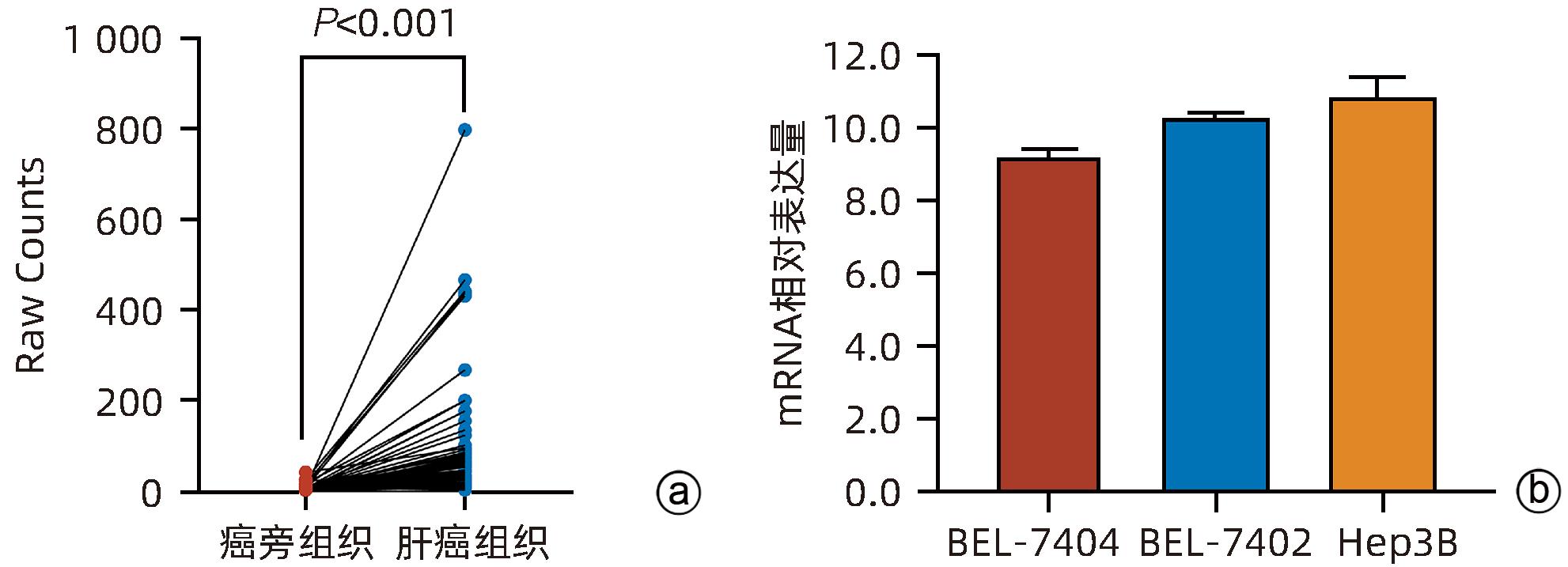
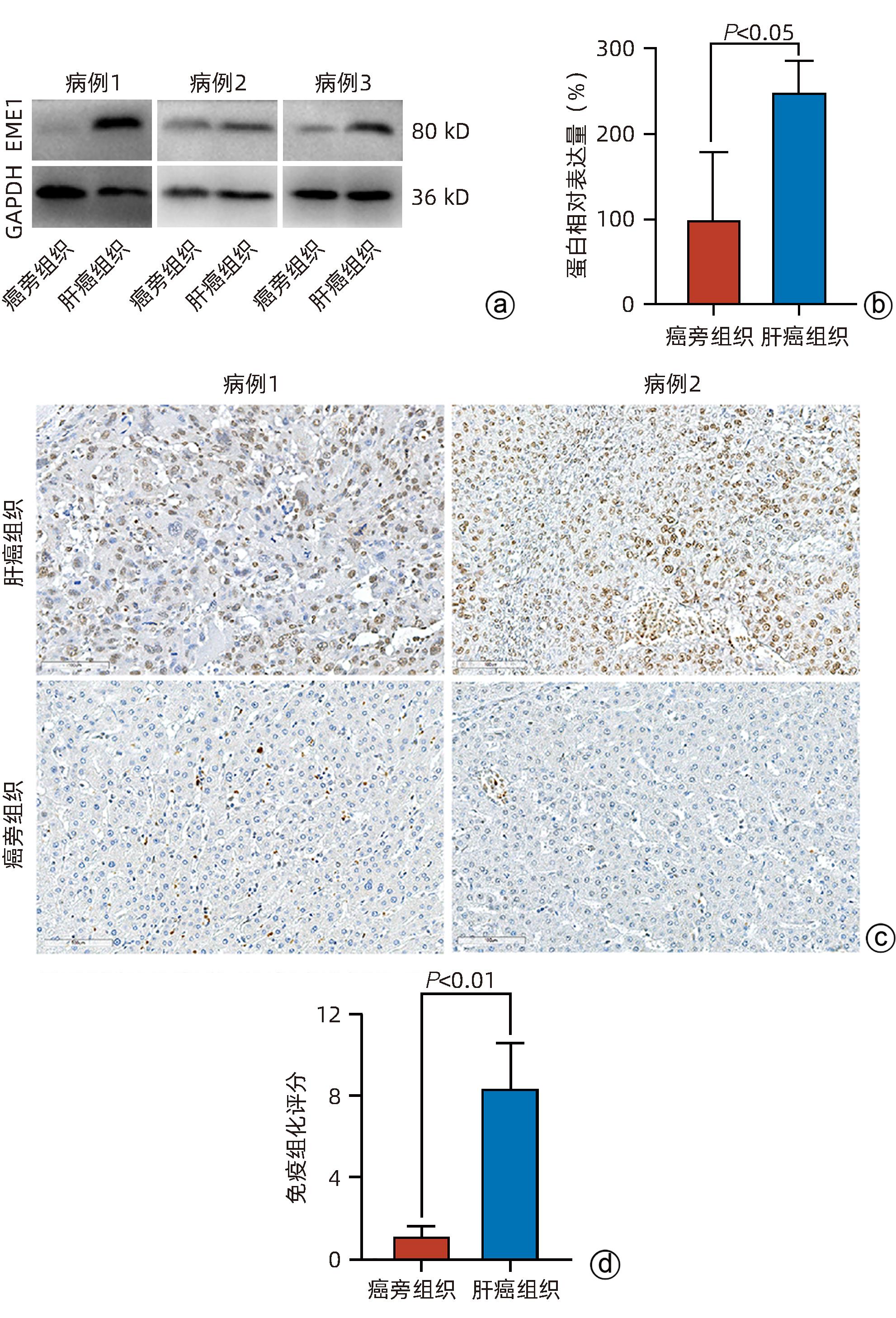
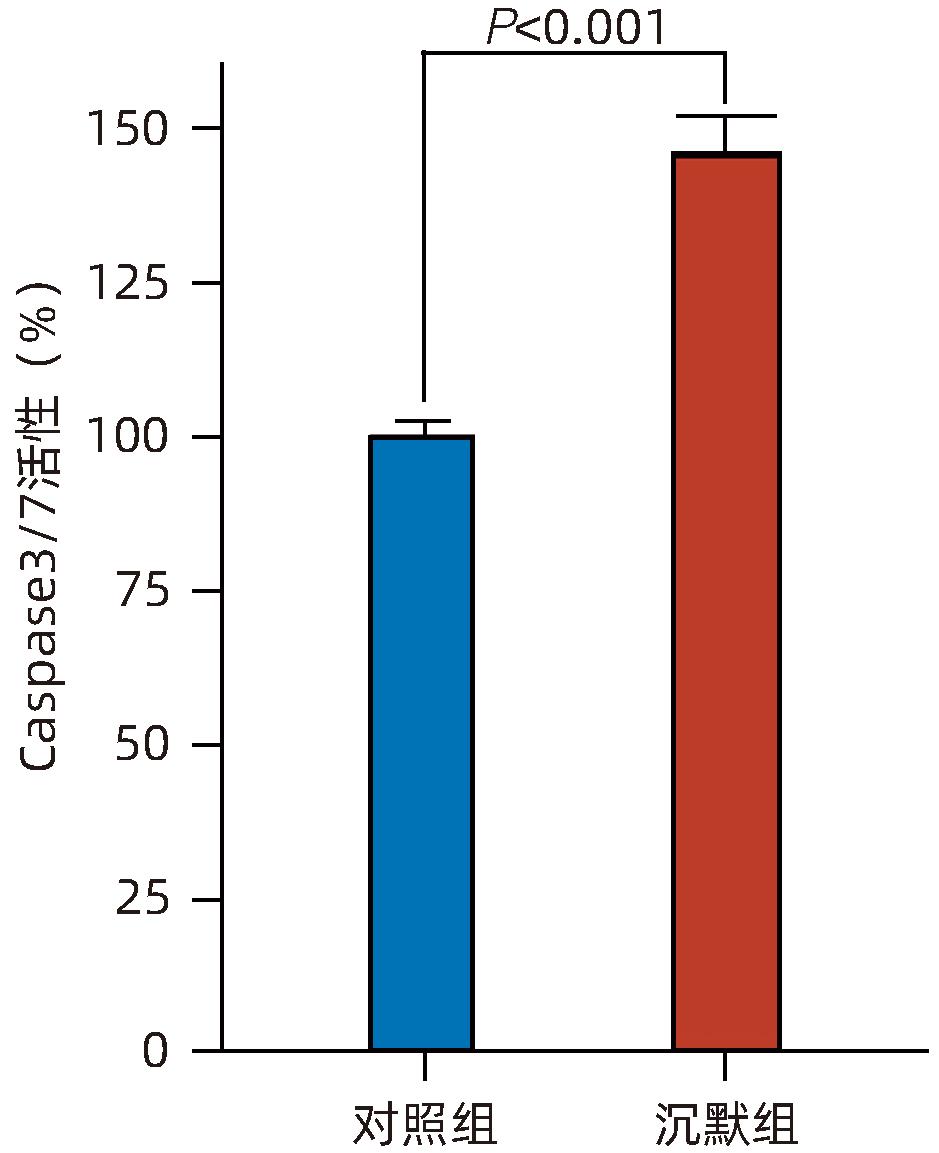
  •   Objective  To investigate the expression of essential meiotic endonuclease 1 (EME1) in liver cancer tissue and its effect on the biological behavior of hepatoma cells.  Methods  The TCGA database was used to identify the differentially expressed genes between liver cancer tissue and paracancerous tissue. Immunohistochemistry and Western Blot were used to measure the expression abundance of EME1 in liver cancer tissue. A lentivirus was constructed by short hairpin RNA, and BEL-7404 cells were transfected with the lentivirus to interfere with the expression of the EME1 gene; the cells were divided into silencing group (shEME1 group) and control group (shCtrl group). Quantitative real-time PCR and Western Blot were used to measure the mRNA and protein expression levels of EME1; Celigo Image Cytometer and MTT assay were used to measure cell proliferation rate; flow cytometry was used to observe cell cycle; Caspase 3/7 activity was used to measure cell apoptosis. The independent-samples t-test was used for comparison between two groups.  Results  TCGA results showed that the mRNA expression level of EME1 in liver cancer tissue was 18.9 times that in paracancerous tissue (t=5.00, P<0.001), and the protein expression level of EME1 in liver cancer tissue was 7.0 times (based on immunohistochemistry: 8.4±2.6 vs 1.2±0.4, t=7.55, P<0.001) or 2.5 times (based on Western Blot: 249.0%±35.5% vs 100.0%±77.8%, t=3.02, P<0.05) that in paracancerous tissue. After lentivirus infection, compared with the shCtrl group, the shEME1 group had an mRNA expression level of EME1 reduced by 29.9% (29.9%±0.9% vs 100.0%±3.6%, t=32.82, P<0.001), a protein expression level of EME1 reduced by 35.7% (35.7%±14.9% vs 100.0%±28.9%, t=3.42, P<0.05), and a level of cell counting reduced by 45.1% (4 053±167 vs 8 988±477, t=16.91, P<0.001), as well as a level of cell activity reduced to 66.9% (0.518±0.046 vs 0.774±0.022, t=8.74, P<0.001) and a level of colony forming ability reduced to 29.0% (75±6 vs 260±9, t=28.92, P<0.001). Compared with the shCtrl group, the shEME1 group had a significant increase in the proportion of cells in G1 phase (49.9% vs 44.0%, t=8.96, P<0.001) and significant reductions in the proportion of cells in G2/M phase (15.9% vs 17.9%, t=9.13, P<0.001) and S phase (34.2% vs 38.1%, t=6.91, P<0.001), while Caspase 3/7 activity was enhanced by 1.5 times (145.8%±5.9% vs 100.0%±2.3%, t=12.50, P<0.001).  Conclusion  EME1 is highly expressed in liver cancer tissue, and silencing the EME1 gene can inhibit the proliferation of hepatoma cells and promote cell apoptosis.


  • 目前,代谢相关脂肪性肝病(metabolism-associated fatty liver disease,MAFLD)已成为我国慢性肝病的第一大原因,患病率高达30%,且预计仍会进一步升高1。MAFLD与代谢性疾病发病密切相关,如肥胖、高血压、2型糖尿病以及心血管疾病等2

    脂质代谢异常是MAFLD的关键问题之一3,肝脏的脂肪酸大多来自脂肪中甘油三酯分解,通过血液循环至肝脏吸收4。胆汁酸由胆固醇代谢产生,可以通过激活肝脏内的法尼醇X受体(farnesoid X receptor,FXR),调节脂肪酸合成、氧化和运输等过程,从而影响脂肪代谢5。笔者预实验通过质谱分析发现猪去氧胆酸(hyodeoxycholic acid,HDCA)在MAFLD患者的血液和粪便样本中含量明显高于健康人,但HDCA在MAFLD发病中的作用及机制尚不清楚。本课题组采用棕榈酸(palmitic acid, PA)诱导L02细胞构建脂肪变性肝细胞模型,分析HDCA对脂肪变性肝细胞活性的影响;并构建FXR低表达肝细胞株,研究HDCA是否通过FXR-PI3K/AKT途径对脂肪变性肝细胞的活性发挥作用,从而探讨HDCA在MAFLD发生发展中的作用及其机制,为MAFLD的预防和治疗提供新途径。

    L02细胞(HL7702)购自上海佰晔生物科技公司。DMEM高糖完全培养基包含10%胎牛血清(FBS)和1%青链霉素混合液(×100),L02细胞接种于25 cm2底面积的培养瓶中,置于37 ℃、5% CO2的细胞培养箱内培养。0.2 mmol/L的PA诱导L02细胞脂肪变性。

    取25.642 mg PA加入至1 mL无水乙醇中水浴加热,配得浓度为100 mmol/L的PA溶液①。另取50 mg无脂肪酸牛血清白蛋白(BSA)加入至10 mL 1640培养基中并混匀,配得BSA浓度为0.5%。PA溶液①取0.2 mL加入至9.7 mL的BSA与1640培养基的混合液中,配得浓度为2 mmol/L的PA溶液②。然后将PA溶液②置于55 ℃水浴锅中水浴30 min。细菌滤过器过滤除菌两次。过滤后的PA溶液②与完全培养基按照体积比1∶9混合稀释10倍配置成0.2 mmol/L的PA混合液。剩余的PA溶液②按照每次实验预估用量分装,-20 ℃冰箱储存。

    20 mg的HDCA溶于100 μL的二甲基亚砜(DMSO),震荡混匀直至完全溶解,配置成浓度5×10⁶ μmol/L的HDCA溶液①。HDCA溶液①取8 μL,加入992 μL含10% FBS的DMEM高糖完全培养基,配置成浓度为4 000 μmol/L的HDCA溶液②。HDCA溶液②与完全培养基按照体积比1∶9稀释10倍配置成浓度400 μmol/L的HDCA溶液③。100、200、300 μmol/L的HDCA溶液从HDCA溶液③逐步稀释。400 μmol/L的HDCA溶液中DMSO的含量为0.08%,低于DMSO对细胞安全界限0.1%,故本实验不设立DMSO对照组。剩余的HDCA溶液③,标记时间和药品名称,密封放入-20 ℃冰箱冻存,避免反复冻融。

    用CCK8试剂测定细胞活性,每100 μL DMEM完全培养基加入10 μL CCK8试剂混匀。吸弃原液,每孔加入100 μL的混合液,将96孔板置于37 ℃细胞培养箱中孵育0.5~2 h,酶标仪测量450/620 nm处的光密度(OD)值。细胞活性计算公式如下:细胞活性(%)=[OD(加药组)-OD(空白组)]/[OD(正常组)-OD空白组)]×100%。

    将L02细胞接种于6孔板,PA诱导24 h后,HDCA处理24 h。通过qRT-PCR检测L02细胞中FXR、增殖细胞核抗原(PCNA)、周期蛋白D1(Cyclin D1)、磷脂酰肌醇-3-激酶(PI3K)和蛋白激酶B(AKT)的mRNA表达。引物(表1)大部分源于Primer Bank6,部分引物参考的编码序列设计来源于NCBI基因(存在多个转录本时,可对各转录本的保守区域进行引物设计),并以GAPDH作为内参基因。所有引物均在Primer-Blast中验证了其特异性。合成由长沙鼎国生物科技有限公司完成。

    表  1  qRT-PCR引物序列
    Table  1.  qRT-PCR primer sequences
    引物 序列(5'-3')
    FXR 上游:AACCATACTCGCAATACAGCAA
    下游:ACAGCTCATCCCCTTTGATCC
    PCNA 上游:CCTGCTGGGATATTAGCTCCA
    下游:CAGCGGTAGGTGTCGAAGC
    Cyclin D1 上游:GCTGCGAAGTGGAAACCATC
    下游:CCTCCTTCTGCACACATTTGAA
    PI3K 上游:TATTTGGACTTTGCGACAAGACT
    下游:TCGAACGTACTGGTCTGGATAG
    AKT-F 上游:AGCGACGTGGCTATTGTGAAG
    下游:GCCATCATTCTTGAGGAGGAAGT
    下载: 导出CSV 
    | 显示表格

    处理后的L02脂肪变性肝细胞用含有蛋白酶抑制剂复合物的冷RIPA缓冲液裂解。收集上清液,12 000×g离心15 min,使用BCA检测试剂盒测定总蛋白浓度。主要使用的抗体包括FXR(1∶1 500)、PCNA(1∶2 000)、Cyclin D1(1∶2 000)、PI3K(1∶2 000)、p-PI3K(1∶1 500)、AKT(1∶2 000)、p-AKT(1∶1 500)和GAPDH(1∶2 000)。条带采用Image J软件进行密度分析。

    FXR siRNA依据其基因序列(序列来源参考NCBI GeneBank数据库7)在广州市锐博生物科技有限公司设计并合成3条特异性干扰链。细胞密度达到50%,进行转染。配置混合转染试剂:5 μL Lipofectamine 3000转染试剂+5 μL FXR siRNA混匀,室温静置30 min。混合的转染试剂加入L02细胞,转染时间24~48 h。弃去原有的培养液,PBS洗涤并更换新的不含双抗的培养基。提取6孔板细胞的总RNA,qRT-PCR方法检测3条FXR siRNA干扰链作用下,FXR mRNA的表达情况并分析表达差异。选择FXR mRNA降低最明显的一条作为实验干扰组。

    所有实验数据均使用仙桃学术进行统计分析。计量资料以x¯±s表示,服从正态分布且方差齐时多组间比较采用单因素方差分析,进一步两两比较采用Tukey HSD检验;服从正态分布但方差不齐时采用Welch方差分析,进一步两两比较采用Games-Howell检验。两组间比较采用成组t检验。P<0.05为差异有统计学意义。

    采用甘油三酯含量测定和油红O染色方法观察肝细胞脂肪变性模型是否构建成功。结果显示油红O染色后,显微镜下观察到L02细胞组细胞质中没有大的红色脂滴,在细胞膜上存在小而均匀的红色脂滴;PA诱导的L02细胞组中细胞质内存在明显的大而深红色的脂滴(图1);与0 h比较,L02细胞在PA诱导后不同时间段甘油三酯水平有不同程度的升高(图2),表明模型构建稳定,将构建成功的脂肪变性肝细胞定义为M组进行后续研究。CCK8实验检测显示,PA诱导L02细胞24 h后细胞活性明显下降(1.000±0.084 vs 0.668±0.022,t=-18.663,P<0.001)。

    注: a,PA诱导前油红O染色;b,PA诱导后24 h油红O染色。
    图  1  油红O染色结果(×20)
    Figure  1.  Results of the oil red O staining
    图  2  PA诱导L02细胞后甘油三酯水平随时间的变化
    Figure  2.  Changes in triglyceride levels over time after PA induction in L02 cells

    为了研究HDCA对脂肪变性肝细胞的影响,将不同浓度的HDCA加入L02细胞和脂肪变性肝细胞。CCK8实验检测结果显示,与L02细胞组相比,L02+100 μmol/L HDCA组和L02+200 μmol/L HDCA组中L02细胞活性没有改变,L02+300 μmol/L HDCA组和L02+400 μmol/L HDCA组中L02细胞活性明显下降(P值均<0.001)(图3a)。与M组相比,M+100 μmol/L HDCA组和M+200 μmol/L HDCA组细胞活性无明显改变,M+300 μmol/L HDCA组和M+400 μmol/L HDCA组细胞活性均显著降低(P值均<0.001)(图3b)。以上实验证明300 μmol/L的HDCA抑制了L02细胞和脂肪变性肝细胞活性。为了观察随着300 μmol/L HDCA处理时间的延长,脂肪变性肝细胞的活性是否发生改变,通过CCK8检测结果显示,M+300 μmol/L HDCA组细胞活性随时间的变化而发生改变(P值均<0.001)(图3c)。后续实验HDCA浓度均采用300 μmol/L。

    注: a,不同浓度HDCA对L02细胞活性的影响;b,不同浓度HDCA对PA诱导的脂肪变性肝细胞活性的影响;c,HDCA(300 μmol/L)不同时间对PA诱导的脂肪变性肝细胞活性的影响。
    图  3  HDCA对正常肝细胞及脂肪变性肝细胞活性的影响
    Figure  3.  Effect of HDCA on the activity of normal hepatocytes and steatosis hepatocytes

    为了进一步研究HDCA(300 μmol/L)抑制脂肪变性肝细胞活性的机制,通过Western Blot检测发现,M+HDCA组中FXR蛋白的表达高于M组(t=4.492,P<0.05)(图4)。qRT-PCR检测M组和M+HDCA组的FXR、PCNA、Cyclin D1以及PI3K/AKT通路关键分子mRNA的表达变化,结果显示,相比于M组,M+HDCA组中FXR mRNA表达升高,PCNA、Cyclin D1、PI3K和AKT mRNA表达均明显下降(P值均<0.05)(表2)。

    注: a,FXR蛋白印迹图;b,FXR蛋白相对表达量。
    图  4  HDCA对脂肪变性肝细胞FXR表达的影响
    Figure  4.  Effect of HDCA on the expression of FXR in steatotic hepatocytes
    表  2  HDCA对脂肪变性肝细胞FXR、PI3K、AKT、PCNA和Cyclin D1 mRNA表达的影响
    Table  2.  Effect of HDCA on the mRNA expression of FXR, PI3K, AKT, PCNA, and Cyclin D1 in steatotic hepatocytes
    指标 M组 M+HDCA组 t P
    FXR mRNA 0.485±0.162 1.010±0.013 5.576 0.005
    PCNA mRNA 1.710±0.052 1.034±0.054 -15.679 <0.001
    Cyclin D1 mRNA 1.435±0.124 1.041±0.053 -5.083 0.007
    PI3K mRNA 1.951±0.500 1.008±0.009 -3.266 0.031
    AKT mRNA 2.373±0.316 1.030±0.050 -7.266 0.002
    下载: 导出CSV 
    | 显示表格

    为了验证HDCA(300 μmol/L)是否通过FXR抑制脂肪变性肝细胞活性,选择干扰FXR受体,在3条FXR siRNA(FXR siRNA 1、2、3)中筛选出干扰效果最强的干扰链。结果如图5a所示,相比于M组,M+FXR siRNA 3的干扰效果最好(P<0.01)。qRT-PCR检测FXR siRNA 3干扰链对HDCA刺激脂肪变性肝细胞FXR mRNA表达的干扰效果,结果显示,相比于M+HDCA组,M+FXR siRNA 3+HDCA组的FXR mRNA表达水平明显降低(P<0.05)(图5b)。

    注: a,3条FXR siRNA干扰链对脂肪变性肝细胞中FXR mRNA表达的干扰效果;b,FXR siRNA 3对HDCA刺激的干扰效果。
    图  5  FXR siRNA干扰效果及HDCA对脂肪变性肝细胞FXR表达的影响
    Figure  5.  Effect of FXR siRNA interference and the effect of HDCA on FXR expression in lipid-denatured hepatocytes

    应用Western Blot检测FXR、PCNA、Cyclin D1、PI3K、p-PI3K、AKT和p-AKT的蛋白表达水平。结果显示,相比于M+HDCA组,M+FXR siRNA 3+HDCA组中FXR蛋白表达下降,PCNA、PI3K、p-PI3K、AKT和p-AKT蛋白表达增加,差异均有统计学意义(P值均<0.05)(图6)。

    注: a,FXR、PCNA、Cyclin D1、PI3K、p-PI3K、AKT和p-AKT的蛋白印迹图;b,FXR蛋白的相对表达水平;c,PCNA蛋白的相对表达水平;d,Cyclin D1蛋白的相对表达水平;e,PI3K蛋白的相对表达水平;f,p-PI3K蛋白的相对表达水平;g,AKT蛋白的相对表达水平;h,p-AKT蛋白的相对表达水平。
    图  6  抑制FXR表达后脂肪变性肝细胞FXR-PI3K/AKT通路关键分子及PCNA和Cyclin D1蛋白表达的变化
    Figure  6.  Changes in the expression of key molecules of FXR-PI3K/AKT pathway and PCNA and Cyclin D1 proteins in steatosis hepatocytes after inhibition of FXR expression

    MAFLD发展包括单纯脂肪肝(以往称非酒精性脂肪肝)、脂肪性肝炎(以往称非酒精性脂肪性肝炎)、脂肪性肝纤维化、肝硬化及肝癌等阶段8。MAFLD一旦进入脂肪性肝炎阶段,肝细胞脂毒性损伤以及炎症因子的大量浸润,会加快肝纤维化、肝硬化和肝癌的进展速度9-10,因此脂肪性肝炎阶段早期防治尤为重要。

    胆汁酸是由肝脏合成的双性分子。在肝细胞胞质和微粒体中,胆固醇通过经典和替代两种途径生成初级胆汁酸11。初级胆汁酸分泌进入肠道后,在肠内细菌的作用下,转化为次级胆汁酸。HDCA是一种次级胆汁酸,存在于猪、鼠和人的胆汁中,在猪和鼠的胆汁中含量较高12,其对动物机体的脂质代谢有着重要的调控作用13。Sehayek等14发现在野生型C57BL/6小鼠中,饲喂HDCA可以减少饮食中胆固醇从肠道吸收,对治疗小鼠血浆胆固醇升高和动脉粥样硬化有较好的效果。Shih等15研究显示,饲粮中添加1.25%的HDCA可通过改善高密度脂蛋白功能来抑制低密度脂蛋白受体基因敲除小鼠动脉粥样硬化的形成。目前,HDCA对MAFLD发生发展的影响未见文献报道。本研究发现浓度低于200 μmol/L的HDCA对正常肝细胞及脂肪变性肝细胞活性无明显影响,300 μmol/L及以上浓度的HDCA处理的L02肝细胞和L02脂肪变性肝细胞活性明显下降,同时PCNA、Cyclin D1的mRNA表达降低,表明较高浓度的HDCA对脂肪变性肝细胞增殖活性具有抑制作用。

    FXR由Forman等16于1995年在大鼠中发现。FXR是胆汁酸代谢的重要受体,通过调节胆汁酸在肝脏和肠道的合成、结合、摄取和转运以维持胆汁酸的动态平衡。疏水性胆汁酸鹅去氧胆酸对FXR的激活作用最强17,其次是脱氧胆酸和石胆酸,而亲水性胆汁酸熊去氧胆酸是最弱的FXR激动剂18。GW4064、奥贝胆酸和Fexaramine等人工合成的FXR激动剂对FXR的激活作用更强19-20。牛磺-β-鼠胆酸和甘氨熊脱氧胆酸是FXR的拮抗剂21-22。本研究发现300 μmol/L的HDCA处理24 h后,脂肪变性肝细胞FXR蛋白表达明显增加,提示HDCA对FXR具有激活作用,能够上调肝细胞FXR的表达。

    FXR在各种代谢过程中发挥调节作用,包括脂肪代谢、胆汁酸平衡、葡萄糖平衡和细胞活性23。在肝脏中,FXR通过激活FXR信号通路,促进脂肪酸的β氧化和胆汁酸的合成,减少脂肪酸的合成和蓄积,改善肝脂肪代谢异常24-25。FXR还与肝细胞的生长、凋亡以及炎症等生物学过程密切相关26-27。此外,FXR通过调节肠道菌群的组成和代谢产物的生成,参与肥胖、糖尿病等代谢性疾病的发生和发展28-29。FXR发挥各种代谢调节作用与PI3K/AKT信号通路有关。Xu等30研究发现激活FXR活性,能够抑制PI3K/AKT信号通路的活化,降低脂肪分解,增加脂肪生成,进而增加鱼体内的脂质积累。本研究结果显示,干扰FXR表达后,脂肪变性肝细胞PI3K/AKT信号通路关键分子,如PI3K、p-PI3K、AKT和p-AKT的蛋白表达明显增加,提示FXR对PI3K/AKT信号通路关键分子表达具有抑制作用;同时还发现,干扰脂肪变性肝细胞FXR表达后,反映细胞增殖活性的PCNA蛋白表达增加,提示FXR对脂肪变性肝细胞增殖具有抑制作用。以上结果表明HDCA通过上调FXR表达抑制PI3K/AKT信号通路,从而造成脂肪变性肝细胞活性下降。

  • [1]
    SUNG H, FERLAY J, SIEGEL RL, et al. Global cancer statistics 2020: GLOBOCAN estimates of incidence and mortality worldwide for 36 cancers in 185 countries[J]. CA Cancer J Clin, 2021, 71( 3): 209- 249. DOI: 10.3322/caac.21660.
    [2]
    LLOVET JM, KELLEY RK, VILLANUEVA A, et al. Hepatocellular carcinoma[J]. Nat Rev Dis Primers, 2021, 7: 6. DOI: 10.1038/s41572-020-00240-3.
    [3]
    CHEN SH, WANG CY, GUO C, et al. Establishment of a nomogram model for predicting the survival of hepatitis B virus-related hepatocellular carcinoma[J]. J Clin Hepatol, 2022, 38( 7): 1566- 1571. DOI: 10.3969/j.issn.1001-5256.2022.07.020.

    陈松海, 王春艳, 郭畅, 等. 预测HBV相关肝细胞癌生存的列线图模型的建立[J]. 临床肝胆病杂志, 2022, 38( 7): 1566- 1571. DOI: 10.3969/j.issn.1001-5256.2022.07.020.
    [4]
    WANG JJ, WANG KX, CHEN C, et al. Survival analysis and development of a prognostic nomogram for patients with hepatitis B virus-associated hepatocellular carcinoma[J]. Heliyon, 2023, 9( 10): e20850. DOI: 10.1016/j.heliyon.2023.e20850.
    [5]
    JI D, CHEN Y, BI JF, et al. Entecavir plus Biejia-Ruangan compound reduces the risk of hepatocellular carcinoma in Chinese patients with chronic hepatitis B[J]. J Hepatol, 2022, 77( 6): 1515- 1524. DOI: 10.1016/j.jhep.2022.07.018.
    [6]
    GWON GH, JO A, BAEK K, et al. Crystal structures of the structure-selective nuclease Mus81-Eme1 bound to flap DNA substrates[J]. EMBO J, 2014, 33( 9): 1061- 1072. DOI: 10.1002/embj.201487820.
    [7]
    HAN YY, ZHENG QY, TIAN Y, et al. Identification of a nine-gene panel as a prognostic indicator for recurrence with muscle-invasive bladder cancer[J]. J Surg Oncol, 2019, 119( 8): 1145- 1154. DOI: 10.1002/jso.25446.
    [8]
    VÉQUAUD E, DESPLANQUES G, JÉZÉQUEL P, et al. Survivin contributes to DNA repair by homologous recombination in breast cancer cells[J]. Breast Cancer Res Treat, 2016, 155( 1): 53- 63. DOI: 10.1007/s10549-015-3657-z.
    [9]
    PENG L, LIANG J, WANG Q, et al. A DNA damage repair gene signature associated with immunotherapy response and clinical prognosis in clear cell renal cell carcinoma[J]. Front Genet, 2022, 13: 798846. DOI: 10.3389/fgene.2022.798846.
    [10]
    GUO ZG, LIANG EB, LI W, et al. Essential meiotic structure-specific endonuclease1(EME1) promotes malignant features in gastric cancer cells via the Akt/GSK3B/CCND1 pathway[J]. Bioengineered, 2021, 12( 2): 9869- 9884. DOI: 10.1080/21655979.2021.1999371.
    [11]
    WANG YX, HUANG XL, SU ZH, et al. The Glu69Asp polymorphism of EME1 gene is associated with an increased risk of hepatocellular carcinoma in Guangxi population, China[J]. Int J Gen Med, 2022, 15: 7855- 7866. DOI: 10.2147/IJGM.S383261.
    [12]
    ZHAO JW, LIU L, ZHANG AQ, et al. Effect of EME1 exon variant Ile350Thr on risk and early onset of breast cancer in southern Chinese women[J]. J Biomed Res, 2013, 27( 3): 193- 201. DOI: 10.7555/JBR.27.20130013.
    [13]
    SUZUKI T, YASUI M, HONMA M. Mutator phenotype and DNA double-strand break repair in BLM helicase-deficient human cells[J]. Mol Cell Biol, 2016, 36( 23): 2877- 2889. DOI: 10.1128/MCB.00443-16.
    [14]
    SHAMMAS MA, SHMOOKLER REIS RJ, KOLEY H, et al. Dysfunctional homologous recombination mediates genomic instability and progression in myeloma[J]. Blood, 2009, 113( 10): 2290- 2297. DOI: 10.1182/blood-2007-05-089193.
    [15]
    FULDA S. Tumor resistance to apoptosis[J]. Int J Cancer, 2009, 124( 3): 511- 515. DOI: 10.1002/ijc.24064.
    [16]
    SZAKAL B, BRANZEI D. Premature Cdk1/Cdc5/Mus81 pathway activation induces aberrant replication and deleterious crossover[J]. EMBO J, 2013, 32( 8): 1155- 1167. DOI: 10.1038/emboj.2013.67.
    [17]
    PALMA A, PUGLIESE GM, MURFUNI I, et al. Phosphorylation by CK2 regulates MUS81/EME1 in mitosis and after replication stress[J]. Nucleic Acids Res, 2018, 46( 10): 5109- 5124. DOI: 10.1093/nar/gky280.
    [18]
    GUO YG, ZHU ZH, HUANG ZY, et al. CK2-induced cooperation of HHEX with the YAP-TEAD4 complex promotes colorectal tumorigenesis[J]. Nat Commun, 2022, 13( 1): 4995. DOI: 10.1038/s41467-022-32674-6.
    [19]
    KWON J, ZHANG JM, MOK B, et al. CK2-mediated phosphorylation upregulates the stability of USP13 and promotes ovarian cancer cell proliferation[J]. Cancers, 2022, 15( 1): 200. DOI: 10.3390/cancers15010200.
    [20]
    PITRODA SP, PASHTAN IM, LOGAN HL, et al. DNA repair pathway gene expression score correlates with repair proficiency and tumor sensitivity to chemotherapy[J]. Sci Transl Med, 2014, 6( 229): 229ra42. DOI: 10.1126/scitranslmed.3008291.
    [21]
    XU YH, LI J, ZHU KK, et al. FIBP interacts with transcription factor STAT3 to induce EME1 expression and drive radioresistance in lung adenocarcinoma[J]. Int J Biol Sci, 2023, 19( 12): 3816- 3829. DOI: 10.7150/ijbs.83134.
    [22]
    WEINANDY A, PIROTH MD, GOSWAMI A, et al. Cetuximab induces eme1-mediated DNA repair: A novel mechanism for cetuximab resistance[J]. Neoplasia, 2014, 16( 3): 207- 220, 220.e1- 220. e4. DOI: 10.1016/j.neo.2014.03.004.
  • Relative Articles

    [1]Zhiru WANG, Wenjing ZHAO, Xi CHEN. Effect and mechanism of gallic acid on the proliferation, migration, invasion, and apoptosis of human hepatocellular carcinoma HepG2 cells[J]. Journal of Clinical Hepatology, 2025, 41(3): 493-498. doi: 10.12449/JCH250315
    [2]Xieer LIANG, Zhihong LIU, Jinlin HOU. New drugs for the functional cure of hepatitis B: Focusing on antisense oligonucleotides and small interfering RNAs[J]. Journal of Clinical Hepatology, 2025, 41(1): 7-14. doi: 10.12449/JCH250102
    [3]Teng LIU, Xiujuan CHANG, Quanwei HE, Ran XU, Yongping YANG. Efficacy and safety of cryoablation combined with lenvatinib and anti-PD-1 monoclonal antibody in treatment of unresectable hepatocellular carcinoma[J]. Journal of Clinical Hepatology, 2024, 40(3): 539-549. doi: 10.12449/JCH240317
    [4]Xiaomeng YAO, Keke SUN, Yunkai LIN, Hui WANG, Liwei DONG, Lei CHEN, Heping HU. Molecular mechanism of lenvatinib resistance in hepatocellular carcinoma[J]. Journal of Clinical Hepatology, 2024, 40(12): 2524-2530. doi: 10.12449/JCH241225
    [5]Junzhe CAI, Songbai LIU, Xiaobin FEI, Peng LIU, Changhao ZHU, Xing WANG, Yaozhen PAN. Effect of ankyrin-repeat domain-containing protein 22 on human hepatoma cells and its mechanism[J]. Journal of Clinical Hepatology, 2024, 40(5): 989-996. doi: 10.12449/JCH240519
    [6]Jiannan QIU, Peng WANG, Yin CAO, Zhongxia WANG, Junhua WU, Chunping JIANG. Effect of kinesin family member 15 on the proliferation of hepatocellular carcinoma cells and its mechanism of action[J]. Journal of Clinical Hepatology, 2024, 40(2): 327-334. doi: 10.12449/JCH240217
    [7]Yuan DUAN, Ting ZHANG, Jing ZHANG, Guiwen GUAN, Jingzhou WANG, Xiangmei CHEN. Expression of AU-rich element RNA-binding factor 1 in hepatocellular carcinoma and its value in prognostic evaluation[J]. Journal of Clinical Hepatology, 2024, 40(9): 1833-1839. doi: 10.12449/JCH240918
    [8]Changqing WANG, Ling CHEN, Ping XU, Xiaojuan ZHU, Zheng LIU. Expression of miR-409-3p in hepatoma HepG2 cells and its mechanism in cell proliferation[J]. Journal of Clinical Hepatology, 2023, 39(8): 1895-1902. doi: 10.3969/j.issn.1001-5256.2023.08.019
    [9]Xiao ErHui, Ning HuiBin, Kang YueHua, Yin Hui, Ma Li, Mao ZhongShan, Zhao Yan, Shang Jia. Downregulation of miR-196b in regulating the growth and apoptosis of hepatoma cells by targeting nuclear apoptosis-inducing factor 1[J]. Journal of Clinical Hepatology, 2020, 36(10): 2230-2235. doi: 10.3969/j.issn.1001-5256.2020.10.014
    [10]ZHONG FuRui, CHENG HuanLi, ZHANG Hao, DU YiChao, HU QiHui, FU WenGuang, XIA XianMing. Effect of kaempferol on the proliferation,migration,invasion,and apoptosis of human hepatoma Bel-7402 cells[J]. Journal of Clinical Hepatology, 2020, 36(12): 2725-2729. doi: 10.3969/j.issn.1001-5256.2020.12.017
    [11]Wang ZeXin, Guan LiJun, Li JianMing, Wang ZhiChao, Xue MengRuo, Qin XiaoJun. Effect of targeted regulation of the Wnt2 gene by microRNA( microRNA-21) on the proliferation and migration of HepG2 hepatoma cells[J]. Journal of Clinical Hepatology, 2020, 36(4): 803-807. doi: 10.3969/j.issn.1001-5256.2020.04.019
    [12]Zhang Yang, Chen ShuYing, Xu GuangRu. Effect of extracellular ubiquitin on the proliferation,invasion,and migration of hepatoma cells[J]. Journal of Clinical Hepatology, 2020, 36(6): 1289-1292. doi: 10.3969/j.issn.1001-5256.2020.06.020
    [13]Lu Qian, Cheng Xin, Xia ZhenGuo, Chen Zhong. Effect of proteasome subunit beta type 4 on the proliferation and viability of human liver cancer SMMC7721 cells[J]. Journal of Clinical Hepatology, 2020, 36(3): 592-595. doi: 10.3969/j.issn.1001-5256.2020.03.024
    [14]Li YingYing, Zhan LiFen, Ceng ZheChao, Xu Shen. Effect of lobaplatin on proliferation and apoptosis of hepatocellular carcinoma HepG2 cells and related mechanism of action[J]. Journal of Clinical Hepatology, 2018, 34(4): 784-788. doi: 10.3969/j.issn.1001-5256.2018.04.018
    [15]Zhu YueKun, Zhao XianQi, Wang DaWei, Zhang Wei, Tian MaoLin, Li ZhengTian, Zhao Long, Li Chao, Jiang HongChi. Effect of cyclooxygenase-2 antisense RNA combined with celecoxib on the proliferation and apoptosis of hepatoma cells[J]. Journal of Clinical Hepatology, 2018, 34(12): 2614-2618. doi: 10.3969/j.issn.1001-5256.2018.12.021
    [16]Geng YaJun, Li ShuDong, Liu ShenZheng, Ren JianJun, Zhang Tong. Inhibitory effect of microRNA-149-5p in the proliferation and migration of HepG2 and Bel-7402 hepatoma cells[J]. Journal of Clinical Hepatology, 2017, 33(6): 1126-1130. doi: 10.3969/j.issn.1001-5256.2017.06.022
    [17]Ci DanWangJiu, Chang ZhiHui, Zhao XiangXuan, Lin Kun, Zhang Qiang, Lu ZaiMing, Wang XiaoMing. Effect of continuous irradiation with ~(125)I radioactive seeds in inhibiting HepG2 cell proliferation and related mechanisms[J]. Journal of Clinical Hepatology, 2016, 32(10): 1920-1924. doi: 10.3969/j.issn.1001-5256.2016.10.019
    [18]Sun LeiQing, Xu Jing, Chen YongQiang, Zheng Lu, Lu: XiaoTing, Liu JunQuan, Chen FuXing, Zhou ZhongHai. Influence of extracellular signal-regulated kinase 1/2 pathway inhibitor PD98059 on proliferation and killing function of γδT cells cultured in vitro[J]. Journal of Clinical Hepatology, 2016, 32(7): 1388-1391. doi: 10.3969/j.issn.1001-5256.2016.07.038
    [19]Tai BoJun, Yao Min, Gu Xing, Shi Yun, Yu DanDan, Chen Jie, Zheng WenJie, Yao DengFu, Lu ShaoLin. Effects of silencing glypican- 3 gene transcription on proliferation and apoptosis of hepatoma cells[J]. Journal of Clinical Hepatology, 2013, 29(11): 863-866. doi: 10.3969/j.issn.1001-5256.2013.10.015
    [20]Yang Yang, Yang Liu, Fan HaiNing. Effect of H2S on intracellular Ca2+ concentration in HSCs under H2O2-induced oxidative stress conditions [J]. Journal of Clinical Hepatology, 2012, 28(11): 852-855.
  • 加载中

Catalog

    通讯作者: 陈斌, bchen63@163.com
    • 1. 

      沈阳化工大学材料科学与工程学院 沈阳 110142

    1. 本站搜索
    2. 百度学术搜索
    3. 万方数据库搜索
    4. CNKI搜索

    Figures(7)  / Tables(2)

    Article Metrics

    Article views (435) PDF downloads(28) Cited by()
    Proportional views
    Related

    /

    DownLoad:  Full-Size Img  PowerPoint
    Return
    Return