中文English
ISSN 1001-5256 (Print)
ISSN 2097-3497 (Online)
CN 22-1108/R
Volume 40 Issue 5
May  2024
Turn off MathJax
Article Contents

Effect of the protein kinase RNA-like endoplasmic reticulum kinase pathway in endoplasmic reticulum stress on hepatic stellate cell activation and collagen type I expression

DOI: 10.12449/JCH240516
Research funding:

Guangxi Natural Science Foundation Project (2023GXNSFBA026056);

The People’s Hospital of Guangxi Zhuang Autonomous Region Youth Fund project (QN2020-1)

More Information
  • Corresponding author: ZHANG Guo, zhangguogx@hotmail.com (ORCID: 0000-0001-7755-443X)
  • Received Date: 2023-10-12
  • Accepted Date: 2023-11-30
  • Published Date: 2024-05-25
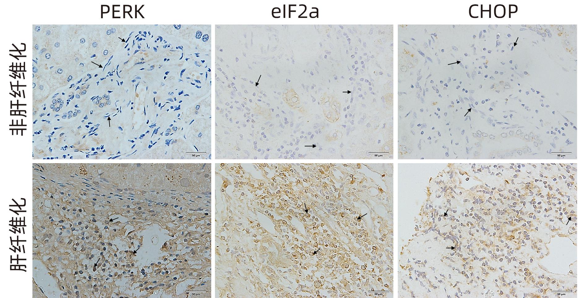
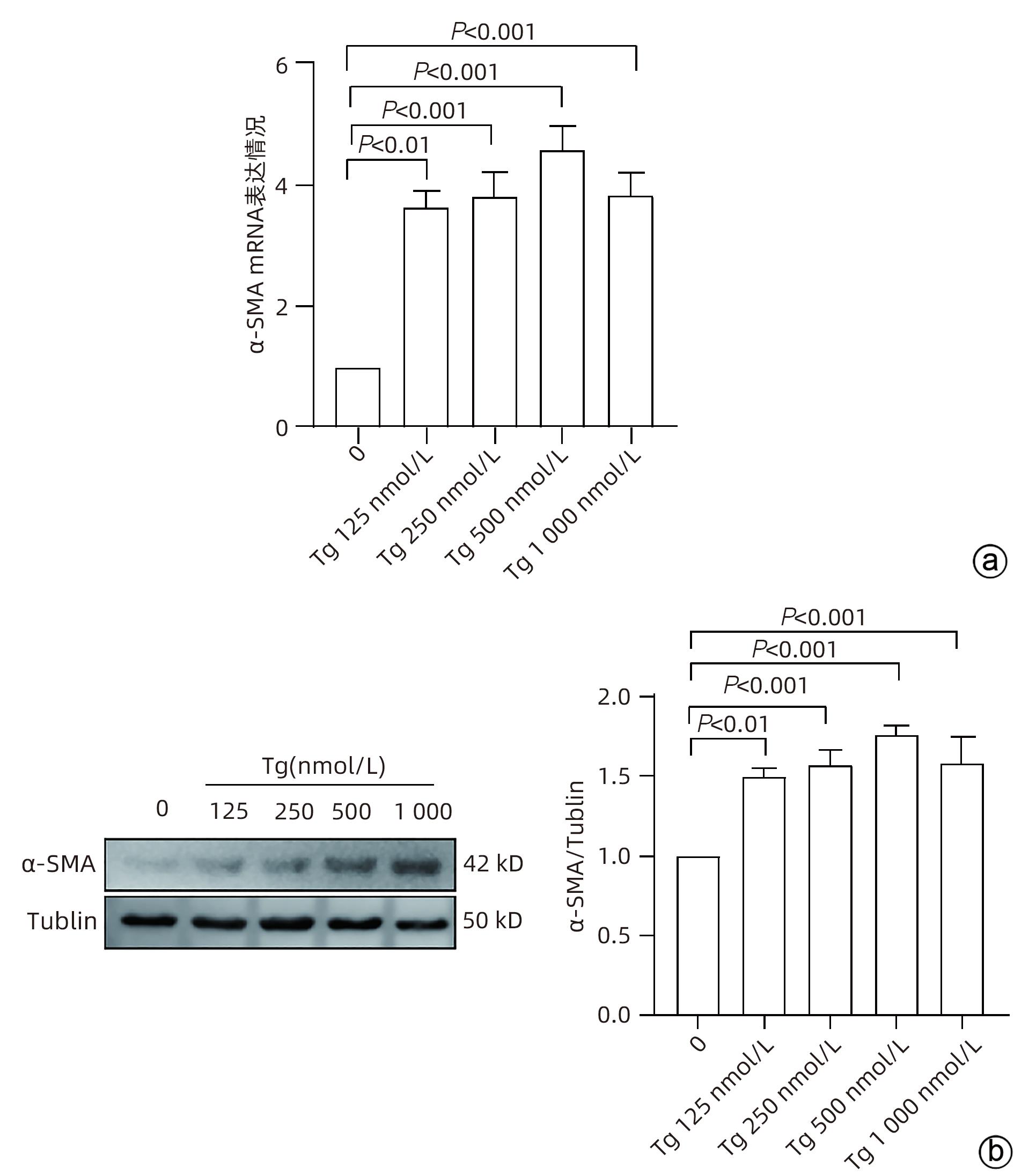
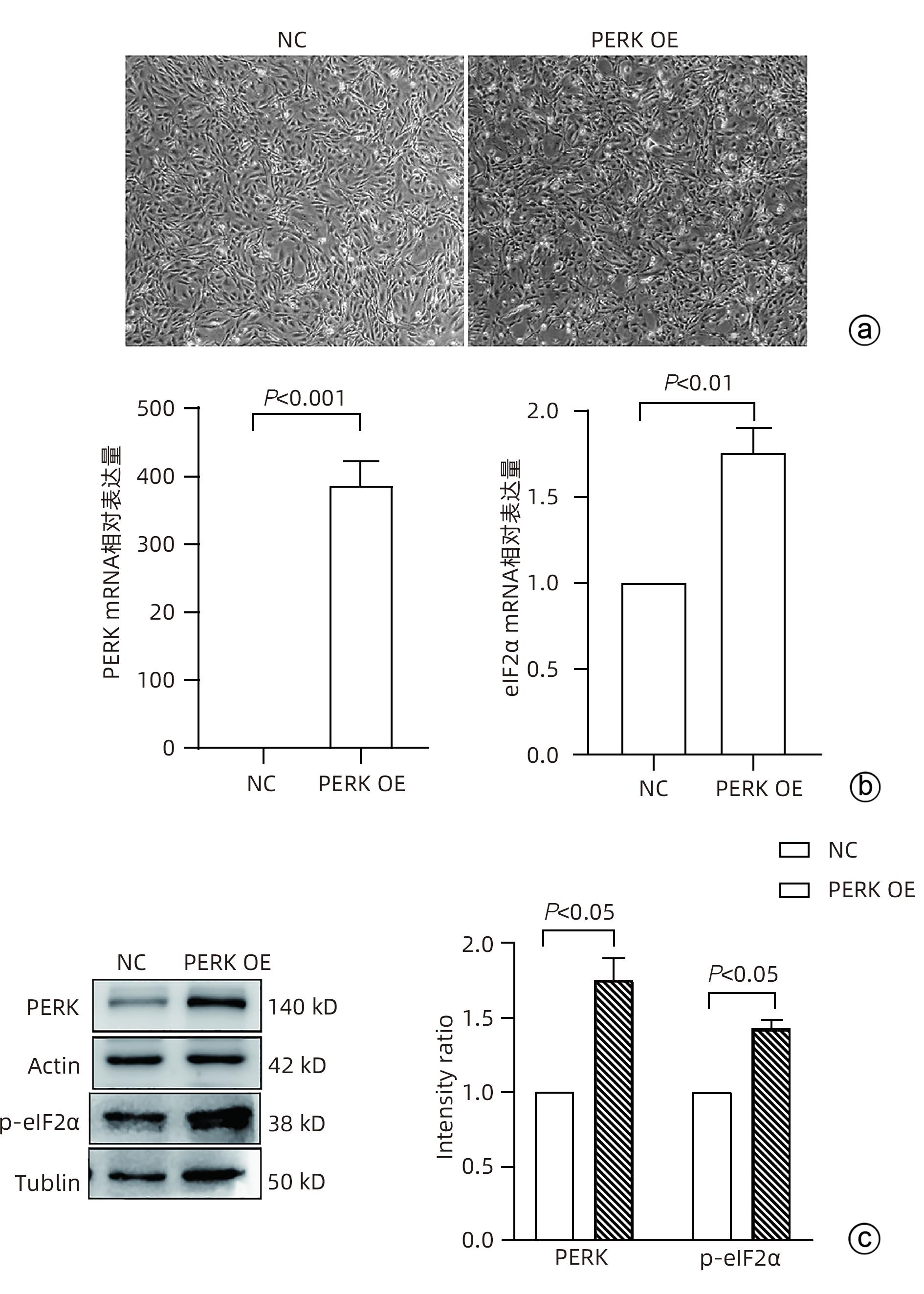
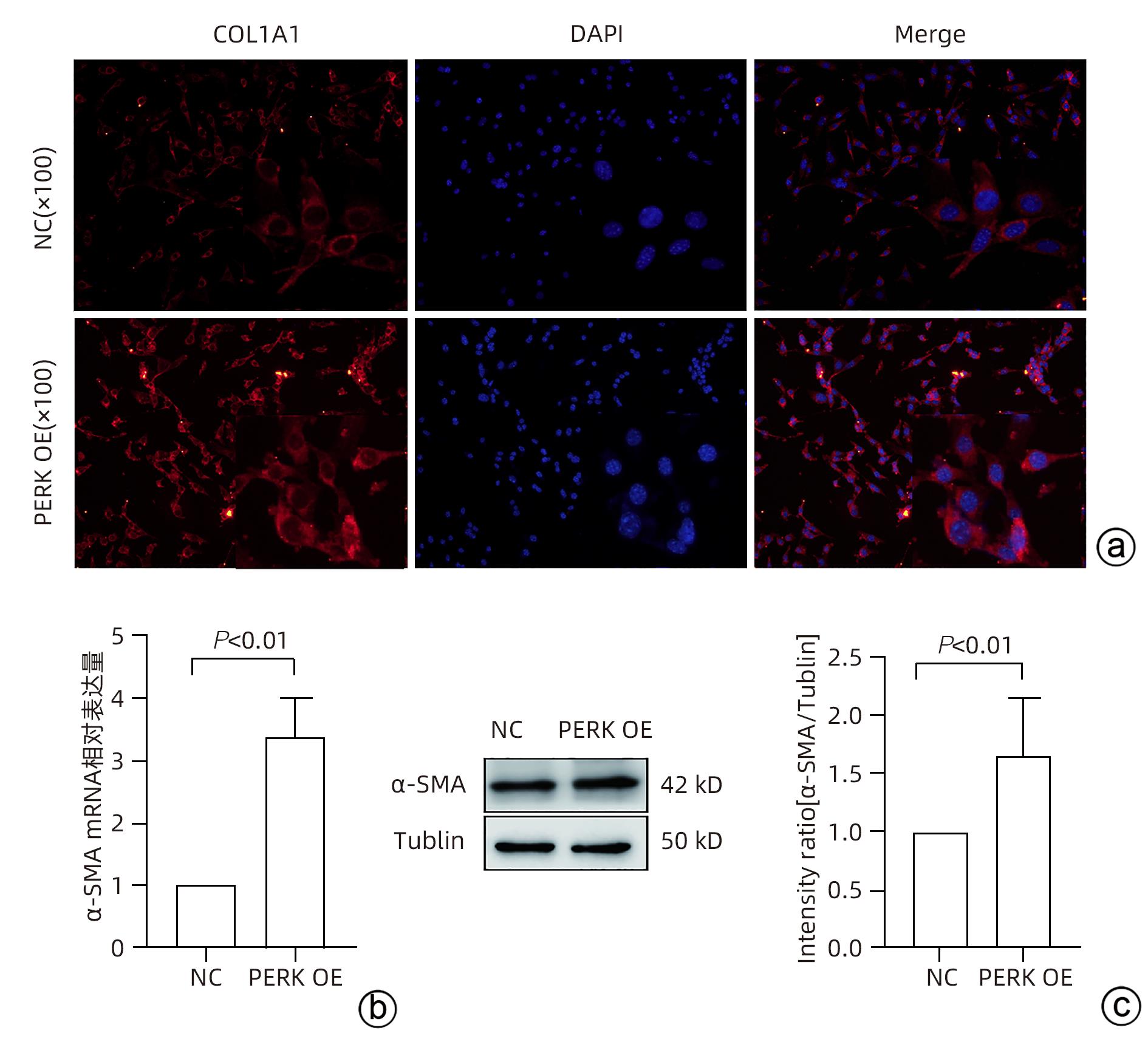
  •   Objective  To investigate the effect of the protein kinase RNA-like endoplasmic reticulum kinase (PERK)/eukaryotic initiation factor 2α (eIF2α) pathway in endoplasmic reticulum stress on the activation of hepatic stellate cells (HSC).  Methods  Pathological sections of normal liver tissue after surgery were collected from 11 patients with hepatic fibrosis (S1-S4) and 9 patients with hepatic hemangioma and hepatic adenoma confirmed by liver biopsy, and immunohistochemistry was used to measure the protein expression levels of PERK, eIF2α, and C/EBP homologous protein (CHOP). Human HSC-LX2 cells were treated with different concentrations of the endoplasmic reticulum stress inducer thapsigargin (0, 125, 250, 500, and 1 000 nmol/L), and qRT-PCR was used to measure the mRNA expression level of PERK, while Western blot was used to measure the protein expression levels of PERK, inositol requiring protein 1 (IRE1), activating transcription factor 6 (ATF6), CHOP, and α-smooth muscle actin (α-SMA). The method of lentivirus transfection was used to construct a PERK stable overexpression LX-2 group and a control group; qRT-PCR was used to measure the mRNA expression levels of PERK, eIF2α, and α-SMA, Western blot was used to measure the protein expression levels of PERK, phosphorylated eIF2α (p-eIF2α), and α-SMA, and immunofluorescence assay was used to measure the expression of collagen type I alpha 1 (COL1A1). The independent samples t-test was used for comparison of normally distributed continuous data between two groups; a one-way analysis of variance was used for comparison between multiple groups, and the least significant difference t-test was used for further comparison between two groups. The Mann-Whitney U test was used for comparison of non-normally distributed continuous data between two groups.  Results  Compared with normal liver tissue, the liver tissue of patients with hepatic fibrosis had significantly higher expression levels of PERK, eIF2α, and CHOP (Z=-3.56, t=-5.75, Z=-3.52, all P<0.001). Compared with the solvent group, the groups treated with different concentrations of thapsigargin had significant increases in the expression levels of the endoplasmic reticulum-associated proteins PERK, CHOP, IRE1, ATF6, and α-SMA (all P<0.05). Compared with the control group, the PERK stable overexpression group had significant increases in the mRNA expression levels of PERK, eIF2α, and α-SMA and the protein expression levels of PERK, p-eIF2α, and α-SMA (all P<0.05), and immunofluorescence assay showed a significant increase in the expression level of COL1A1 in the PERK stable overexpression group (P<0.05).  Conclusion  PERK overexpression can induce the expression of α-SMA and COL1A1 in LX-2 cells, suggesting that the PERK signaling pathway might be one of the important mechanisms of HSC activation.


  • loading
  • [1]
    HE YH, YAN YJ, ZHANG SF. Quantitative liver surface nodularity score based on imaging for assessment of early cirrhosis in patients with chronic liver disease: A protocol for systematic review and meta-analysis[J]. Medicine, 2021, 100( 4): e23636. DOI: 10.1097/MD.0000000000023636.
    [2]
    GBD 2017 Cirrhosis Collaborators. The global, regional, and national burden of cirrhosis by cause in 195 countries and territories, 1990-2017: A systematic analysis for the Global Burden of Disease Study 2017[J]. Lancet Gastroenterol Hepatol, 2020, 5( 3): 245- 266. DOI: 10.1016/S2468-1253(19)30349-8.
    [3]
    ZHANG J, GUO JF, YANG NN, et al. Endoplasmic reticulum stress-mediated cell death in liver injury[J]. Cell Death Dis, 2022, 13( 12): 1051. DOI: 10.1038/s41419-022-05444-x.
    [4]
    XIA SW, WANG ZM, SUN SM, et al. Endoplasmic reticulum stress and protein degradation in chronic liver disease[J]. Pharmacol Res, 2020, 161: 105218. DOI: 10.1016/j.phrs.2020.105218.
    [5]
    SHAN S, ZHAO LH, MA H, et al. Definition, etiology, and epidemiology of liver cirrhosis[J]. J Clin Hepatol, 2021, 37( 1): 14- 16. DOI: 10.3969/j.issn.1001-5256.2021.01.003.

    单姗, 赵连晖, 马红, 等. 肝硬化的定义、病因及流行病学[J]. 临床肝胆病杂志, 2021, 37( 1): 14- 16. DOI: 10.3969/j.issn.1001-5256.2021.01.003.
    [6]
    XU LY, WEI ST, DONG Y, et al. Regulatory effect of lncRNA MALAT1 on activation of hepatic stellate cell and its mechanism[J]. J Jilin Univ Med Ed, 2023, 49( 3): 697- 705. DOI: 10.13481/j.1671-587X.20230319.

    徐菱遥, 魏书堂, 董勇, 等. lncRNA MALAT1对肝星状细胞活化的调节作用及其机制[J]. 吉林大学学报(医学版), 2023, 49( 3): 697- 705. DOI: 10.13481/j.1671-587X.20230319.
    [7]
    TSUCHIDA T, FRIEDMAN SL. Mechanisms of hepatic stellate cell activation[J]. Nat Rev Gastroenterol Hepatol, 2017, 14( 7): 397- 411. DOI: 10.1038/nrgastro.2017.38.
    [8]
    SUN DL, GUO JB, WANG DD, et al. Effect of exosomes derived from mesenchymal stem cells on the proliferation and activation of hepatic stellate cells in vitro[J]. J Pract Hepatol, 2023, 26( 1): 11- 14. DOI: 10.3969/j.issn.1672-5069.2023.01.004.

    孙东磊, 郭金波, 王丹丹, 等. 间充质干细胞外泌体对体外肝星状细胞增殖和活化的影响[J]. 实用肝脏病杂志, 2023, 26( 1): 11- 14. DOI: 10.3969/j.issn.1672-5069.2023.01.004.
    [9]
    FABRE B, LIVNEH I, ZIV T, et al. Identification of proteins regulated by the proteasome following induction of endoplasmic reticulum stress[J]. Biochem Biophys Res Commun, 2019, 517( 2): 188- 192. DOI: 10.1016/j.bbrc.2019.07.040.
    [10]
    PETERKOVÁ L, KMONÍČKOVÁ E, RUML T, et al. Sarco/endoplasmic reticulum calcium ATPase inhibitors: Beyond anticancer perspective[J]. J Med Chem, 2020, 63( 5): 1937- 1963. DOI: 10.1021/acs.jmedchem.9b01509.
    [11]
    IBRAHIM IM, ABDELMALEK DH, ELFIKY AA. GRP78: A cell's response to stress[J]. Life Sci, 2019, 226: 156- 163. DOI: 10.1016/j.lfs.2019.04.022.
    [12]
    KOO JH, LEE HJ, KIM W, et al. Endoplasmic reticulum stress in hepatic stellate cells promotes liver fibrosis via PERK-mediated degradation of HNRNPA1 and up-regulation of SMAD2[J]. Gastroenterology, 2016, 150( 1): 181- 193. e 8. DOI: 10.1053/j.gastro.2015.09.039.
    [13]
    MAIERS JL, MALHI H. Endoplasmic reticulum stress in metabolic liver diseases and hepatic fibrosis[J]. Semin Liver Dis, 2019, 39( 2): 235- 248. DOI: 10.1055/s-0039-1681032.
    [14]
    LIAO X, ZHAN W, LI R, et al. Irisin ameliorates endoplasmic reticulum stress and liver fibrosis through inhibiting PERK-mediated destabilization of HNRNPA1 in hepatic stellate cells[J]. Biol Chem, 2021, 402( 6): 703- 715. DOI: 10.1515/hsz-2020-0251.
  • 加载中

Catalog

    通讯作者: 陈斌, bchen63@163.com
    • 1. 

      沈阳化工大学材料科学与工程学院 沈阳 110142

    1. 本站搜索
    2. 百度学术搜索
    3. 万方数据库搜索
    4. CNKI搜索

    Figures(5)  / Tables(2)

    Article Metrics

    Article views (457) PDF downloads(33) Cited by()
    Proportional views
    Related

    /

    DownLoad:  Full-Size Img  PowerPoint
    Return
    Return