中文English
ISSN 1001-5256 (Print)
ISSN 2097-3497 (Online)
CN 22-1108/R
Volume 42 Issue 7
Jul.  2026
Turn off MathJax
Article Contents

Impact of metabolic risk factor control on mortality risk in patients with metabolic dysfunction-associated steatotic liver disease

DOI: 10.12449/JCH260714
Research funding:

Basic Scientific Research Operating Expenses Project of Universities Directly Under Inner Mongolia Autonomous Region “Young Teachers’ Scientific Research Innovation Capability Enhancement Program” (GXKY23Z044)

More Information
  • Corresponding author: Ge Hongyan, gehongyan_1999@126.com (ORCID: 0000-0003-3691-2805)
  • Received Date: 2025-12-15
  • Accepted Date: 2026-03-12
  • Published Date: 2026-07-25
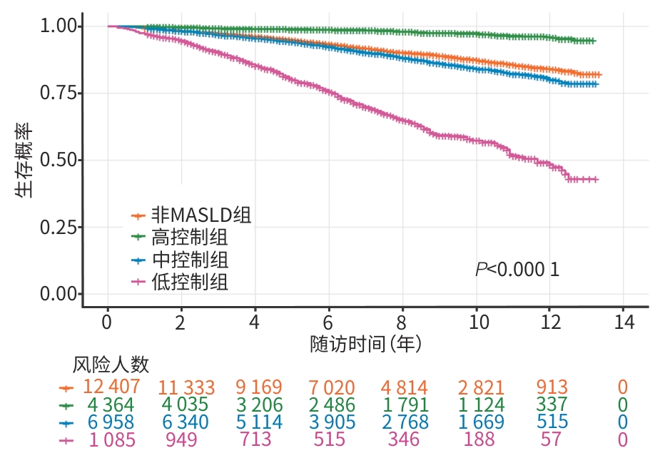
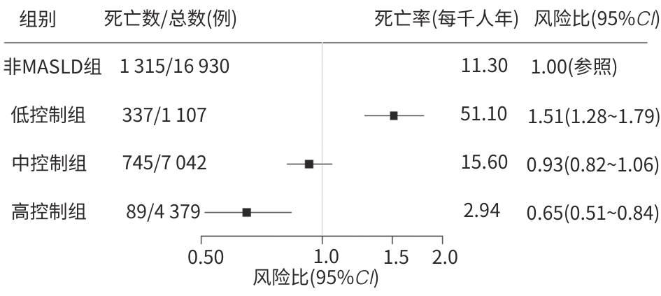
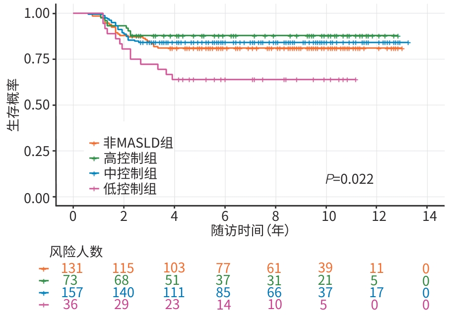
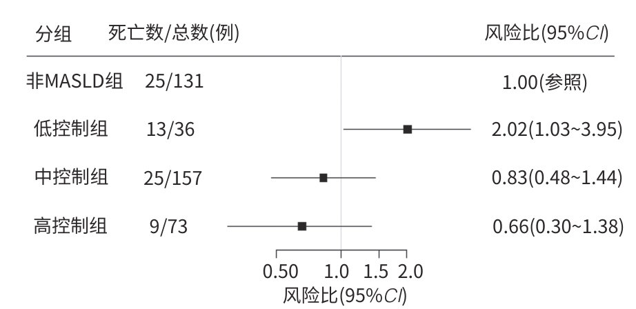
  •   Objective  To investigate the association of comprehensive control of multiple metabolic risk factors with the risk of all-cause mortality in patients with metabolic dysfunction-associated steatotic liver disease (MASLD) and its quantitative effect.  Methods  A retrospective cohort study was conducted based on the data from National Health and Nutrition Examination Survey (NHANES) in 2003—2018. A total of 29 458 participants were enrolled, including 12 528 patients with MASLD and 16 930 non-MASLD controls. The control status of nine risk factors was assessed, i.e., hypertension, diabetes, liver fibrosis (assessed by fibrosis-4 index [FIB-4]), aspartate aminotransferase (AST)/alanine aminotransferase (ALT) ratio, serum albumin, renal function (assessed by serum creatinine), systemic inflammation (assessed by neutrophil count), smoking, and physical activity. According to the status of comprehensive risk factor control, the patients with MASLD were further divided into low-control group with 1 107 patients, moderate-control group with 7 042 patients, and high-control group with 4 379 patients. A total of 397 participants were enrolled in the external validation cohort, among whom there were 266 participants with MASLD and 131 participants without MASLD. The Kruskal-Wallis H test was used for comparison of continuous data between multiple groups, and the chi-square test was used for comparison of categorical data between groups. The Cox proportional hazards model incorporating complex sampling weights for NHANES data were used to investigate the association between the degree of risk factor control and all-cause mortality, and the Kaplan-Meier curve, the log-rank test, restricted mean survival time (RMST), and population attributable fraction (PAF) were used for further assessment. The robustness of the findings was validated in an independent clinical cohort (n=397).  Results  The cohort study based on the NHANES database showed that during the median follow-up time of 6.7 years, 1 171 deaths occurred among the 12 528 patients with MASLD. There was a significant reduction in mortality risk with the increase in the number of risk factors controlled (trend test: χ2 =65.06, P<0.001). Compared with the low-control group, the mortality risk decreased successively in the moderate-control group (hazard ratio [HR]=0.59, 95% confidence interval [CI]: 0.51 — 0.69) and the high-control group (HR=0.36, 95%CI: 0.27 — 0.48), and the mortality risk was reduced by 22% for each additional risk factor controlled (HR=0.78, 95%CI: 0.74 — 0.82). The PAF analysis showed that AST/ALT ratio control (PAF=19.2%) and physical activity (PAF=18.9%) were the largest contributors. The survival analysis of the sub-cohort constructed based on nearest neighbor matching (with a maximum follow-up time of 13.3 years) showed that there was a significant difference in cumulative survival rate between the MASLD groups with different control levels and the non-MASLD group (P<0.001). The RMST analysis further quantified the survival benefit of each group, and the high-control group had the longest 10-year RMST (9.88 years), which was superior to that of the non-MASLD group (9.43 years), the moderate-control group (9.33 years), and the low-control group (7.96 years). The multivariable-adjusted model analysis of the complete cohort showed that compared with the non-MASLD population, the MASLD patients in the low-control group had a significant increase in mortality risk, the moderate-control group had a comparable mortality risk, and the high-control group had a significantly lower mortality risk. The subgroup analysis showed a significant protective effect of comprehensive control in both male and female individuals (P<0.05), and the interaction analysis suggested a potentially stronger protective effect in female individuals (Pfor interaction=0.031). The protective effect was statistically significant in patients aged ≥60 years (P<0.05). There was a significant difference in protective effect across the groups stratified based on waist circumference (P<0.05), but there was no significant interaction between groups (Pfor interaction=0.821). In the external validation cohort, among the 397 participants, there were 72 cases of all-cause mortality (18.1%). The survival analysis showed a significant gradient increase in the survival rate of MASLD patients with the increase in the level of control (P=0.022). The RMST analysis showed that the moderate- and high-control groups had a better RMST than the non-MASLD group at 5 years (4.47 years vs 4.56 years vs 4.40 years), with a more significant survival advantage at 10 years (8.67 years vs 8.95 years vs 8.44 years). The Cox regression analysis showed that, with the low-control MASLD group as the reference, there was a significant reduction in mortality risk in the moderate-control group (HR=0.41, 95%CI: 0.21 — 0.81) and the high-control group (HR=0.32, 95%CI: 0.14 — 0.71); with the non-MASLD population as the reference, there was a significant increase in mortality risk in the low-control group (HR=2.02, 95%CI: 1.03 — 3.95), with a comparable mortality risk in the moderate-control group (HR=0.83, 95%CI: 0.48 — 1.44) and the high-control group (HR=0.66, 95%CI: 0.30 — 1.38), and the point estimates of the effects were highly consistent with the main study.  Conclusion  Among MASLD patients, better control of metabolic risk factors is associated with a lower mortality risk. Achieving optimal control can eliminate the excess mortality risk associated with MASLD, thereby helping patients achieve a better survival prognosis than the general population.


  • loading
  • [1]
    Rinella M E, Lazarus J V, Ratziu V, et al. A multisociety Delphi consensus statement on new fatty liver disease nomenclature[J]. Hepatology, 2023, 78( 6): 1966- 1986. DOI: 10.1097/HEP.0000000000000520.
    [2]
    Kaylan K B, Paul S. NAFLD No more: A review of current guidelines in the diagnosis and evaluation of metabolic dysfunction-associated steatotic liver disease(MASLD)[J]. Curr Diab Rep, 2024, 25( 1): 5. DOI: 10.1007/s11892-024-01558-y.
    [3]
    Riazi K, Azhari H, Charette J H, et al. The prevalence and incidence of NAFLD worldwide: A systematic review and meta-analysis[J]. Lancet Gastroenterol Hepatol, 2022, 7( 9): 851- 861. DOI: 10.1016/S2468-1253(22)00165-0.
    [4]
    Ekstedt M, Hagström H, Nasr P, et al. Fibrosis stage is the strongest predictor for disease-specific mortality in NAFLD after up to 33 years of follow-up[J]. Hepatology, 2015, 61( 5): 1547- 1554. DOI: 10.1002/hep.27368.
    [5]
    Le M H, Devaki P, Ha N B, et al. Prevalence of non-alcoholic fatty liver disease and risk factors for advanced fibrosis and mortality in the United States[J]. PLoS One, 2017, 12( 3): e0173499. DOI: 10.1371/journal.pone.0173499.
    [6]
    Wang T T, Xi Y Z, Raji A, et al. Overall and subgroup prevalence of non-alcoholic fatty liver disease and prevalence of advanced fibrosis in the United States: An updated national estimate in National Health and Nutrition Examination Survey(NHANES) 2011-2018[J]. Ann Hepatol, 2024, 29( 1): 101154. DOI: 10.1016/j.aohep.2023.101154.
    [7]
    Chinese Society of Hepatology, Chinese Medical Association. Guidelines for the prevention and treatment of metabolic dysfunction-associated(non-alcoholic) fatty liver disease(version 2024)[J]. J Prac Hepatol, 2024, 27( 4): 494- 510. DOI: 10.3760/cma.j.cn501113-20240327-00163.

    中华医学会肝病学分会. 代谢相关(非酒精性)脂肪性肝病防治指南(2024年版)[J]. 实用肝脏病杂志, 2024, 27( 4): 494- 510. DOI: 10.3760/cma.j.cn501113-20240327-00163.
    [8]
    Bedogni G, Bellentani S, Miglioli L, et al. The Fatty Liver Index: A simple and accurate predictor of hepatic steatosis in the general population[J]. BMC Gastroenterol, 2006, 6: 33. DOI: 10.1186/1471-230X-6-33.
    [9]
    Musso G, Gambino R, Cassader M, et al. Meta-analysis: Natural history of non-alcoholic fatty liver disease(NAFLD) and diagnostic accuracy of non-invasive tests for liver disease severity[J]. Ann Med, 2011, 43( 8): 617- 649. DOI: 10.3109/07853890.2010.518623.
    [10]
    Aneni E C, Oni E T, Martin S S, et al. Blood pressure is associated with the presence and severity of nonalcoholic fatty liver disease across the spectrum of cardiometabolic risk[J]. J Hypertens, 2015, 33( 6): 1207- 1214. DOI: 10.1097/HJH.0000000000000532.
    [11]
    Ciardullo S, Monti T, Sala I, et al. Nonalcoholic fatty liver disease and advanced fibrosis in US adults across blood pressure categories[J]. Hypertension, 2020, 76( 2): 562- 568. DOI: 10.1161/HYPERTENSIONAHA.120.15220.
    [12]
    National Center for Health Statistics. Biochemistry profile-creatinine( 2019-2020)[R/OL]. National Health and Nutrition Examination Survey, 2019. https://wwwn.cdc.gov/nchs/data/nhanes/public/2019/labmethods/BIOPRO-K-MET-Creatinine-508.pdf. https://wwwn.cdc.gov/nchs/data/nhanes/public/2019/labmethods/BIOPRO-K-MET-Creatinine-508.pdf
    [13]
    Mohammadi M, Aminian K, Joukar F, et al. Diagnostic value of serologic biomarkers for the detection of liver fibrosis in non-alcoholic fatty liver disease[J]. Caspian J Intern Med, 2025, 16( 2): 275- 283. DOI: 10.22088/cjim.16.2.275.
    [14]
    Rinella M E, Neuschwander-tetri B A, Siddiqui M S, et al. AASLD practice guidance on the clinical assessment and management of nonalcoholic fatty liver disease[J]. Hepatology, 2023, 77( 5): 1797- 1835. DOI: 10.1097/HEP.0000000000000323.
    [15]
    National Center for Health Statistics. Complete blood count-whole blood( 2017-2018)[R/OL]. National Health and Nutrition Examination Survey, 2019. https://wwwn.cdc.gov/nchs/data/nhanes/public/2017/labmethods/CBC-J-MET-508.pdf. https://wwwn.cdc.gov/nchs/data/nhanes/public/2017/labmethods/CBC-J-MET-508.pdf
    [16]
    Tahir N, Zahra F. Neutrophilia[EB/OL]. Treasure Island(FL): StatPearls Publishing, 2023. https://www.ncbi.nlm.nih.gov/books/NBK570571/. https://www.ncbi.nlm.nih.gov/books/NBK570571/
    [17]
    World Health Organization. Global recommendations on physical activity for health[R]. Geneva: World Health Organization, 2010.
    [18]
    Akinbami L J, Chen T C, Davy O, et al. National health and nutrition examination survey, 2017-March 2020 prepandemic file: Sample design, estimation, and analytic guidelines[J]. Vital Health Stat 1, 2022( 190): 1- 36.
    [19]
    Nguyen V K, Middleton L Y M, Huang L, et al. Harmonized US National Health and Nutrition Examination Survey 1988—2018 for high throughput exposome-health discovery[J]. Environ Int, 2023, 181: 108259. DOI: 10.1016/j.envint.2023.108259.
    [20]
    National Center for Health Statistics. Demographic variables and sample weights( 2015-2016)[R/OL]. National Health and Nutrition Examination Survey, 2017. https://wwwn.cdc.gov/nchs/nhanes/Search/DataPage.aspx?Component=Demographics&CycleBeginYear=2015. https://wwwn.cdc.gov/nchs/nhanes/Search/DataPage.aspx?Component=Demographics&CycleBeginYear=2015
    [21]
    Hakeem A, Van berlo J, Revelo X S. The cardiohepatic axis in metabolic disease liver to heart[J]. JACC Basic Transl Sci, 2025, 10( 7): 101309. DOI: 10.1016/j.jacbts.2025.05.004.
    [22]
    Han E, Woo S Y, Jeon J Y, et al. Impact of physical activities in metabolic dysfunction associated steatotic liver disease, sarcopenia, and cardiovascular disease[J]. Diabetes Res Clin Pract, 2025, 224: 112209. DOI: 10.1016/j.diabres.2025.112209.
    [23]
    Song Q R, Liu S L, Ling Q H, et al. Severity of nonalcoholic fatty liver disease is associated with cardiovascular outcomes in patients with prehypertension or hypertension: A community-based cohort study[J]. Front Endocrinol, 2022, 13: 942647. DOI: 10.3389/fendo.2022.942647.
    [24]
    Han Y, Tang J, Wu N, et al. Association between the Life’s essential 8 health behaviors score and all-cause mortality in patients with metabolic dysfunction-associated steatotic liver disease[J]. Metabolism, 2025, 163: 156096. DOI: 10.1016/j.metabol.2024.156096.
    [25]
    Cho Y K, Kim M J, Kim E H, et al. Comparison of diagnostic criteria for fatty liver disease in assessing cardiac dysfunction: A cross-sectional study[J]. Hepatol Int, 2026, 20( 1): 81- 90. DOI: 10.1007/s12072-025-10924-1.
    [26]
    Celsa C, Pennisi G, Tulone A, et al. Risk of hepatic and extrahepatic outcomes associated with metabolic dysfunction-associated steatotic liver disease and metabolic dysfunction and alcohol-associated steatotic liver disease: A systematic review and meta-analysis[J]. Lancet Gastroenterol Hepatol, 2025, 10( 11): 998- 1012. DOI: 10.1016/S2468-1253(25)00188-8.
    [27]
    Younossi Z M, Koenig A B, Abdelatif D, et al. Global epidemiology of nonalcoholic fatty liver disease-Meta-analytic assessment of prevalence, incidence, and outcomes[J]. Hepatology, 2016, 64( 1): 73- 84. DOI: 10.1002/hep.28431.
    [28]
    Sakai K, Shima T, Oya H, et al. Association between liver fibrosis and cause-specific mortality in Japanese patients with biopsy-confirmed metabolic dysfunction-associated steatotic liver disease: A prospective cohort study/liver fibrosis and mortality in Japanese MASLD[J]. Hepatol Res, 2026, 56( 1): 33- 49. DOI: 10.1111/hepr.70034.
    [29]
    Angulo P, Kleiner D E, Dam-larsen S, et al. Liver fibrosis, but no other histologic features, is associated with long-term outcomes of patients with nonalcoholic fatty liver disease[J]. Gastroenterology, 2015, 149( 2): 389- 397.e10. DOI: 10.1053/j.gastro.2015.04.043.
    [30]
    Liu Zhengyi, Zhuang Yingjie, Dong Xu, et al. Construction of a non-invasive diagnostic model for liver fibrosis in metabolic dysfunction-associated steatotic liver disease(MASLD)[J]. Clin J Med Offic, 2024, 52( 7): 688- 691. DOI: 10.16680/j.1671-3826.2024.07.07.

    刘正一, 庄颖洁, 董旭, 等. 非酒精性脂肪性肝病肝纤维化无创诊断模型构建[J]. 临床军医杂志, 2024, 52( 7): 688- 691. DOI: 10.16680/j.1671-3826.2024.07.07.
    [31]
    Machado M V, Cortez-pinto H. Non-invasive diagnosis of non-alcoholic fatty liver disease. A critical appraisal[J]. J Hepatol, 2013, 58( 5): 1007- 1019. DOI: 10.1016/j.jhep.2012.11.021.
    [32]
    Vilar-gomez E, Chalasani N. Non-invasive assessment of non-alcoholic fatty liver disease: Clinical prediction rules and blood-based biomarkers[J]. J Hepatol, 2018, 68( 2): 305- 315. DOI: 10.1016/j.jhep.2017.11.013.
  • 加载中

Catalog

    通讯作者: 陈斌, bchen63@163.com
    • 1. 

      沈阳化工大学材料科学与工程学院 沈阳 110142

    1. 本站搜索
    2. 百度学术搜索
    3. 万方数据库搜索
    4. CNKI搜索

    Figures(4)  / Tables(4)

    Article Metrics

    Article views (70) PDF downloads(17) Cited by()
    Proportional views
    Related

    /

    DownLoad:  Full-Size Img  PowerPoint
    Return
    Return