中文English
ISSN 1001-5256 (Print)
ISSN 2097-3497 (Online)
CN 22-1108/R
Volume 42 Issue 3
Mar.  2026
Turn off MathJax
Article Contents

Effect and mechanism of Wnt5a knockdown on the efficacy of M1 bone marrow-derived macrophage in treatment of liver cirrhosis

DOI: 10.12449/JCH260317
Research funding:

General Project of National Natural Science Foundation of China (81874390);

Sponsored by Natural Science Foundation of Shanghai (21ZR1464100);

Special Project for Biomedical Science and Technology Support of the “Science and Technology Innovation Action Plan” of Shanghai Science and Technology Commission in 2022 (22S11901700);

Shanghai Key Specialty of Traditional Chinese Clinical Medicine (shslczdzk01201)

More Information
  • Corresponding author: MU Yongping, ypmu8888@126.com (ORCID: 0000-0002-6808-8243)
  • Received Date: 2025-08-04
  • Accepted Date: 2025-11-07
  • Published Date: 2026-03-25
  •   Objective  To observe the effect of M1 bone marrow-derived macrophages (M1-BMDM) with Wnt5a knockdown on liver fibrosis and regeneration in a rat model of liver cirrhosis, and to investigate its gain-of-function effect compared with unmodified M1-BMDM.  Methods  Primary bone marrow-derived macrophages were isolated from rats and were polarized to M1 phenotype to construct M1-BMDMWnt5a-KD cells. A rat model of liver cirrhosis induced by CCl4/2-AAF was established, and at the end of week 8, rats were randomly divided into model group, M1-BMDM group, M1-BMDM Wnt5a-knockdown empty vector group (M1-BMDMKD-EV group), and M1-BMDM Wnt5a-knockdown group (M1-BMDMWnt5a-KD group), with 6 rats in each group. On the first day of week 9, the rats in each group were given a single injection of the corresponding cells via the caudal vein, along with an intraperitoneal injection of a CCR2 inhibitor. Six rats without any treatment were used as normal control group. Samples were collected at the end of week 12 to assess liver histopathology, serum liver function parameters, hepatic stellate cell activation, and the expression levels of mature hepatocyte markers. A one-way analysis of variance was used for comparison of continuous data between multiple groups, and the least significant difference t-test was used for further comparison between two groups.  Results  Compared with the model group, all cell treatment groups had significant alleviation of liver inflammatory response and significant reductions in the activities of alanine aminotransferase and aspartate aminotransferase (AST) in serum (all P<0.01), and the M1-BMDMWnt5a-KD group had a significantly lower serum level of AST than the M1-BMDM group (P<0.05). The semi-quantitative analysis based on immunohistochemical staining showed that compared with the model group, all cell treatment groups had a significant reduction in the percentage of CD68-positive area (all P<0.05), and compared with the M1-BMDMKD-EV group, the M1-BMDMWnt5a-KD group had a significant reduction in the percentage of CD68-positive area and a significant increase in the percentage of CD163-positive area (both P<0.05). Compared with the model group, all cell treatment groups had significant reductions in the mRNA expression levels of CD68 and tumor necrosis factor-α (all P<0.05) and the protein expression level of CD68 (all P<0.01); compared with the M1-BMDMKD-EV group, the M1-BMDMWnt5a-KD group had significant increases in the protein and mRNA expression levels of CD163 (both P<0.05), significant reductions in the protein and mRNA expression levels of CD68 (both P<0.05), and a significant reduction in the protein expression level of tumor necrosis factor-α (P<0.01). Sirius Red collagen staining and alpha-smooth muscle actin (α-SMA) immunohistochemical staining showed that compared with the model group, all cell treatment groups had significant alleviation of liver collagen deposition and α-SMA-positive area, with the most significant changes in the M1-BMDMWnt5a-KD group, and compared with the M1-BMDMKD-EV group, the M1-BMDMWnt5a-KD group had significantly smaller Sirius Red-positive area and α-SMA-positive area and a significantly lower content of hydroxyproline in liver tissue (all P<0.05). Compared with the M1-BMDMKD-EV group, the M1-BMDMWnt5a-KD group had significant reductions in the protein and mRNA expression levels of α-SMA and the mRNA expression level of COL-I and TGF-β (all P<0.05). Compared with the model group, all cell treatment groups had a significant increase in the protein expression level of HNF-4α in liver tissue (all P<0.05), and the M1-BMDMWnt5a-KD group had significantly higher protein and mRNA expression levels of HNF-4α and hepatocyte specific antigen than the M1-BMDMKD-EV group (both P<0.05). The M1-BMDMWnt5a-KD group had a significantly higher serum level of albumin than the M1-BMDMKD-EV group (P<0.01). Immunofluorescence co-staining showed that compared with the model group, all cell treatment groups had a significant increase in the number of cells stained positive for HNF and HNF-4α and Ki67 (all P<0.01), and the M1-BMDMWnt5a-KD group had a significantly higher number of such cells than the M1-BMDMKD-EV group (P<0.05).  Conclusion  Inhibition ofWnt5aexpression enhances the therapeutic effect of M1-BMDM on rats with liver cirrhosis induced by CCl4/2-AAF, which provides new ideas for enhancing the anti-cirrhotic effect of M1-BMDM through genetic modification.


  • loading
  • [1]
    OAKLEY F. Interrogating mechanisms of liver fibrosis with omics[J]. Nat Rev Gastroenterol Hepatol, 2022, 19( 2): 89- 90. DOI: 10.1038/s41575-021-00567-6.
    [2]
    LIAO ZH, XIE ZY. Research progress in molecular mechanism of hepatic fibrosis and related therapeutic targets[J]. J Jilin Univ: Med Edit, 2024, 50( 5): 1450- 1456. DOI: 10.13481/j.1671-587X.20240532.

    廖昭辉, 谢正元. 肝纤维化发病的分子机制及其相关治疗靶点的研究进展[J]. 吉林大学学报(医学版), 2024, 50( 5): 1450- 1456. DOI: 10.13481/j.1671-587X.20240532.
    [3]
    LI L, CUI L, LIN P, et al. Kupffer-cell-derived IL-6 is repurposed for hepatocyte dedifferentiation via activating progenitor genes from injury-specific enhancers[J]. Cell Stem Cell, 2023, 30( 3): 283- 299.e9. DOI: 10.1016/j.stem.2023.01.009.
    [4]
    THOMAS JA, POPE C, WOJTACHA D, et al. Macrophage therapy for murine liver fibrosis recruits host effector cells improving fibrosis, regeneration, and function[J]. Hepatology, 2011, 53( 6): 2003- 2015. DOI: 10.1002/hep.24315.
    [5]
    MA PF, GAO CC, YI J, et al. Cytotherapy with M1-polarized macrophages ameliorates liver fibrosis by modulating immune microenvironment in mice[J]. J Hepatol, 2017, 67( 4): 770- 779. DOI: 10.1016/j.jhep.2017.05.022.
    [6]
    ZONG MY, JIAN X, WANG DY, et al. Effect of Yiguan Decoction on the efficacy of M1 bone marrow-derived macrophages in treatment of liver cirrhosis rats and its mechanism[J]. J Clin Hepatol, 2024, 40( 8): 1612- 1619. DOI: 10.12449/JCH240817.

    宗梦瑶, 简迅, 王丹阳, 等. 一贯煎对M1型骨髓巨噬细胞治疗肝硬化大鼠模型效果的影响及其机制分析[J]. 临床肝胆病杂志, 2024, 40( 8): 1612- 1619. DOI: 10.12449/JCH240817.
    [7]
    CAO MZ, CHAN RWS, CHENG FHC, et al. Myometrial cells stimulate self-renewal of endometrial mesenchymal stem-like cells through WNT5A/β-catenin signaling[J]. Stem Cells, 2019, 37( 11): 1455- 1466. DOI: 10.1002/stem.3070.
    [8]
    LIN QQ, WU LM, CHATLA S, et al. Hematopoietic stem cell regeneration through paracrine regulation of the Wnt5a/Prox1 signaling axis[J]. J Clin Invest, 2022, 132( 12): e155914. DOI: 10.1172/JCI155914.
    [9]
    ZHAO CG, QIN J, LI J, et al. LINGO-1 regulates Wnt5a signaling during neural stem and progenitor cell differentiation by modulating miR-15b-3p levels[J]. Stem Cell Res Ther, 2021, 12( 1): 372. DOI: 10.1186/s13287-021-02452-0.
    [10]
    TIAN Y, CHEN JC, HUANG WS, et al. Myeloid-derived Wnts play an indispensible role in macrophage and fibroblast activation and kidney fibrosis[J]. Int J Biol Sci, 2024, 20( 6): 2310- 2322. DOI: 10.7150/ijbs.94166.
    [11]
    JIAN X, WANG DY, XU YN, et al. Effect of polarized bone marrow-derived macrophage transplantation on the progression of CCl4-induced liver fibrosis in rats[J]. J Clin Hepatol, 2021, 37( 12): 2830- 2837. DOI: 10.3969/j.issn.1001-5256.2021.12.020.

    简迅, 王丹阳, 许燕楠, 等. 极化骨髓巨噬细胞移植对CCl4诱导的肝纤维化大鼠模型的影响[J]. 临床肝胆病杂志, 2021, 37( 12): 2830- 2837. DOI: 10.3969/j.issn.1001-5256.2021.12.020.
    [12]
    HAAG SM, MURTHY A. Murine monocyte and macrophage culture[J]. Bio Protoc, 2021, 11( 6): e3928. DOI: 10.21769/BioProtoc.3928.
    [13]
    JAMALL IS, FINELLI VN, QUE HEE SS. A simple method to determine nanogram levels of 4-hydroxyproline in biological tissues[J]. Anal Biochem, 1981, 112( 1): 70- 75. DOI: 10.1016/0003-2697(81)90261-x.
    [14]
    GUILLOT A, TACKE F. Spatial dimension of macrophage heterogeneity in liver diseases[J]. eGastroenterology, 2023, 1( 1): e000003. DOI: 10.1136/egastro-2023-000003.
    [15]
    FLORES MOLINA M, ABDELNABI MN, MAZOUZ S, et al. Distinct spatial distribution and roles of Kupffer cells and monocyte-derived macrophages in mouse acute liver injury[J]. Front Immunol, 2022, 13: 994480. DOI: 10.3389/fimmu.2022.994480.
    [16]
    NA YR, KIM SW, SEOK SH. A new era of macrophage-based cell therapy[J]. Exp Mol Med, 2023, 55( 9): 1945- 1954. DOI: 10.1038/s12276-023-01068-z.
    [17]
    CHEN T, CHEN Y. Research progress in mechanism of fibrosis regulated by macrophage polarization[J]. J Jilin Univ: Med Edit, 2024, 50( 5): 1465- 1473. DOI: 10.13481/j.1671-587X.20240534.

    陈潭, 陈艳. 巨噬细胞极化调控纤维化机制的研究进展[J]. 吉林大学学报(医学版), 2024, 50( 5): 1465- 1473. DOI: 10.13481/j.1671-587X.20240534.
    [18]
    SHAO Y, ZHENG QQ, WANG W, et al. Biological functions of macrophage-derived Wnt5a, and its roles in human diseases[J]. Oncotarget, 2016, 7( 41): 67674- 67684. DOI: 10.18632/oncotarget.11874.
    [19]
    MENCK K, KLEMM F, GROSS JC, et al. Induction and transport of Wnt 5a during macrophage-induced malignant invasion is mediated by two types of extracellular vesicles[J]. Oncotarget, 2013, 4( 11): 2057- 2066. DOI: 10.18632/oncotarget.1336.
    [20]
    LIU Q, SONG JL, PAN Y, et al. Wnt5a/CaMKII/ERK/CCL2 axis is required for tumor-associated macrophages to promote colorectal cancer progression[J]. Int J Biol Sci, 2020, 16( 6): 1023- 1034. DOI: 10.7150/ijbs.40535.
    [21]
    MA X, HUANG TD, CHEN XZ, et al. Molecular mechanisms in liver repair and regeneration: From physiology to therapeutics[J]. Signal Transduct Target Ther, 2025, 10( 1): 63. DOI: 10.1038/s41392-024-02104-8.
    [22]
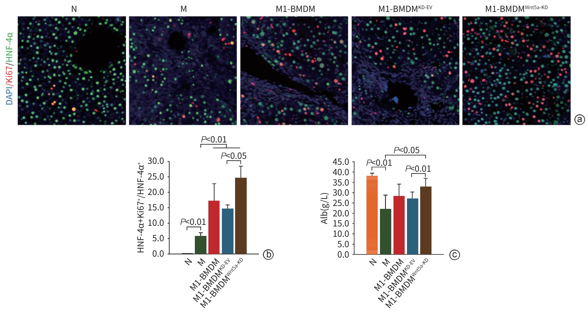
    HUCK I, GUNEWARDENA S, ESPANOL-SUNER R, et al. Hepatocyte nuclear factor 4 alpha activation is essential for termination of liver regeneration in mice[J]. Hepatology, 2019, 70( 2): 666- 681. DOI: 10.1002/hep.30405.
    [23]
    SONG ZM, LIN SR, WU XW, et al. Hepatitis B virus-related intrahepatic cholangiocarcinoma originates from hepatocytes[J]. Hepatol Int, 2023, 17( 5): 1300- 1317. DOI: 10.1007/s12072-023-10556-3.
    [24]
    SCHOLZEN T, GERDES J. The Ki-67 protein: From the known and the unknown[J]. J Cell Physiol, 2000, 182( 3): 311- 322. DOI: 3.0.CO;2-9">10.1002/(SICI)1097-4652(200003)182: 3<311:: AID-JCP1>3.0.CO;2-9.
    [25]
    SUN DQ, ZHOU XL, WU T, et al. Quercetin promotes the M1-to-M2 macrophage phenotypic switch during liver fibrosis treatment by modulating the JAK2/STAT3 signaling pathway[J]. Recent Pat Anticancer Drug Discov, 2026, 21( 1): 31- 46. DOI: 10.2174/0115748928318948240920044716.
    [26]
    MORONI F, DWYER BJ, GRAHAM C, et al. Safety profile of autologous macrophage therapy for liver cirrhosis[J]. Nat Med, 2019, 25( 10): 1560- 1565. DOI: 10.1038/s41591-019-0599-8.
    [27]
    BRENNAN PN, MACMILLAN M, MANSHIP T, et al. Autologous macrophage therapy for liver cirrhosis: A phase 2 open-label randomized controlled trial[J]. Nat Med, 2025, 31( 3): 979- 987. DOI: 10.1038/s41591-024-03406-8.
  • 加载中

Catalog

    通讯作者: 陈斌, bchen63@163.com
    • 1. 

      沈阳化工大学材料科学与工程学院 沈阳 110142

    1. 本站搜索
    2. 百度学术搜索
    3. 万方数据库搜索
    4. CNKI搜索

    Figures(7)

    Article Metrics

    Article views (23) PDF downloads(12) Cited by()
    Proportional views
    Related

    /

    DownLoad:  Full-Size Img  PowerPoint
    Return
    Return