中文English
ISSN 1001-5256 (Print)
ISSN 2097-3497 (Online)
CN 22-1108/R
Volume 42 Issue 4
Apr.  2026
Turn off MathJax
Article Contents

Construction and evaluation of a novel KPC mouse model of type 2 diabetes mellitus comorbid with spontaneous pancreatic cancer

DOI: 10.12449/JCH260420
Research funding:

National Natural Science Foundation of China (81860508)

More Information
  • Corresponding author: QIN Wen, fenglingcao1980@163.com (ORCID: 0009-0002-8282-7388)
  • Received Date: 2025-10-14
  • Accepted Date: 2025-12-26
  • Published Date: 2026-04-25
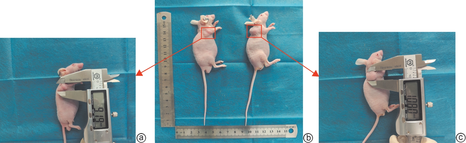
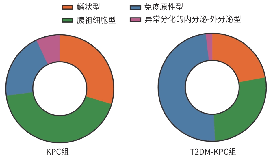
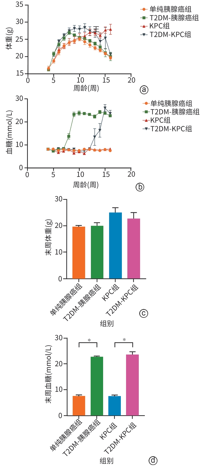
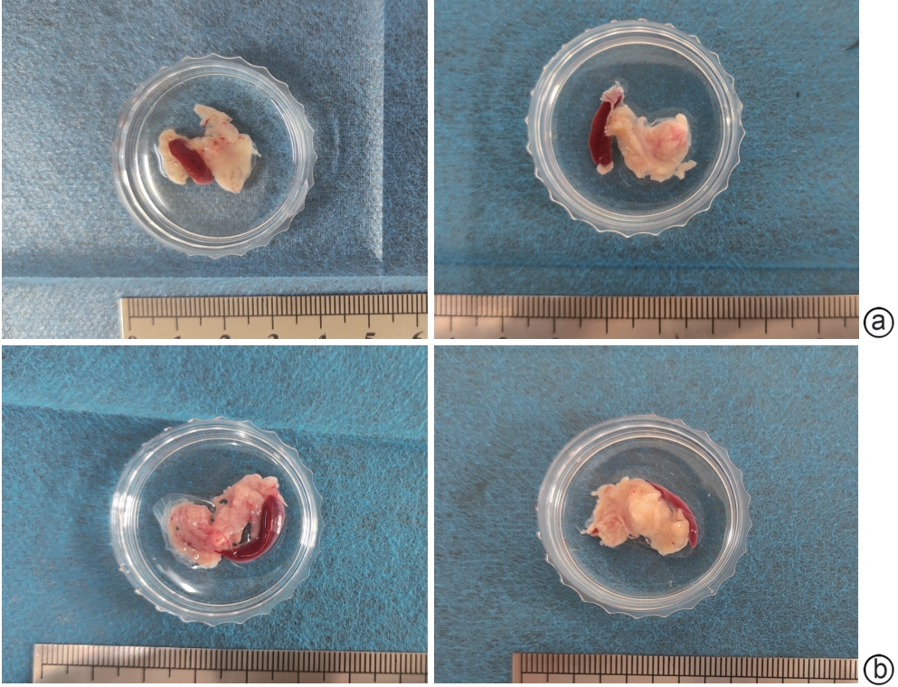
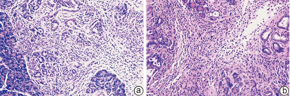
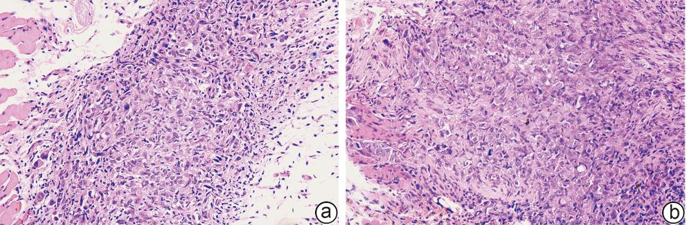
  •   Objective  To construct a novel KPC mouse model of type 2 diabetes mellitus (T2DM) comorbid with spontaneous pancreatic cancer based on the gene editing-metabolic intervention dual-driven strategy, and to compare it with traditional models.  Methods  A total of 14 male KPC mice were randomly divided into novel model group (T2DM-KPC group with 7 mice) and control group (KPC group with 7 mice), and 14 male BALB/c-nu nude mice were randomly divided into traditional model group (T2DM-pancreatic cancer group with 7 mice) and control group (pancreatic cancer group with 7 mice). The mice in the KPC group and the pancreatic cancer group were fed with normal diet, and those in the T2DM-KPC group and the T2DM-pancreatic cancer group were fed with a high-fat diet. After 4 weeks, the mice in the T2DM-KPC group and the T2DM-pancreatic cancer group were given intraperitoneal injection of streptozotocin. Subsequently, the mice in the KPC group and the T2DM-KPC group developed primary pancreatic tumor spontaneously over time, while those in the T2DM-pancreatic cancer group and the pancreatic cancer group were inoculated with tumor cells to form subcutaneous tumor xenograft at 2 weeks after stabilization of blood glucose. The 4 groups were observed in terms of tumor formation rate, tumor formation time, body weight, and the change in blood glucose; RNA sequencing was performed for tumors from the KPC group and the T2DM-KPC group, and then molecular subtyping was performed; HE staining, Masson staining, and immunohistochemical staining were used to assess the histopathological features and tumor microenvironment of pancreatic tumor from the T2DM-KPC group, which were compared with those of the T2DM-pancreatic cancer group. A one-way analysis of variance was used for comparison of continuous data between multiple groups, and the least significant difference t-test was used for further comparison between two groups; the Fisher’s exact test was used for comparison of categorical data between multiple groups.  Results  The T2DM-KPC group had a tumor formation rate of 85.71% and a tumor formation time of 104.40±2.87 days, while the T2DM-pancreatic cancer group had a tumor formation rate of 71.43% and a tumor formation time of 95.20±9.47 days, and there were no significant differences between the two groups in tumor formation rate, tumor formation time, body weight, and blood glucose (all P>0.05). Molecular subtyping showed that the model in the KPC group highly resembled the pancreatic progenitor subtype of human pancreatic ductal adenocarcinoma (PDAC), and the model in the T2DM-KPC group highly resembled the immunogenic subtype of PDAC. HE staining showed that tumor cells in the T2DM-KPC group were arranged into glandular tubular structures of varying shapes, exhibiting significant cellular atypia, and this model faithfully recapitulated the pathological features of primary pancreatic cancer and showed greater invasiveness than the KPC group. Immunohistochemical staining and Masson staining showed that compared with the T2DM-pancreatic cancer group, the T2DM-KPC group had significantly higher degrees of tumor proliferation (assessed by Ki-67 expression) and fibrosis (assessed by α-SMA and Masson) (all P<0.05), suggesting that the mouse model in the T2DM-KPC group could better recapitulate the features of hyperproliferation and pronounced desmoplasia in human pancreatic cancer.  Conclusion  A novel KPC mouse model of T2DM comorbid with spontaneous pancreatic cancer is successfully constructed in this study. This model can accurately mimic the histopathological architecture and stromal microenvironment of T2DM comorbid with pancreatic cancer, realize the longitudinal simulation of the progression of pancreatic tissue from intraepithelial neoplasia to invasive carcinoma and metastasis in the presence of T2DM, and support the translational research on immunotherapy, thereby providing a novel experimental carrier for in vivo studies on spontaneous pancreatic cancer in T2DM.


  • loading
  • [1]
    LUO WH, TAO JX, ZHENG LF, et al. Current epidemiology of pancreatic cancer: Challenges and opportunities[J]. Chin J Cancer Res, 2020, 32( 6): 705- 719. DOI: 10.21147/j.issn.1000-9604.2020.06.04.
    [2]
    HAN BF, ZHENG RS, ZENG HM, et al. Cancer incidence and mortality in China, 2022[J]. J Natl Cancer Cent, 2024, 4( 1): 47- 53. DOI: 10.1016/j.jncc.2024.01.006.
    [3]
    SIEGEL RL, MILLER KD, WAGLE NS, et al. Cancer statistics, 2023[J]. CA Cancer J Clin, 2023, 73( 1): 17- 48. DOI: 10.3322/caac.21763.
    [4]
    CHAUDHRY ZW, HALL E, KALYANI RR, et al. Diabetes and pancreatic cancer[J]. Curr Probl Cancer, 2013, 37( 5): 287- 292. DOI: 10.1016/j.currproblcancer.2013.10.006.
    [5]
    CHEN HM, ZHAO JF, JIANG NN, et al. Hyperglycemia promotes pancreatic cancer initiation and progression by activating the Wnt/β-catenin signaling pathway[J]. Anticancer Agents Med Chem, 2021, 21( 18): 2592- 2602. DOI: 10.2174/1871520621666210201095613.
    [6]
    RUZE R, SONG JL, YIN XP, et al. Mechanisms of obesity- and diabetes mellitus-related pancreatic carcinogenesis: A comprehensive and systematic review[J]. Signal Transduct Target Ther, 2023, 8( 1): 139. DOI: 10.1038/s41392-023-01376-w.
    [7]
    BAILEY P, CHANG DK, NONES K, et al. Genomic analyses identify molecular subtypes of pancreatic cancer[J]. Nature, 2016, 531( 7592): 47- 52. DOI: 10.1038/nature16965.
    [8]
    RACINE KC, IGLESIAS-CARRES L, HERRING JA, et al. The high-fat diet and low-dose streptozotocin type-2 diabetes model induces hyperinsulinemia and insulin resistance in male but not female C57BL/6J mice[J]. Nutr Res, 2024, 131: 135- 146. DOI: 10.1016/j.nutres.2024.09.008.
    [9]
    XU YN, HUANG XH, TANG ZP, et al. Establishment and in vivo imaging observation of a nude mouse model of type 2 diabetes mellitus and pancreatic cancer[J]. J Clin Hepatol, 2024, 40( 6): 1231- 1239. DOI: 10.12449/JCH240625.

    许永宁, 黄雪桓, 唐芷盼, 等. 2型糖尿病胰腺癌裸鼠模型的建立及活体成像观察[J]. 临床肝胆病杂志, 2024, 40( 6): 1231- 1239. DOI: 10.12449/JCH240625.
    [10]
    Cancer Genome Atlas Research Network. Integrated genomic characterization of pancreatic ductal adenocarcinoma[J]. Cancer Cell, 2017, 32( 2): 185- 203. DOI: 10.1016/j.ccell.2017.07.007.
    [11]
    LI J, QIAN WK, QIN T, et al. Mouse-derived allografts: A complementary model to the KPC mice on researching pancreatic cancerin vivo[J]. Comput Struct Biotechnol J, 2019, 17: 498- 506. DOI: 10.1016/j.csbj.2019.03.016.
    [12]
    NIKNAFS N, ZHONG Y, MORAL JA, et al. Characterization of genetic subclonal evolution in pancreatic cancer mouse models[J]. Nat Commun, 2019, 10( 1): 5435. DOI: 10.1038/s41467-019-13100-w.
    [13]
    KELLER RB, MAZOR T, SHOLL L, et al. Programmatic precision oncology decision support for patients with gastrointestinal cancer[J]. JCO Precis Oncol, 2023, 7: e2200342. DOI: 10.1200/PO.22.00342.
    [14]
    ROYAL RE, LEVY C, TURNER K, et al. Phase 2 trial of single agent Ipilimumab(anti-CTLA-4) for locally advanced or metastatic pancreatic adenocarcinoma[J]. J Immunother, 2010, 33( 8): 828- 833. DOI: 10.1097/CJI.0b013e3181eec14c.
    [15]
    MINAEI E, JOYCE P, WADE SJ, et al. Gut microbial diversity is preserved through localised chemo-immunotherapy delivery in a KPC mouse model of pancreatic cancer[J]. J Control Release, 2025, 386: 114143. DOI: 10.1016/j.jconrel.2025.114143.
    [16]
    MA Y, LI J, WANG HM, et al. Combination of PD-1 inhibitor and OX40 agonist induces tumor rejection and immune memory in mouse models of pancreatic cancer[J]. Gastroenterology, 2020, 159( 1): 306- 319. DOI: 10.1053/j.gastro.2020.03.018.
    [17]
    LI KY, WANG JK, ZHANG R, et al. Overcome the challenge for intratumoral injection of STING agonist for pancreatic cancer by systemic administration[J]. J Hematol Oncol, 2024, 17( 1): 62. DOI: 10.1186/s13045-024-01576-z.
    [18]
    DAY CP, MERLINO G, VAN DYKE T. Preclinical mouse cancer models: A maze of opportunities and challenges[J]. Cell, 2015, 163( 1): 39- 53. DOI: 10.1016/j.cell.2015.08.068.
    [19]
    WANG JY, HUANG R, LU Y, et al. Construction of pancreatic cancer organoids and their sensitivity to chemotherapy drugs[J]. J Clin Hepatol, 2024, 40( 9): 1853- 1858. DOI: 10.12449/JCH240921.

    王靖宇, 黄容, 卢艳, 等. 胰腺癌类器官模型的构建及其对化疗药物的敏感性试验[J]. 临床肝胆病杂志, 2024, 40( 9): 1853- 1858. DOI: 10.12449/JCH240921.
    [20]
    FERNANDEZ JL, ÅRBOGEN S, SADEGHINIA MJ, et al. A comparative analysis of orthotopic and subcutaneous pancreatic tumour models: Tumour microenvironment and drug delivery[J]. Cancers, 2023, 15( 22): 5415. DOI: 10.3390/cancers15225415.
    [21]
    KUWATA T, YANAGIHARA K, IINO Y, et al. Establishment of novel gastric cancer patient-derived xenografts and cell lines: Pathological comparison between primary tumor, patient-derived, and cell-line derived xenografts[J]. Cells, 2019, 8( 6): 585. DOI: 10.3390/cells8060585.
  • 加载中

Catalog

    通讯作者: 陈斌, bchen63@163.com
    • 1. 

      沈阳化工大学材料科学与工程学院 沈阳 110142

    1. 本站搜索
    2. 百度学术搜索
    3. 万方数据库搜索
    4. CNKI搜索

    Figures(11)

    Article Metrics

    Article views (43) PDF downloads(8) Cited by()
    Proportional views
    Related

    /

    DownLoad:  Full-Size Img  PowerPoint
    Return
    Return