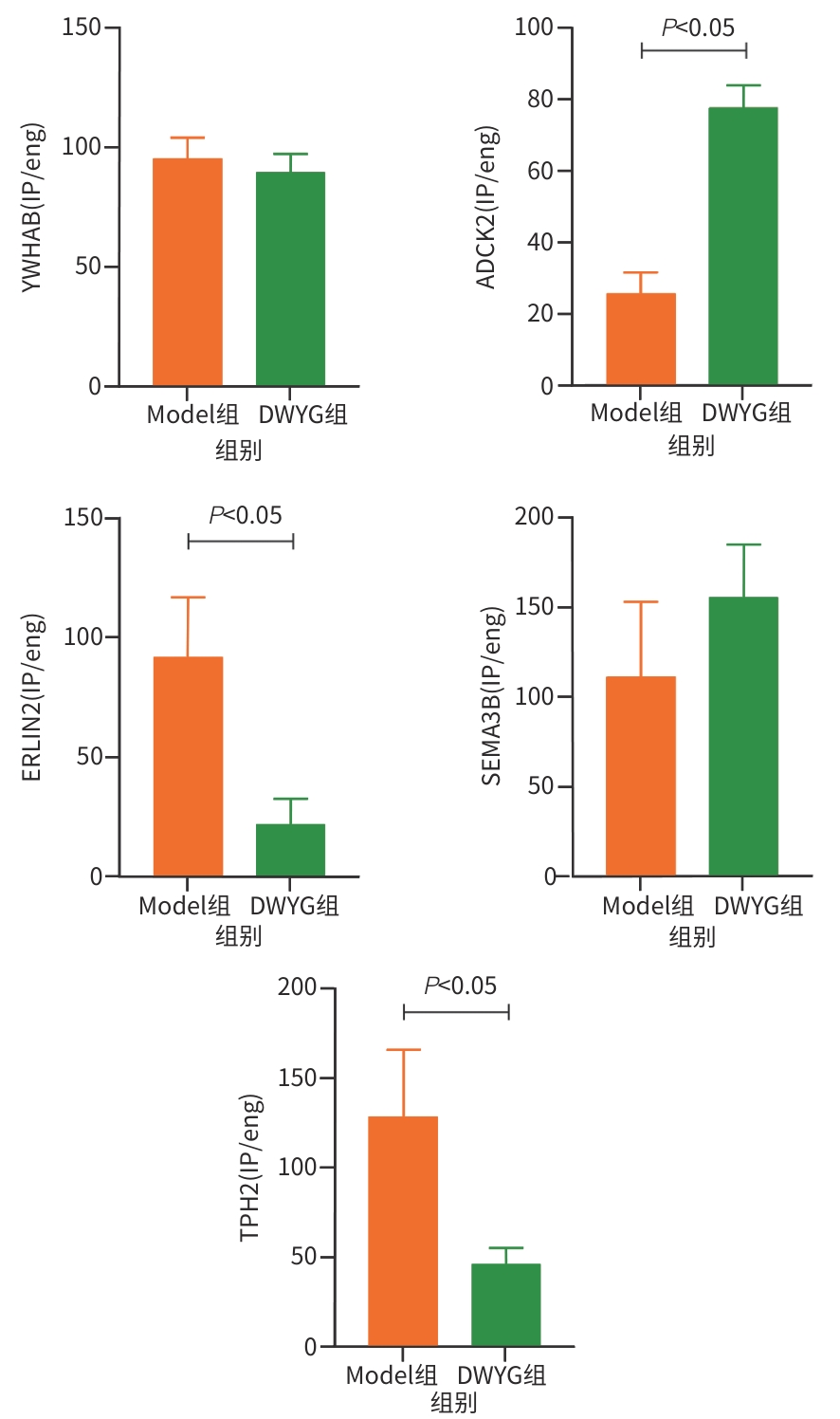
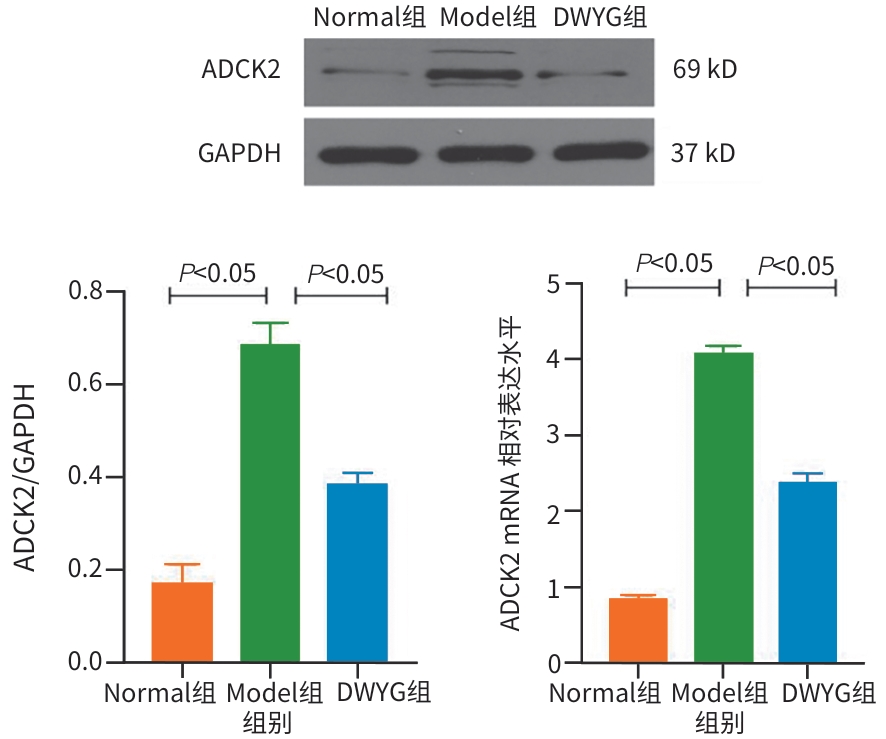
中文English
ISSN 1001-5256 (Print)
ISSN 2097-3497 (Online)
CN 22-1108/R
Volume 42 Issue 2
Feb.  2026
Turn off MathJax
Article Contents

Epigenetic mechanism of Diwu Yanggan Capsule in improving liver regeneration microenvironment in a rat model of liver cancer

DOI: 10.12449/JCH260216
Research funding:

National Natural Science Foundation of China (81973669);

National Natural Science Foundation of China (81774280);

National Natural Science Foundation of China (81703912);

National Natural Science Foundation of China (81603484);

National Natural Science Foundation of China (81573815);

National Natural Science Foundation of China (81274147);

Key Disease Research Project of the National Clinical Research Base of Traditional Chinese Medicine (Hubei) (JDZX2015172);

Li Hanmin’s Hubei Famous Traditional Chinese Medicine Master’s Transmission Studio Funding Project (Hubei Health and Family Planning Commission Document (2018) No.32);

Guangxi Zhuang Autonomous Region Traditional Chinese Medicine Administration Department’s Multi-Disciplinary Cross-Interactive Innovation Team Project for Severe Liver Diseases (GZKJ2306);

Guangxi Science and Technology Project (Guike AD17129001)

More Information
  • Corresponding author: LI Hanmin, lihanmin69@126.com (ORCID: 0000-0003-1168-0876)
  • Received Date: 2025-07-17
  • Accepted Date: 2025-11-04
  • Published Date: 2026-02-25
  •   Objective  To investigate the epigenetic mechanism of Diwu Yanggan Capsule in improving liver regeneration microenvironment in a rat model of liver cancer by regulating DNA methylation, and to provide a basis for scientific clinical medication.  Methods  A total of 48 specific pathogen-free Sprague-Dawley rats were divided into normal group, model group, and Diwu Yanggan Capsule group using a random number table, with 16 rats in each group. The Solt-Farber two-step method was used to establish a rat model of liver cancer. The rats in the Diwu Yanggan Capsule group were given Diwu Yanggan Capsule at a dose of 750 mg/kg/d by gavage, and those in the normal group and the model group were given an equal volume of normal saline by gavage. Liver tissue samples were collected from each group of rats after 16 weeks of continuous intervention; DNA methylation chips were used to analyze the change in DNA methylation in liver tissue, and gene ontology (GO) and Kyoto Encyclopedia of Genes and Genomes (KEGG) analyses were used for data analysis. In addition, the MeDIP-PCR technique was used to detect the changes in candidate differentially methylated genes such as YWHAB, ADCK2, ERLIN2, SEMA3B, and TPH2 in the liver tissue of rats, and Western blot and RT-qPCR were used to verify the expression of key methylated genes. The independent-samples t test was used for comparison of continuous data between two groups, and a one-way analysis of variance was used for comparison between multiple groups, while the least significant difference t-test was used for further comparison between two groups.  Results  The DNA methylation chip analysis showed that compared with the normal group, the model group had significant methylation changes in the promoter region of 2 422 genes in liver tissue of rats. The GO functional enrichment analysis and the KEGG pathway enrichment analysis showed that these differentially methylated genes were significantly enriched in metabolic pathways such as steroid hormone biosynthesis and drug metabolism-cytochrome P450. Compared with the model group, the Diwu Yanggan Capsule group had significant reversal of promoter methylation in 1 650 genes, and the KEGG enrichment analysis showed that these genes were mainly involved in the pathways closely associated with cell proliferation, apoptosis, and microenvironment regulation, such as the calcium ion signaling pathway, the cAMP signaling pathway, and the extracellular factor signaling pathway. Compared with the model group, the Diwu Yanggan Capsule group had a significant increase in the promoter methylation level of the ADCK2 gene (P<0.05) and significant reductions in the promoter methylation levels of the ERLIN2 and TPH2 genes (all P<0.05). Compared with the model group, the Diwu Yanggan Capsule group had significant reductions in the mRNA expression levels and the protein expression levels of the ADCK2 (all P<0.05).  Conclusion  Abnormal DNA methylation in liver tissue participates in the development and progression of liver cancer. The effect of Diwu Yanggan Capsule on DNA methylation level is an important epigenetic mechanism for its effect in the prevention and treatment of liver cancer.


  • loading
  • [1]
    BROWN ZJ, TSILIMIGRAS DI, RUFF SM, et al. Management of hepatocellular carcinoma: A review[J]. JAMA Surg, 2023, 158( 4): 410- 420. DOI: 10.1001/jamasurg.2022.7989.
    [2]
    SUNG H, FERLAY J, SIEGEL RL, et al. Global cancer statistics 2020: GLOBOCAN estimates of incidence and mortality worldwide for 36 cancers in 185 countries[J]. CA Cancer J Clin, 2021, 71( 3): 209- 249. DOI: 10.3322/caac.21660.
    [3]
    ZHOU J, SUN HC, WANG Z, et al. Guidelines for the diagnosis and treatment of primary liver cancer(2022 edition)[J]. Liver Cancer, 2023, 12( 5): 405- 444. DOI: 10.1159/000530495.
    [4]
    RICH NE, YOPP AC, SINGAL AG. Medical management of hepatocellular carcinoma[J]. J Oncol Pract, 2017, 13( 6): 356- 364. DOI: 10.1200/JOP.2017.022996.
    [5]
    CHEN WQ. Cancer statistics: Updated cancer burden in China[J]. Chin J Cancer Res, 2015, 27( 1): 1. DOI: 10.3978/j.issn.1000-9604.2015.02.07.
    [6]
    TAHMASEBI BIRGANI M, CARLONI V. Tumor microenvironment, a paradigm in hepatocellular carcinoma progression and therapy[J]. Int J Mol Sci, 2017, 18( 2): 405. DOI: 10.3390/ijms18020405.
    [7]
    SAS Z, CENDROWICZ E, WEINHÄUSER I, et al. Tumor microenvironment of hepatocellular carcinoma: Challenges and opportunities for new treatment options[J]. Int J Mol Sci, 2022, 23( 7): 3778. DOI: 10.3390/ijms23073778.
    [8]
    CLEVERS H. The cancer stem cell: Premises, promises and challenges[J]. Nat Med, 2011, 17( 3): 313- 319. DOI: 10.1038/nm.2304.
    [9]
    WU K, DING J, CHEN C, et al. Hepatic transforming growth factor beta gives rise to tumor-initiating cells and promotes liver cancer development[J]. Hepatology, 2012, 56( 6): 2255- 2267. DOI: 10.1002/hep.26007.
    [10]
    LI HM, ZHANG LS. Liver regeneration microenvironment of hepatocellular carcinoma for prevention and therapy[J]. Oncotarget, 2017, 8( 1): 1805- 1813. DOI: 10.18632/oncotarget.12101.
    [11]
    LI HM. Microcirculation of liver cancer, microenvironment of liver regeneration, and the strategy of Chinese medicine[J]. Chin J Integr Med, 2016, 22( 3): 163- 167. DOI: 10.1007/s11655-016-2460-y.
    [12]
    LI HM, YE ZH. Microenvironment of liver regeneration in liver cancer[J]. Chin J Integr Med, 2017, 23( 7): 555- 560. DOI: 10.1007/s11655-017-2806-0.
    [13]
    WANG MG, YE QL, MAO DW, et al. Research progress in liver-regenerating microenvironment and DNA methylation in hepatocellular carcinoma: The role of traditional Chinese medicine[J]. Med Sci Monit, 2020, 26: e920310. DOI: 10.12659/MSM.920310.
    [14]
    LI Y, LI HM, LI ZC, et al. Ingredients, anti-liver cancer effects and the possible mechanism of DWYG formula based on network prediction[J]. Onco Targets Ther, 2020, 13: 4213- 4227. DOI: 10.2147/OTT.S238901.
    [15]
    LI HM, YE ZH, GAO X, et al. Diwu Yanggan capsule improving liver histological response for patients with HBeAg-negative chronic hepatitis B: A randomized controlled clinical trial[J]. Am J Transl Res, 2018, 10( 5): 1511- 1521.
    [16]
    ZHAO BB, YE ZH, GAO X, et al. Diwu Yanggan modulates the Wnt/β-catenin pathway and inhibits liver carcinogenesis signaling in 2-AAF/PH model rats[J]. Curr Med Sci, 2019, 39( 6): 913- 919. DOI: 10.1007/s11596-019-2123-2.
    [17]
    YE ZH, GAO X, ZHAO BB, et al. Diwu Yanggan capsule inhibits the occurrence and development of liver cancer in the Solt-Farber rat model by regulating the Ras/Raf/Mek/Erk signaling pathway[J]. Am J Transl Res, 2018, 10( 11): 3797- 3805.
    [18]
    ZHAO BB, LI HM, GAO X, et al. The herbal compound“Diwu Yanggan” modulates liver regeneration by affecting the hepatic stem cell microenvironment in 2-acetylaminofluorene/partial hepatectomy rats[J]. Evid Based Complement Alternat Med, 2015, 2015: 468303. DOI: 10.1155/2015/468303.
    [19]
    VIERTHALER M, SUN Q, WANG YM, et al. ADCK2 knockdown affects the migration of melanoma cells via MYL6[J]. Cancers, 2022, 14( 4): 1071. DOI: 10.3390/cancers14041071.
    [20]
    VÁZQUEZ-FONSECA L, SCHAEFER J, NAVAS-ENAMORADO I, et al. ADCK2 haploinsufficiency reduces mitochondrial lipid oxidation and causes myopathy associated with CoQ deficiency[J]. J Clin Med, 2019, 8( 9): 1374. DOI: 10.3390/jcm8091374.
    [21]
    ZHANG JZ, LIU J, XU YX, et al. Identification of the mitochondrial protein ADCK2 as a therapeutic oncotarget of NSCLC[J]. Int J Biol Sci, 2022, 18( 16): 6163- 6175. DOI: 10.7150/ijbs.78354.
    [22]
    HERNÁNDEZ-CAMACHO JD, FERNÁNDEZ-AYALA DJM, VICENTE-GARCÍA C, et al. Calorie restriction rescues mitochondrial dysfunction in Adck2-deficient skeletal muscle[J]. Front Physiol, 2022, 13: 898792. DOI: 10.3389/fphys.2022.898792.
    [23]
    XU Y, WANG L, ZHANG H, et al. ADCK2 regulates protein phosphorylation and membrane composition in mammalian cells[J]. Biochem Biophys Res Commun, 2020, 529( 4): 987- 993. DOI: 10.1016/j.bbrc.2020.07.054.
    [24]
    SCHOOLMEESTERS A, BROWN DD, FEDOROV Y. Kinome-wide functional genomics screen reveals a novel mechanism of TNFα-induced nuclear accumulation of the HIF-1α transcription factor in cancer cells[J]. PLoS One, 2012, 7( 2): e31270. DOI: 10.1371/journal.pone.0031270.
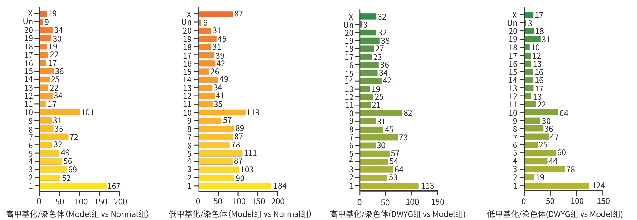
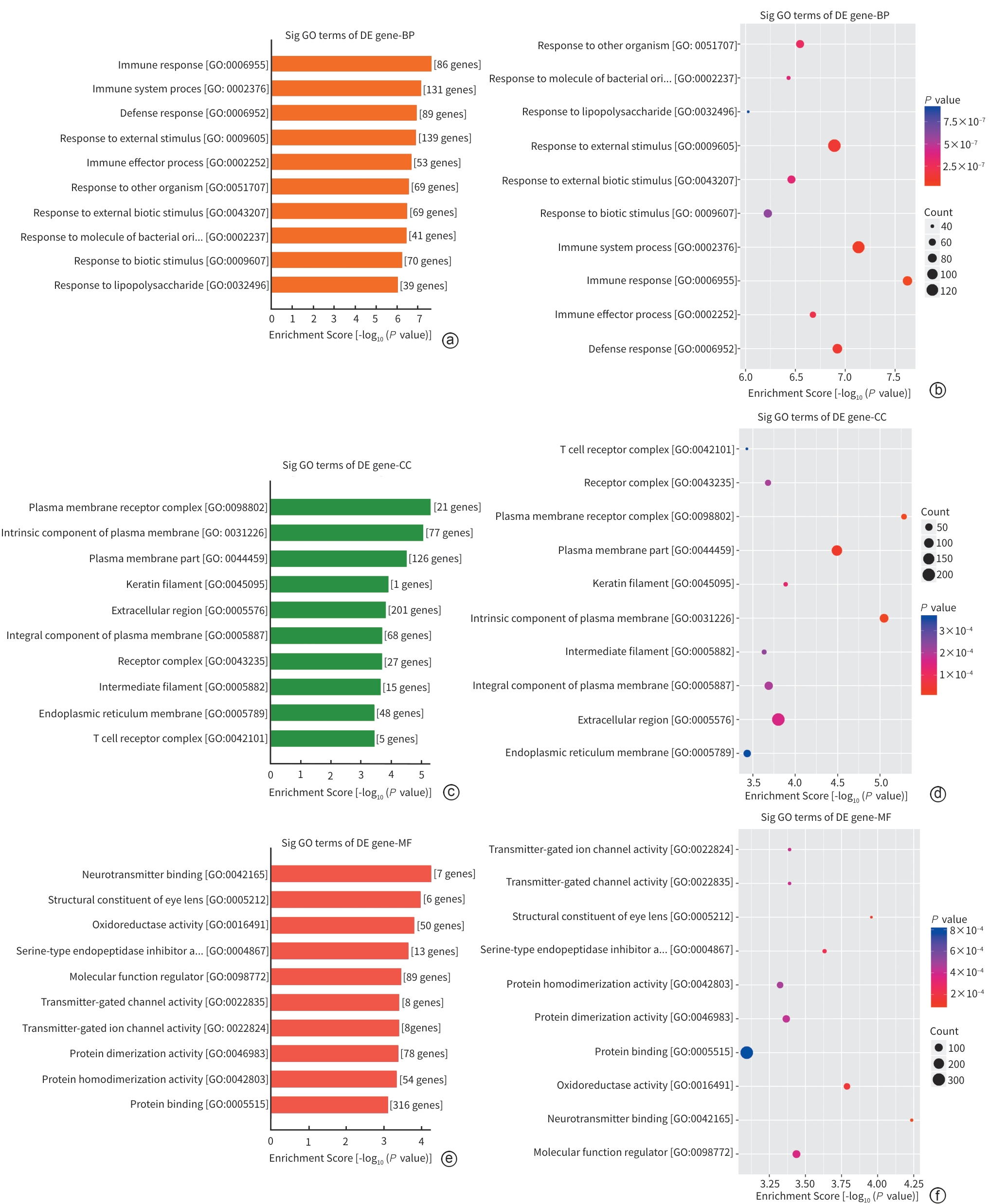
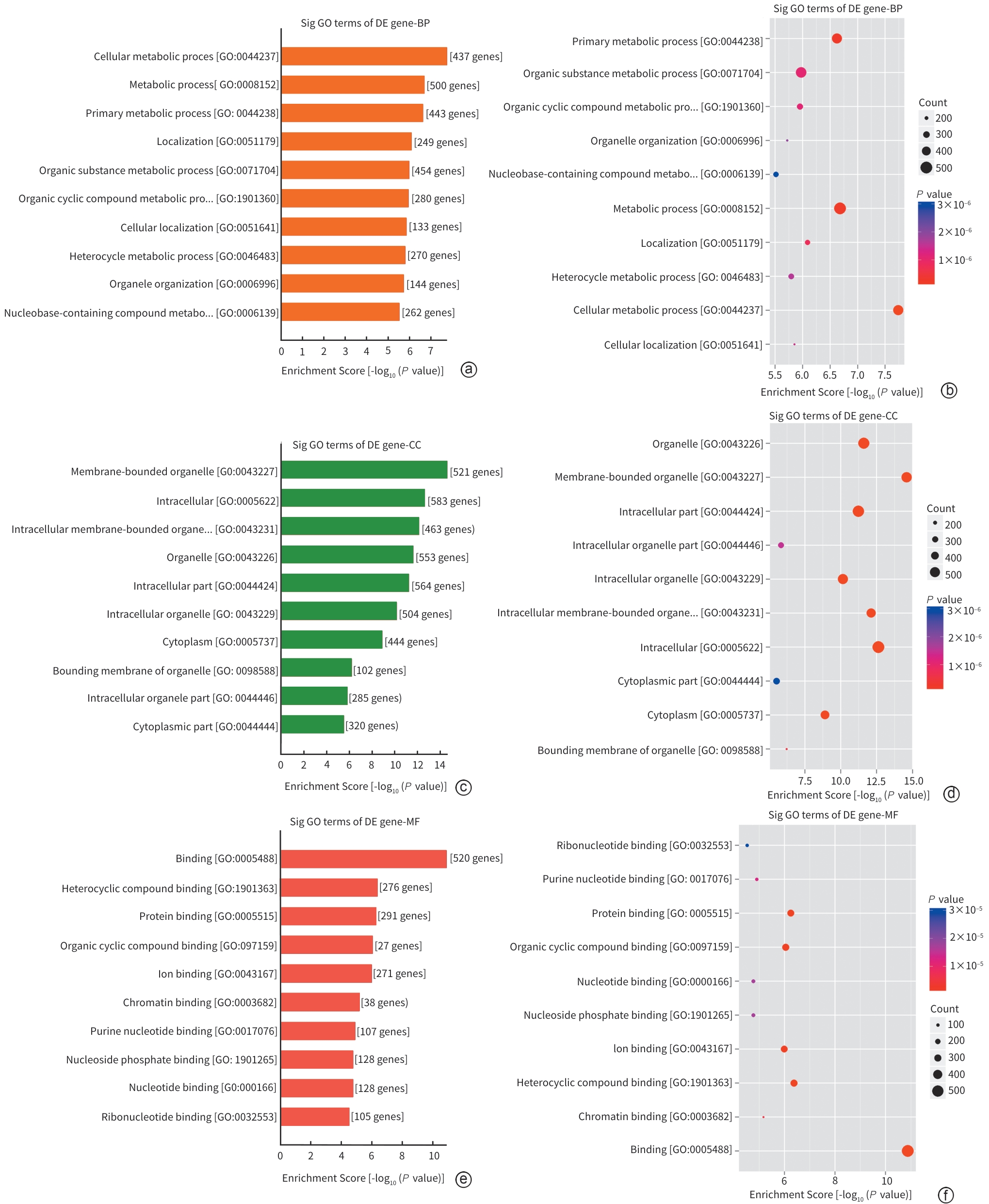
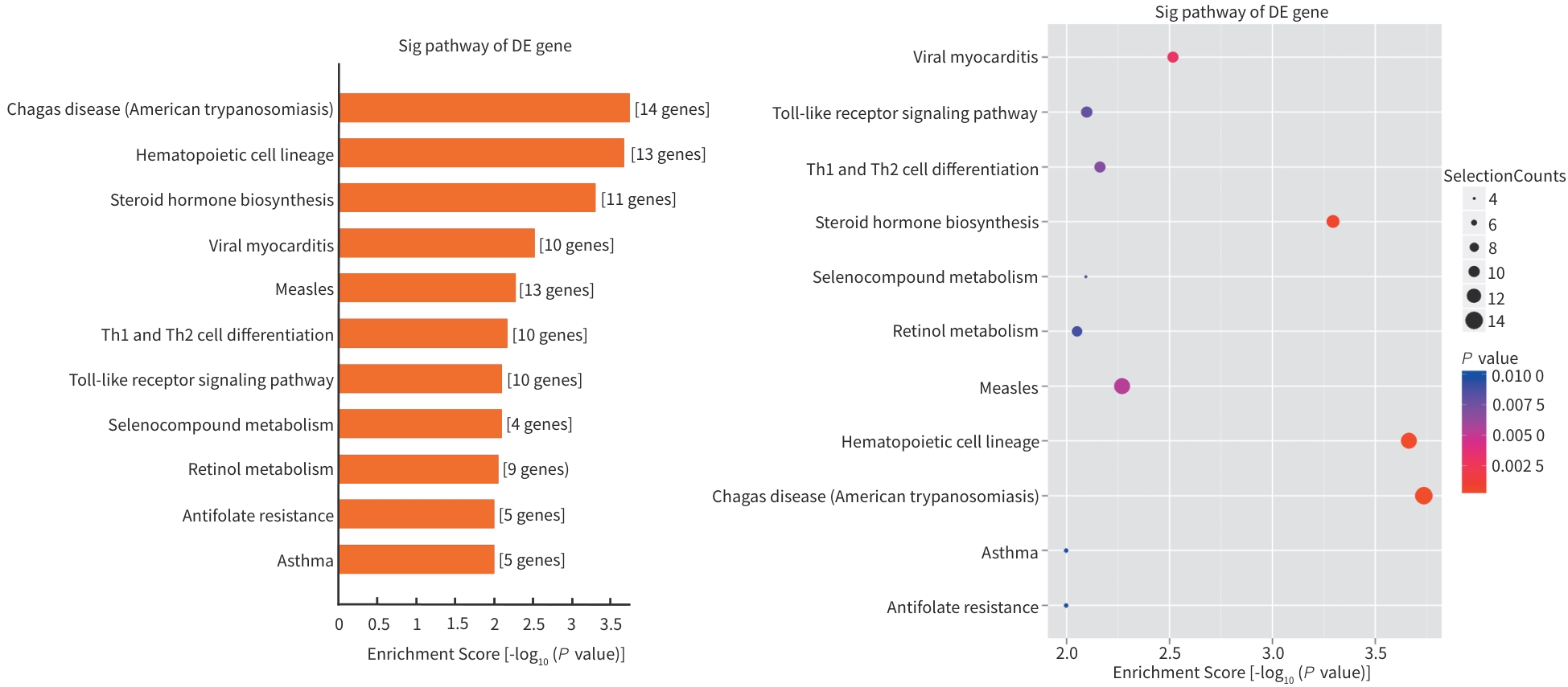
  • 加载中

Catalog

    通讯作者: 陈斌, bchen63@163.com
    • 1. 

      沈阳化工大学材料科学与工程学院 沈阳 110142

    1. 本站搜索
    2. 百度学术搜索
    3. 万方数据库搜索
    4. CNKI搜索

    Figures(8)  / Tables(1)

    Article Metrics

    Article views (24) PDF downloads(7) Cited by()
    Proportional views
    Related

    /

    DownLoad:  Full-Size Img  PowerPoint
    Return
    Return