

 PDF下载 ( 1071 KB)

O-连接β-N-乙酰葡萄糖胺糖基化修饰在代谢相关脂肪性肝病中的作用
DOI: 10.12449/JCH260623
利益冲突声明:本文不存在任何利益冲突。
作者贡献声明:刘素彤负责撰写论文及图片整理;张丽慧、赵晴等负责修改论文;马唯琛、朱万怡等负责文献检索及整理;赵文霞、刘鸣昊等负责拟定写作思路及最后定稿。
Role of O-linked β-N-acetylglucosamine modification in metabolic associated fatty liver disease
-
摘要: 代谢相关脂肪性肝病(MAFLD)作为全球范围内发病率迅速增长的慢性肝病,其复杂的发病机制与O-连接β-N-乙酰葡萄糖胺糖基化(O-GlcNAc)修饰密切相关。O-GlcNAc修饰作为一种动态可逆的蛋白质翻译后修饰,主要由O-GlcNAc转移酶和O-GlcNAc糖苷酶调控。O-GlcNAc修饰能驱动肝脂肪变性、加重胰岛素抵抗以及损害线粒体功能,加剧代谢紊乱,促进炎症反应,推动MAFLD向代谢相关脂肪性肝炎及肝纤维化发展。本文系统综述了O-GlcNAc修饰在MAFLD发生发展中的最新研究进展,旨在为深入理解MAFLD的病理机制及开发有效治疗策略提供理论支持和研究方向。
-
关键词:
- 代谢相关脂肪性肝病 /
- O-连接β-N-乙酰葡萄糖胺糖基化 /
- 蛋白质加工, 转译后
Abstract: Metabolic associated fatty liver disease (MAFLD) is a chronic liver disease with a rapidly increasing incidence rate worldwide, and its complex pathogenesis is closely associated with O-linked β-N-acetylglucosamine (O-GlcNAc) modification. As a dynamic and reversible post-translational modification of proteins, O-GlcNAc modification is mainly regulated by O-GlcNAc transferase and O-GlcNAcase. O-GlcNAc modification can drive hepatic steatosis, exacerbate insulin resistance, and impair mitochondrial function, thereby leading to the aggravation of metabolic disorders, promoting inflammation response, and driving the progression of MAFLD to metabolic associated steatohepatitis and hepatic fibrosis. This article systematically reviews the latest research advances in the role of O-GlcNAc modification in the development and progression of MAFLD, in order to provide theoretical support and research direction for a deeper understanding of the pathological mechanism of MAFLD and the development of effective therapeutic strategies. -
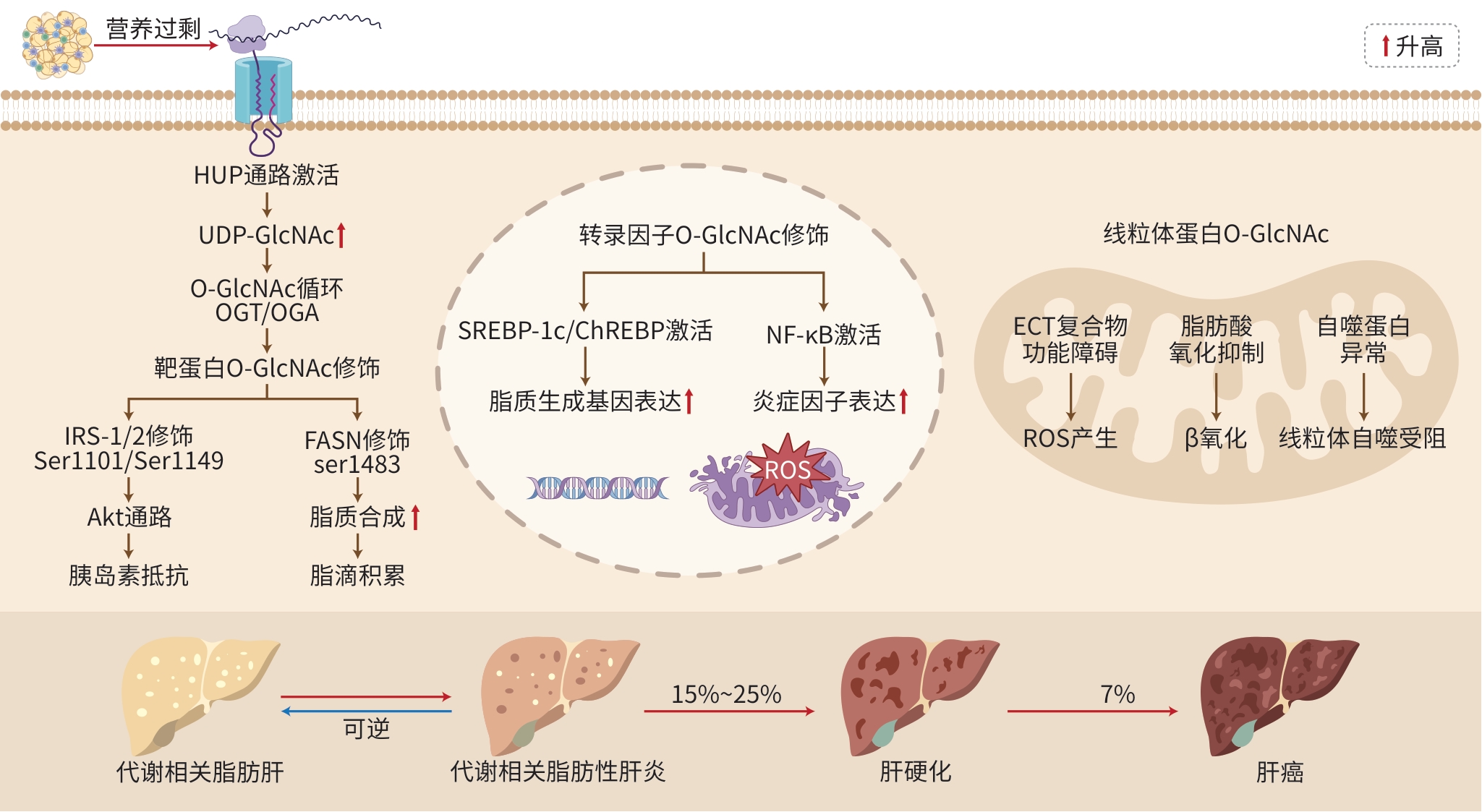
注: HUP,己糖胺生物合成途径;UDP-GlcNAc,尿苷二磷酸-N-乙酰葡糖胺;O-GlcNAc,O-连接β-N-乙酰葡萄糖胺糖基化;OGT,O-GlcNAc转移酶;OGA,O-GlcNAc糖苷酶;IRS-1/2,胰岛素受体底物1/2;Akt,蛋白激酶B;FASN,脂肪酸合酶;SREBP-1c,甾醇调节元件结合蛋白1c;ChREBP,碳水化合物应答元件结合蛋白;NF-κB,核因子κB;ECT,电子传递链;ROS,活性氧。
图 1 O-GlcNAc修饰在MAFLD中的机制变化
Figure 1. The mechanism changes of O-GlcNAc glycosylation modification in metabolic associated fatty liver disease
-
[1] XIE ZY, XIE T, LIU JY, et al. Emerging role of protein O-GlcNAcylation in liver metabolism: Implications for diabetes and NAFLD[J]. Int J Mol Sci, 2023, 24( 3): 2142. DOI: 10.3390/ijms24032142. [2] HU YJ, ZHANG X, LV HM, et al. Protein O-GlcNAcylation: The sweet hub in liver metabolic flexibility from a(patho)physiological perspective[J]. Liver Int, 2024, 44( 2): 293- 315. DOI: 10.1111/liv.15812. [3] LU QS, ZHANG XZ, LIANG TB, et al. O-GlcNAcylation: An important post-translational modification and a potential therapeutic target for cancer therapy[J]. Mol Med, 2022, 28( 1): 115. DOI: 10.1186/s10020-022-00544-y. [4] HONG ZZ, LOWE J, JIANG JY. Dissecting the mechanisms underlying substrate recognition and functional regulation of O-GlcNAc cycling enzymes[J]. ACS Chem Biol, 2025, 20( 11): 2534- 2546. DOI: 10.1021/acschembio.5c00633. [5] MA JF, HOU CY, WU C. Demystifying the O-GlcNAc code: A systems view[J]. Chem Rev, 2022, 122( 20): 15822- 15864. DOI: 10.1021/acs.chemrev.1c01006. [6] ZAHRA F, ZACHARA NE. OGT’s inner circle: Protein interactions and functional impact[J]. Adv Biol Regul, 2025: 101120. DOI: 10.1016/j.jbior.2025.101120. [7] LEI Q, YU HB, CHEN F, et al. Tissue-specific profiling of O-GlcNAcylated proteins in Drosophila using TurboID-CpOGA(M)[J]. Bio Protoc, 2025, 15( 5): e5234. DOI: 10.21769/BioProtoc.5234. [8] ZHU WZ, PALAZZO T, ZHOU MW, et al. First comprehensive identification of cardiac proteins with putative increased O-GlcNAc levels during pressure overload hypertrophy[J]. PLoS One, 2022, 17( 10): e0276285. DOI: 10.1371/journal.pone.0276285. [9] MORALES MM, PRATT MR. The post-translational modification O-GlcNAc is a sensor and regulator of metabolism[J]. Open Biol, 2024, 14( 10): 240209. DOI: 10.1098/rsob.240209. [10] SORIA LR, MAKRIS G, D’ALESSIO AM, et al. O-GlcNAcylation enhances CPS1 catalytic efficiency for ammonia and promotes ureagenesis[J]. Nat Commun, 2022, 13( 1): 5212. DOI: 10.1038/s41467-022-32904-x. [11] KALEEM A, JAVED S, REHMAN N, et al. Phosphorylated and O-GlcNAc modified IRS-1(Ser1101) and-2(Ser1149) contribute to human diabetes type II[J]. Protein Pept Lett, 2021, 28( 3): 333- 339. DOI: 10.2174/0929866527666200813210407. [12] SERMIKLI BP, AYDOGDU G, YILMAZ E. Role of the O-GlcNAc modification on insulin resistance and endoplasmic reticulum stress in 3T3-L1 cells[J]. Mol Biol Rep, 2020, 47( 8): 5927- 5942. DOI: 10.1007/s11033-020-05665-3. [13] ONG Q, LIM LTR, GOH C, et al. Spatiotemporal control of subcellular O-GlcNAc signaling using Opto-OGT[J]. Nat Chem Biol, 2025, 21( 2): 300- 308. DOI: 10.1038/s41589-024-01770-7. [14] GONZALEZ-RELLAN MJ, FONDEVILA MF, FERNANDEZ U, et al. O-GlcNAcylated p53 in the liver modulates hepatic glucose production[J]. Nat Commun, 2021, 12( 1): 5068. DOI: 10.1038/s41467-021-25390-0. [15] ZHOU YC, XU MX, YU PJ, et al. Empagliflozin downregulates AMP-activated protein kinase α O-GlcNAcylation to ameliorate hepatic steatosis[J]. FASEB J, 2025, 39( 20): e71151. DOI: 10.1096/fj.202500538RR. [16] RAAB S, GADAULT A, VERY N, et al. Dual regulation of fatty acid synthase(FASN) expression by O-GlcNAc transferase(OGT) and mTOR pathway in proliferating liver cancer cells[J]. Cell Mol Life Sci, 2021, 78( 13): 5397- 5413. DOI: 10.1007/s00018-021-03857-z. [17] LI XS, ZHANG ZY, ZHANG M, et al. Mechanism of O-GlcNAcylation regulating liver lipid synthesis in mice through FASN[J]. FASEB J, 2025, 39( 4): e70359. DOI: 10.1096/fj.202402451RR. [18] VANAUBERG D, SCHULZ C, RAAB S, et al. O-GlcNAcylation of fatty acid synthase is required for its proper subcellular localization, expression level, and activity[J]. J Biol Chem, 2025, 301( 8): 110497. DOI: 10.1016/j.jbc.2025.110497. [19] PANG YN, XU X, XIANG XJ, et al. High fat activates O-GlcNAcylation and affects AMPK/ACC pathway to regulate lipid metabolism[J]. Nutrients, 2021, 13( 6): 1740. DOI: 10.3390/nu13061740. [20] YANG YF, FU MN, LI MD, et al. O-GlcNAc transferase inhibits visceral fat lipolysis and promotes diet-induced obesity[J]. Nat Commun, 2020, 11( 1): 181. DOI: 10.1038/s41467-019-13914-8. [21] SUN QH, WANG YS, LIU GL, et al. Enhanced O-linked Glcnacylation in Crohn’s disease promotes intestinal inflammation[J]. EBioMedicine, 2020, 53: 102693. DOI: 10.1016/j.ebiom.2020.102693. [22] CHEN HH, SHI YH, YING JY, et al. O-linked N-acetylglucosamine modification induced by lipopolysaccharide is involved in inflammatory signaling pathway in endothelial cells[J]. Chin Crit Care Med, 2023, 35( 2): 164- 169. DOI: 10.3760/cma.j.cn121430-20220314-00242.陈赫赫, 石燕华, 应佳云, 等. 脂多糖诱导内皮细胞O-GlcNAc修饰参与炎症信号通路[J]. 中华危重病急救医学, 2023, 35( 2): 164- 169. DOI: 10.3760/cma.j.cn121430-20220314-00242. [23] MAO Z, MU JP, GAO ZX, et al. Biological functions and potential therapeutic significance of O-GlcNAcylation in hepatic cellular stress and liver diseases[J]. Cells, 2024, 13( 10): 805. DOI: 10.3390/cells13100805. [24] JÓŹWIAK P, CIESIELSKI P, ZAKRZEWSKI PK, et al. Mitochondrial O-GlcNAc transferase interacts with and modifies many proteins and its up-regulation affects mitochondrial function and cellular energy homeostasis[J]. Cancers, 2021, 13( 12): 2956. DOI: 10.3390/cancers13122956. [25] XUE Q, YAN R, JI ST, et al. Regulation of mitochondrial network homeostasis by O-GlcNAcylation[J]. Mitochondrion, 2022, 65: 45- 55. DOI: 10.1016/j.mito.2022.04.007. [26] ALGHUSEN IM, CARMAN MS, WILKINS HM, et al. O-GlcNAc impacts mitophagy via the PINK1-dependent pathway[J]. Front Aging Neurosci, 2024, 16: 1387931. DOI: 10.3389/fnagi.2024.1387931. [27] WANG FX, CHEN L, ZHANG BY, et al. O-GlcNAcylation coordinates glutaminolysis by regulating the stability and membrane trafficking of ASCT2 in hepatic stellate cells[J]. J Clin Transl Hepatol, 2022, 10( 6): 1107- 1116. DOI: 10.14218/JCTH.2021.00413. [28] LI R, ONG Q, WONG CC, et al. O-GlcNAcylation inhibits hepatic stellate cell activation[J]. J Gastroenterol Hepatol, 2021, 36( 12): 3477- 3486. DOI: 10.1111/jgh.15690. [29] ZHANG BC, LAPENTA K, WANG Q, et al. Trefoil factor 2 secreted from damaged hepatocytes activates hepatic stellate cells to induce fibrogenesis[J]. J Biol Chem, 2021, 297( 1): 100887. DOI: 10.1016/j.jbc.2021.100887. [30] YANG F, CHEN Y, ZHENG G, et al. LIMA1 O-GlcNAcylation promotes hepatic lipid deposition through inducing β-catenin-regulated FASn expression in metabolic dysfunction-associated steatotic liver disease[J]. Adv Sci(Weinh), 2025, 12( 15): e2415941. DOI: 10.1002/advs.202415941. [31] ROBARTS DR, KOTULKAR M, PAINE-CABRERA D, et al. The essential role of O-GlcNAcylation in hepatic differentiation[J]. Hepatol Commun, 2023, 7( 11): e0283. DOI: 10.1097/HC9.0000000000000283. [32] LI S, YANG F, CHENG F, et al. Lipotoxic hepatocyte derived LIMA1 enriched small extracellular vesicles promote hepatic stellate cells activation via inhibiting mitophagy[J]. Cell Mol Biol Lett, 2024, 29( 1): 82. DOI: 10.1186/s11658-024-00596-4. [33] MUKHERJEE S, CHAKRABORTY M, ULMASOV B, et al. Pleiotropic actions of IP6K1 mediate hepatic metabolic dysfunction to promote nonalcoholic fatty liver disease and steatohepatitis[J]. Mol Metab, 2021, 54: 101364. DOI: 10.1016/j.molmet.2021.101364. [34] LI S, NI P, SHAO H, et al. Dual-targeted liposomes delivering ginsenoside CK attenuate cerebral ischemia-reperfusion injury by suppressing PANoptosis via O-GlcNAcylation of RIPK1/RIPK3[J]. J Ginseng Res, 2026. DOI: 10.1016/j.jgr.2026.100978.[ Epub ahead of print] [35] ROBARTS DR, MCGREAL SR, UMBAUGH DS, et al. Regulation of liver regeneration by hepatocyte O-GlcNAcylation in mice[J]. Cell Mol Gastroenterol Hepatol, 2022, 13( 5): 1510- 1529. DOI: 10.1016/j.jcmgh.2022.01.014. [36] HU JC, CHEN RY, JIA P, et al. Augmented O-GlcNAc signaling via glucosamine attenuates oxidative stress and apoptosis following contrast-induced acute kidney injury in rats[J]. Free Radic Biol Med, 2017, 103: 121- 132. DOI: 10.1016/j.freeradbiomed.2016.12.032. [37] HODREA J, BALOGH DB, HOSSZU A, et al. Reduced O-GlcNAcylation and tubular hypoxia contribute to the antifibrotic effect of SGLT2 inhibitor dapagliflozin in the diabetic kidney[J]. Am J Physiol Renal Physiol, 2020, 318( 4): F1017- F1029. DOI: 10.1152/ajprenal.00021.2020. [38] CHEN DQ, QIU ZJ, WU YX, et al. Astragalus polysaccharide enhances OGT-mediated O-GlcNAcylation to stabilize PINK1 to induce mitophagy in D-galactose treated C2C12 myoblasts[J]. Int Immunopharmacol, 2025, 166: 115617. DOI: 10.1016/j.intimp.2025.115617. -

 下载:
下载:
 本文二维码
本文二维码
图(1)
计量
- 文章访问数: 414
- HTML全文浏览量: 125
- PDF下载量: 99
- 被引次数: 0




