

 PDF下载 ( 80767 KB)

偶氮鬼臼毒素衍生物SU056对四氯化碳诱导的肝纤维化小鼠模型的影响及其作用机制
DOI: 10.12449/JCH260612
Effect and mechanism of the azo-podophyllotoxin derivative SU056 in a mouse model of carbon tetrachloride-induced liver fibrosis
-
摘要:
目的 探究偶氮鬼臼毒素衍生物SU056治疗四氯化碳(CCl4)诱导的小鼠肝纤维化的效果,并解析相关作用机制。 方法 选取12只小鼠随机分为3组:对照组、模型组(CCl4+生理盐水组)和治疗组(CCl4+SU056),每组4只。通过腹腔注射CCl4构建小鼠肝纤维化模型,并在造模中期开始每日腹腔注射SU056进行治疗。通过苏木素-伊红染色、马松染色、天狼星红染色评估肝组织病理损伤与胶原沉积,并通过检测肝组织羟脯氨酸含量及血清丙氨酸氨基转移酶、天冬氨酸氨基转移酶水平评价肝功能。采用免疫荧光检测平滑肌肌动蛋白α(α-SMA)、Ⅰ型胶原和Y-框结合蛋白1(YB1)的表达。以人肝星状细胞(HSC)系LX-2以及小鼠原代HSC为研究对象,通过细胞计数试剂盒法检测细胞增殖;Transwell实验检测细胞迁移;实时荧光定量逆转录聚合酶链式反应和免疫印迹检测Ⅰ型胶原、Ⅲ型胶原、YB1、磷酸化哺乳动物雷帕霉素靶蛋白(mTOR)和磷酸化S6K的表达,从而验证YB1/mTOR信号轴的功能。计量资料多组间比较采用单因素方差分析或双因素方差分析,进一步两两比较采用LSD-t检验。 结果 在CCl4诱导的肝纤维化小鼠模型中,与模型组相比,治疗组小鼠肝组织炎症细胞浸润、胶原沉积及假小叶形成显著减轻,血清丙氨酸氨基转移酶、天冬氨酸氨基转移酶水平以及肝组织羟脯氨酸水平显著降低(P值均<0.01)。免疫荧光显示,SU056显著抑制了肝组织中α-SMA、Ⅰ型胶原及YB1的异常高表达(P值均<0.01)。体外实验表明,SU056以剂量依赖性方式抑制转化生长因子β1(TGF-β1)诱导的LX-2细胞增殖(P<0.01)、迁移(P<0.05)及Ⅰ型胶原、Ⅲ型胶原转录上调(P值均<0.05);且SU056还可抑制原代HSC的体外自发活化。机制研究表明,TGF-β1可同时上调LX-2细胞中YB1和磷酸化mTOR、磷酸化S6K的表达;两种浓度SU056(10、20 μmol/L)处理均能下调Ⅰ型胶原、YB1、磷酸化mTOR及磷酸化S6K的蛋白水平。特异性敲低YB1或使用mTOR抑制剂雷帕霉素,也能产生与SU056类似的效应。在CCl4模型小鼠肝组织中,SU056同样能抑制α-SMA与磷酸化mTOR的共表达上调(P<0.01)。 结论 SU056在体内外均能有效抑制HSC的活化、增殖、迁移及细胞外基质生成,从而缓解肝纤维化进程,其作用机制是通过阻断YB1/mTOR信号轴的正反馈效应,实现对HSC活化的抑制。 -
关键词:
- 肝纤维化 /
- 肝星状细胞 /
- 药物疗法 /
- 小鼠, 近交C57BL
Abstract:Objective To investigate the effect of SU056, an azo-podophyllotoxin derivative, on carbon tetrachloride (CCl4)-induced liver fibrosis in mice and related mechanisms of action. Methods A total of 12 mice were randomly divided into control group, model group (CCl4+normal saline), and treatment group (CCl4+SU056), with 4 mice in each group. Mice were given intraperitoneal injection of CCl4 to establish a model of liver fibrosis, and during the middle stage of modeling, the mice in the treatment group were given daily intraperitoneal injection of SU056. Liver histopathological injury, collagen deposition, and liver function were assessed based on HE staining, Masson staining, Sirius Red staining, the content of hydroxyproline in liver tissue, and the serum levels of alanine aminotransferase and aspartate aminotransferase, and immunofluorescence assay was used to measure the expression levels of smooth muscle actin α (α-SMA), collagen type Ⅰ, and Y-box binding protein 1 (YB1). The human hepatic stellate cell (HSC) line LX-2 and primary mouse HSC were used, and CCK-8 assay was used to measure cell proliferation; Transwell assay was used to observe cell migration; quantitative reverse transcription-polymerase chain reaction and Western Blot were used to measure the expression levels of collagen type Ⅰ, collagen type Ⅲ, YB1, phosphorylated mammalian target of rapamycin (mTOR), and phosphorylated S6K, so as to validate the function of the YB1/mTOR signaling axis. The one-way or two-way analysis of variance was used for comparison of continuous data between multiple groups, and the least significant difference t-test was used for further comparison between two groups. Results In the mouse model of liver fibrosis induced by CCl4, compared with the model group, the treatment group had significant alleviation of inflammatory cell infiltration, collagen deposition, and pseudolobule formation in liver tissue and significant reductions in the serum levels of alanine aminotransferase and aspartate aminotransferase and the content of hydroxyproline in liver tissue (all P<0.01). Immunofluorescence assay showed that SU056 significantly inhibited the abnormal high expression of α-SMA, collagen type I, and YB1 in liver tissue (all P<0.01). In vitro experiments showed that SU056 inhibited the transforming growth factor-β1-induced proliferation of LX-2 cells (P<0.01), the migration of LX-2 cells (P<0.05), and the transcriptional up-regulation of collagen type Ⅰ and collagen type Ⅲ (all P<0.05) in a dose-dependent manner, and SU056 could inhibit the spontaneous activation of primary HSC in vitro. Mechanistic studies revealed that transforming growth factor-β1 simultaneously upregulated the expression levels of YB1, phosphorylated mTOR, and phosphorylated S6K in LX-2 cells, and treatment with SU056 (10 and 20 µmol/L) could downregulate the protein expression levels of collagen type I, YB1, phosphorylated mTOR, and phosphorylated S6K. Specific knockdown of YB1 or administration of the mTOR inhibitor rapamycin exerted a similar effect as SU056. SU056 also inhibited the co-upregulation of α-SMA and phosphorylated mTOR in liver tissue of model mice (P<0.01). Conclusion SU056 can effectively inhibit HSC activation, proliferation, migration, and extracellular matrix production both in vivo and in vitro and thus delay the progression of liver fibrosis, by disrupting the YB1/mTOR positive feedback signaling axis. -
Key words:
- Hepatic Fibrosis /
- Hepatic Stellate Cells /
- Drug Therapy /
- Mice, Inbred C57BL
-

注: a,肝纤维化组织染色(×200);b,天狼星红染色相对阳性面积及肝功能指标。CCl4,四氯化碳;HE,苏木素-伊红;ALT,丙氨酸氨基转移酶;AST,天冬氨酸氨基转移酶。***P<0.001;**P<0.01。
图 1 SU056对CCl4诱导的肝纤维化及肝损伤的影响
Figure 1. Effect of SU056 on CCl4-induced liver fibrosis and liver function injury
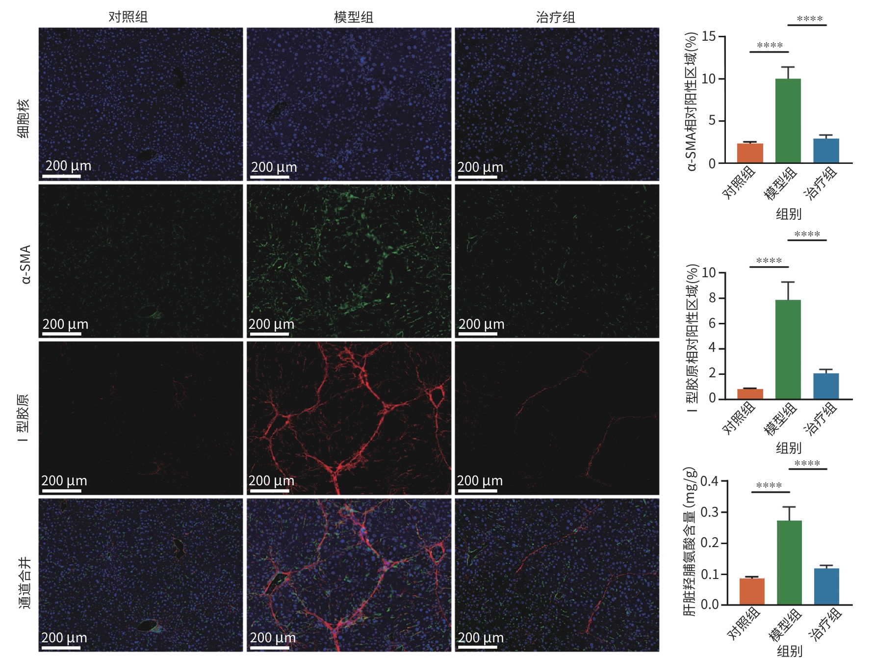
注: CCl4,四氯化碳;α-SMA,平滑肌肌动蛋白α。****P<0.000 1。
图 2 SU056对CCl4引起的肝脏胶原沉积及HSC活化的影响(免疫荧光染色,×200)
Figure 2. Effect of SU056 on CCl4-induced liver collagen deposition and HSC activation (IF staining,×200)

注: a,肝组织免疫荧光染色以及α-SMA、YB1相对阳性区域统计(×400);b,免疫荧光检测小鼠原代HSC体外培养自活化程度以及SU056治疗效果(×400)。CCl4,四氯化碳;HSC,肝星状细胞;α-SMA,平滑肌肌动蛋白α;YB1,Y-框结合蛋白1;DMSO,二甲基亚砜。****P<0.000 1;***P<0.001;**P<0.01。
图 3 SU056对CCl4模型以及体外自活化HSC中的YB1表达的影响
Figure 3. Effect of SU056 on YB1 expression in the CCl4 model and in vitro activated HSC

注: a,CCK-8检测不同浓度SU056抑制活化LX-2细胞增殖的效果;b,qRT-PCR检测不同浓度SU056抑制活化LX-2细胞中Ⅰ型胶原和Ⅲ型胶原基因转录;c,免疫荧光检测SU056抑制活化LX-2细胞表达Ⅰ型胶原蛋白(×200);d,SU056抑制活化LX-2细胞的迁移能力(结晶紫染色,×200);e:SU056抑制活化LX-2细胞表达Ⅰ型胶原蛋白和细胞迁移能力效果。HSC,肝星状细胞;DMSO,二甲基亚砜;TGF-β1,转化生长因子β1;ECM,细胞外基质。****P<0.000 1;***P<0.001;**P<0.01;*P<0.05。
图 4 SU056对HSC的增殖、迁移以及ECM生成的影响
Figure 4. Effect of SU056 on HSC proliferation, migration, and ECM production
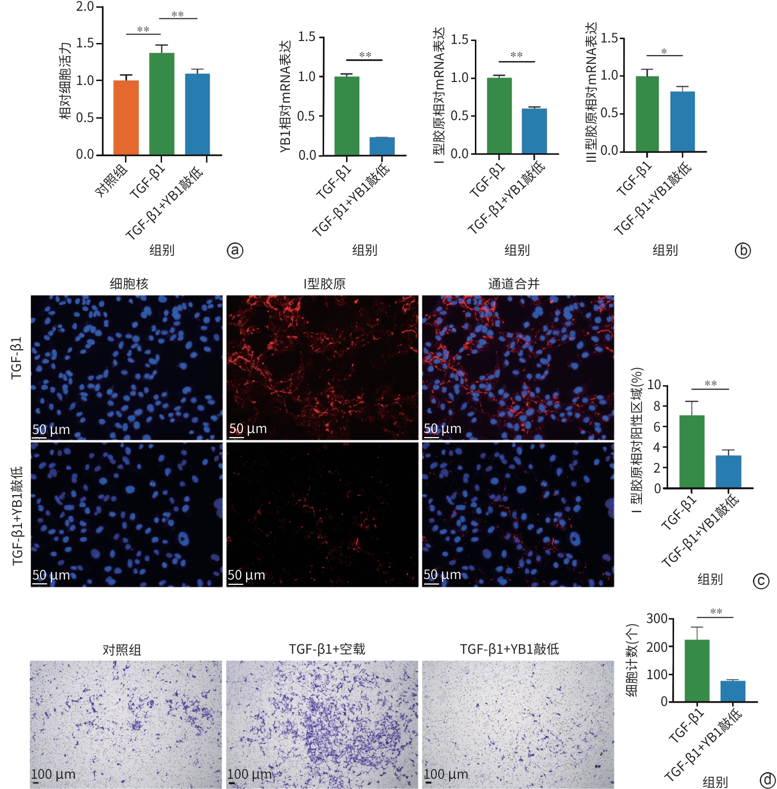
注: a,CCK-8检测YB1敲减抑制活化LX-2细胞增殖的效果;b,qRT-PCR检测YB1敲减抑制活化LX-2细胞中Ⅰ型胶原和Ⅲ型胶原基因转录的效果;c,免疫荧光检测YB1敲减抑制活化LX-2细胞表达Ⅰ型胶原蛋白及统计分析(×200);d,YB1敲减抑制活化LX-2细胞的迁移能力及统计分析(结晶紫染色,×200)。YB1,Y-框结合蛋白1;TGF-β1,转化生长因子β1;ECM,细胞外基质;HSC,肝星状细胞。**P<0.01;*P<0.05。
图 5 YB1调控HSC的增殖、迁移以及ECM生成
Figure 5. YB1 regulates HSC proliferation, migration, and ECM production

注: a,免疫印迹检测SU056抑制活化LX-2细胞中mTOR通路活化;b,免疫印迹检测YB1敲减抑制活化LX-2细胞中mTOR通路活化;c,免疫印迹显示抑制LX-2细胞的mTOR通路可以减少YB1蛋白表达;d,小鼠肝组织免疫荧光染色(×200)以及α-SMA、磷酸化mTOR相对阳性区域统计;e,免疫印迹实验各目的蛋白条带灰度值统计。mTOR,哺乳动物雷帕霉素靶蛋白;YB1,Y-框结合蛋白1;HSC,肝星状细胞;α-SMA,平滑肌肌动蛋白α。****P<0.000 1;***P<0.001;**P<0.01;*P<0.05。
图 6 SU056通过YB1/mTOR信号轴调控HSC活化
Figure 6. SU056 regulates HSC activation through the YB1/mTOR signaling axis
表 1 qPCR引物序列
Table 1. Primers used for qPCR
基因 引物序列 Actin 5'-CACCATTGGCAATGAGCGGTTC-3' 3'-AGGTCTTTGCGGATGTCCACGT-5' Col1a1 5'-CCGCTTCACCTACAGCGTCA-3' 3'-TTGTATTCAATCACTGTCTTGCCC-5' Col3a1 5'-TGGTCTGCAAGGAATGCCTGGA-3' 3'-TCTTTCCCTGGGACACCATCAG-5' YB1 5'-GCAGGAGAACAAGGTAGACCAG-3' 3'-CTTCATTGCCGTCCTCTCTAGG-5' 注:qPCR,实时荧光定量聚合酶链式反应。
 下载: 导出CSV
-
[1] TSUCHIDA T, FRIEDMAN SL. Mechanisms of hepatic stellate cell activation[J]. Nat Rev Gastroenterol Hepatol, 2017, 14( 7): 397- 411. DOI: 10.1038/nrgastro.2017.38. [2] KISSELEVA T, BRENNER D. Molecular and cellular mechanisms of liver fibrosis and its regression[J]. Nat Rev Gastroenterol Hepatol, 2021, 18( 3): 151- 166. DOI: 10.1038/s41575-020-00372-7. [3] GUO YC, ZHANG QQ, LI BH, et al. Biliary YB-1/GLI2 axis facilitates ductular reaction and promotes HSC activation via SPP1/integrin αvβ1 signaling during liver fibrogenesis[J]. Hepatology, 2025. DOI: 10.1097/HEP.0000000000001458.[ Epub ahead of print] [4] TANG ZH, LIN BW, LI W, et al. Y-box binding protein 1 promotes chromatin accessibility to aggravate liver fibrosis[J]. Cell Signal, 2023, 109: 110750. DOI: 10.1016/j.cellsig.2023.110750. [5] ZHANG Y, HE FF, LEI MZ, et al. Hepatic stellate cell-derived IL-11 exacerbates liver fibrosis via interplay between HSCs and macrophages[J]. Int J Biol Sci, 2026, 22( 1): 126- 141. DOI: 10.7150/ijbs.119659. [6] HUANG H, RU SJ, CHEN JM, et al. Quantitative proteomic study reveals amygdalin alleviates liver fibrosis through inhibiting mTOR/PDCD4/JNK pathway in hepatic stellate cells[J]. Drug Des Devel Ther, 2025, 19: 3735- 3749. DOI: 10.2147/DDDT.S500439. [7] WANG JZ, ZHU H, YOU P, et al. Upregulated YB-1 protein promotes glioblastoma growth through a YB-1/CCT4/mLST8/mTOR pathway[J]. J Clin Invest, 2022, 132( 8): e146536. DOI: 10.1172/JCI146536. [8] DHEERAJ A, GARCIA MARQUES FJ, TAILOR D, et al. Inhibition of protein translational machinery in triple-negative breast cancer as a promising therapeutic strategy[J]. Cell Rep Med, 2024, 5( 5): 101552. DOI: 10.1016/j.xcrm.2024.101552. [9] LI BR, XING FL, WANG JY, et al. YBX1 as a therapeutic target to suppress the LRP1-β-catenin-RRM1 axis and overcome gemcitabine resistance in pancreatic cancer[J]. Cancer Lett, 2024, 602: 217197. DOI: 10.1016/j.canlet.2024.217197. [10] MENG HY, MIAO HX, ZHANG YS, et al. YBX1 promotes homologous recombination and resistance to platinum-induced stress in ovarian cancer by recognizing m5C modification[J]. Cancer Lett, 2024, 597: 217064. DOI: 10.1016/j.canlet.2024.217064. [11] LI Y, CAI SM, ZHANG LL, et al. Simultaneous isolation and primary culture of rat hepatocytes, hepatic stellate cells, Kupffer’s cells and hepatic sinus endothelial cells[J]. J South Med Univ, 2014, 34( 4): 532- 537. DOI: 10.3969/j.issn.1673-4254.2014.04.20.李洋, 蔡双明, 张莉莉, 等. 大鼠原代肝细胞、星状细胞、枯否细胞和肝窦内皮细胞的同步分离与培养[J]. 南方医科大学学报, 2014, 34( 4): 532- 537. DOI: 10.3969/j.issn.1673-4254.2014.04.20. [12] PAROLA M, PINZANI M. Liver fibrosis: Pathophysiology, pathogenetic targets and clinical issues[J]. Mol Aspects Med, 2019, 65: 37- 55. DOI: 10.1016/j.mam.2018.09.002. [13] LIU PY, MAO YC, XIE Y, et al. Stem cells for treatment of liver fibrosis/cirrhosis: Clinical progress and therapeutic potential[J]. Stem Cell Res Ther, 2022, 13( 1): 356. DOI: 10.1186/s13287-022-03041-5. [14] YAO LC, HU X, DAI K, et al. Mesenchymal stromal cells: Promising treatment for liver cirrhosis[J]. Stem Cell Res Ther, 2022, 13( 1): 308. DOI: 10.1186/s13287-022-03001-z. [15] DAI HR, ZHU C, HUAI Q, et al. Chimeric antigen receptor-modified macrophages ameliorate liver fibrosis in preclinical models[J]. J Hepatol, 2024, 80( 6): 913- 927. DOI: 10.1016/j.jhep.2024.01.034. [16] ROEHLEN N, CROUCHET E, BAUMERT TF. Liver fibrosis: Mechanistic concepts and therapeutic perspectives[J]. Cells, 2020, 9( 4): 875. DOI: 10.3390/cells9040875. [17] LI BH, LI F, GU TY, et al. Specific knockdown of Y-box binding protein 1 in hepatic progenitor cells inhibits proliferation and alleviates liver fibrosis[J]. Eur J Pharmacol, 2022, 921: 174866. DOI: 10.1016/j.ejphar.2022.174866. [18] XIE GF, XIE YP, XU XC. Multifaceted functions of Y-box binding protein 1 in RNA methylated modifications(Review)[J]. Mol Med Rep, 2025, 32( 6): 1- 10. DOI: 10.3892/mmr.2025.13707. [19] KAMALOV G, VARMA BR, LU L, et al. Expression of the multifunctional Y-box protein, YB-1, in myofibroblasts of the infarcted rat heart[J]. Biochem Biophys Res Commun, 2005, 334( 1): 239- 244. DOI: 10.1016/j.bbrc.2005.06.082. [20] KIM YC, GUAN KL. mTOR: A pharmacologic target for autophagy regulation[J]. J Clin Invest, 2015, 125( 1): 25- 32. DOI: 10.1172/JCI73939. [21] MOSSMANN D, PARK S, HALL MN. mTOR signalling and cellular metabolism are mutual determinants in cancer[J]. Nat Rev Cancer, 2018, 18( 12): 744- 757. DOI: 10.1038/s41568-018-0074-8. [22] ZHAO ZZ, ZHANG YM, ZHANG CY, et al. TGF-β promotes pericyte-myofibroblast transition in subretinal fibrosis through the Smad2/3 and Akt/mTOR pathways[J]. Exp Mol Med, 2022, 54( 5): 673- 684. DOI: 10.1038/s12276-022-00778-0. [23] HUANG GC, YANG XS, YU QY, et al. Overexpression of STX11 alleviates pulmonary fibrosis by inhibiting fibroblast activation via the PI3K/AKT/mTOR pathway[J]. Signal Transduct Target Ther, 2024, 9( 1): 306. DOI: 10.1038/s41392-024-02011-y. [24] ZHAO XJ, YANG WJ, YU TM, et al. Th17 cell-derived amphiregulin promotes colitis-associated intestinal fibrosis through activation of mTOR and MEK in intestinal myofibroblasts[J]. Gastroenterology, 2023, 164( 1): 89- 102. DOI: 10.1053/j.gastro.2022.09.006. [25] ZHANG ZL, SHANG J, YANG QY, et al. Exosomes derived from human adipose mesenchymal stem cells ameliorate hepatic fibrosis by inhibiting PI3K/Akt/mTOR pathway and remodeling choline metabolism[J]. J Nanobiotechnology, 2023, 21( 1): 29. DOI: 10.1186/s12951-023-01788-4. -

 本文二维码
本文二维码
计量
- 文章访问数: 359
- HTML全文浏览量: 77
- PDF下载量: 103
- 被引次数: 0




