PDF下载 ( 1029 KB)

肠-肝轴与肝脏铁过载的交互作用对代谢相关脂肪性肝病的影响及相关干预策略
DOI: 10.12449/JCH260926
利益冲突声明:本文不存在任何利益冲突。
作者贡献声明:梁莹负责文献查阅,初稿写作;张晶晶负责文献查阅;赵文霞负责设计论文框架和思路;刘晓彦负责指导撰写、修改文章并最后定稿。
Impact of the interaction between the gut-liver axis and hepatic iron overload on metabolic dysfunction-associated fatty liver disease and related intervention strategies
-
摘要: 代谢相关脂肪性肝病(MAFLD)是一种主要由营养过剩和胰岛素抵抗导致的慢性、全球性肝脏疾病,其疾病谱广泛,发病率高,可进一步发展为肝炎、肝硬化及肝癌。MAFLD的发病涉及多个环节,但其具体作用机制尚未完全阐明。本文围绕“肠-肝轴紊乱与肝脏铁过载”这一前沿理论,归纳总结二者调控肝内脂毒性损伤、氧化应激、胰岛素抵抗及铁死亡的分子机制,揭示其协同促进MAFLD进展的病理生理过程,同时基于此理论阐述MAFLD的治疗策略,为其临床诊疗提供参考。Abstract: Metabolic dysfunction-associated fatty liver disease (MAFLD) is a chronic liver condition worldwide mainly caused by overnutrition and insulin resistance (IR). It encompasses a broad disease spectrum with a high incidence rate and may further progress to hepatitis, liver cirrhosis, and hepatocellular carcinoma. The pathogenesis of MAFLD involves multiple factors, but the specific mechanism of action has not yet been fully elucidated. This article reviews the cutting-edge theory of “gut-liver axis dysfunction-hepatic iron overload”, summarizes the molecular mechanisms by which these two factors regulate intrahepatic lipotoxicity, oxidative stress, IR, and ferroptosis, reveals the pathophysiological process by which they synergistically promote the progression of MAFLD, and elaborates on the therapeutic strategies for MAFLD on this basis, so as to provide a reference for clinical diagnosis and treatment.
-
[1] Gofton C, Upendran Y, Zheng M H, et al. MAFLD: How is it different from NAFLD[J]. Clin Mol Hepatol, 2023, 29( Suppl): S17- S31. DOI: 10.3350/cmh.2022.0367. [2] Li J, Zou B Y, Yeo Y H, et al. Prevalence, incidence, and outcome of non-alcoholic fatty liver disease in Asia, 1999—2019: A systematic review and meta-analysis[J]. Lancet Gastroenterol Hepatol, 2019, 4( 5): 389- 398. DOI: 10.1016/S2468-1253(19)30039-1. [3] Liu Xuan, Li Chang, Zhang Hongli, et al. Clinical characteristics and influencing factors in type 2 diabetes mellitus patients with MAFLD at moderate-to-high risk of metabolic-associated fatty liver fibrosis[J]. Med J Chin PLA, 2025, 50( 11): 1366- 1373. DOI: 10.11855/j.issn.0577-7402.1948.2025.0811.刘璇, 李畅, 张宏利, 等. 代谢相关脂肪性肝纤维化中高风险2型糖尿病合并代谢相关脂肪性肝病患者的临床特征及其影响因素分析[J]. 解放军医学杂志, 2025, 50( 11): 1366- 1373. DOI: 10.11855/j.issn.0577-7402.1948.2025.0811. [4] Mandato C, Delli Bovi A P, Vajro P. The gut-liver axis as a target of liver disease management[J]. Hepatobiliary Surg Nutr, 2021, 10( 1): 100- 102. DOI: 10.21037/hbsn.2020.03.27. [5] Huang Deng, Zhai Fude, Li Jianwei. Mechanisms and therapeutic strategies of gut microbiota in the immune-suppressive micro-environment of hepatocellular carcinoma[J]. Chin J Dig Surg, 2025, 24( 11): 1451- 1458. DOI: 10.3760/cma.j.cn115610-20251008-00616.黄登, 翟富德, 李建伟. 肠道菌群在肝细胞癌免疫抑制微环境中的作用机制与治疗策略[J]. 中华消化外科杂志, 2025, 24( 11): 1451- 1458. DOI: 10.3760/cma.j.cn115610-20251008-00616. [6] Babuta M, Morel C, de Carvalho Ribeiro M, et al. Neutrophil extracellular traps activate hepatic stellate cells and monocytes via NLRP3 sensing in alcohol-induced acceleration of MASH fibrosis[J]. Gut, 2024, 73( 11): 1854- 1869. DOI: 10.1136/gutjnl-2023-331447. [7] Trebicka J, Macnaughtan J, Schnabl B, et al. The microbiota in cirrhosis and its role in hepatic decompensation[J]. J Hepatol, 2021, 75( Suppl 1): S67- S81. DOI: 10.1016/j.jhep.2020.11.013. [8] Allam-Ndoul B, Castonguay-Paradis S, Veilleux A. Gut microbiota and intestinal trans-epithelial permeability[J]. Int J Mol Sci, 2020, 21( 17): 6402. DOI: 10.3390/ijms21176402. [9] Li Yongqiang, Tang Wenjuan, Zhou Yongjian. Role of intestinal microbiota and metabolites in the development, progression, and treatment of nonalcoholic fatty liver disease[J]. J Clin Hepatol, 2023, 39( 8): 1805- 1810. DOI: 10.3969/j.issn.1001-5256.2023.08.006.李永强, 唐文娟, 周永健. 肠道菌群及其代谢产物在非酒精性脂肪性肝病发生发展及治疗中的作用[J]. 临床肝胆病杂志, 2023, 39( 8): 1805- 1810. DOI: 10.3969/j.issn.1001-5256.2023.08.006. [10] Ren Lu, Liu Na. Characteristics of intestinal flora in metabolism-related fatty liver disease and the correlation with metabolic indicators[J]. Chin J Microecol, 2025, 37( 5): 577- 586. DOI: 10.13381/j.cnki.cjm.202505011.任璐, 刘娜. 代谢相关脂肪性肝病患者肠道菌群特征及其与代谢指标的相关性[J]. 中国微生态学杂志, 2025, 37( 5): 577- 586. DOI: 10.13381/j.cnki.cjm.202505011. [11] Meijnikman A S, Davids M, Herrema H, et al. Microbiome-derived ethanol in nonalcoholic fatty liver disease[J]. Nat Med, 2022, 28( 10): 2100- 2106. DOI: 10.1038/s41591-022-02016-6. [12] Guo J, Wang Y K, Zhang D M, et al. The FMO3/TMAO/HSP90β axis aggravates MAFLD by disrupting mitochondrial protein homeostasis[J]. Cell Signal, 2025, 134: 111960. DOI: 10.1016/j.cellsig.2025.111960. [13] Choi H, Choi J, Go Y, et al. Coenzyme Q and selenium co-supplementation alleviate methionine choline-deficient diet-induced metabolic dysfunction-associated steatohepatitis in mice[J]. Nutrients, 2025, 17( 2): 229. DOI: 10.3390/nu17020229. [14] Wei Herui, Xiao Fan, Wei Hongshan. Research progress on the mechanism of endoplasmic reticulum stress in metabolic dysfunction-associated fatty liver disease[J/CD]. Chin J Liver Dis Electron Version, 2024, 16( 1): 13- 17. DOI: 10.3969/j.issn.1674-7380.2024.01.003.韦何锐, 肖凡, 魏红山. 内质网应激在代谢相关脂肪性肝病中的作用机制研究进展[J/CD]. 中国肝脏病杂志(电子版), 2024, 16( 1): 13- 17. DOI: 10.3969/j.issn.1674-7380.2024.01.003. [15] Kolodziejczyk A A, Zheng D P, Shibolet O, et al. The role of the microbiome in NAFLD and NASH[J]. EMBO Mol Med, 2019, 11( 2): e9302. DOI: 10.15252/emmm.201809302. [16] Guo Ziwei, Wu Dongjie, Liu Jing, et al. Research progress on biological mechanism of bile acid metabolism therapy for metabolism related fatty liver[J]. Chin J Integr Tradit West Med, 2025, 45( 9): 1145- 1152. DOI: 10.7661/j.cjim.20250303.113.郭紫薇, 吴东杰, 刘婧, 等. 胆汁酸代谢治疗代谢相关性脂肪肝生物机制研究进展[J]. 中国中西医结合杂志, 2025, 45( 9): 1145- 1152. DOI: 10.7661/j.cjim.20250303.113. [17] Alhabeeb H, AlFaiz A, Kutbi E, et al. Gut hormones in health and obesity: The upcoming role of short chain fatty acids[J]. Nutrients, 2021, 13( 2): 481. DOI: 10.3390/nu13020481. [18] Pearce S C, Karl J P, Weber G J. Effects of short-chain fatty acids on intestinal function in an enteroid model of hypoxia[J]. Front Physiol, 2022, 13: 1056233. DOI: 10.3389/fphys.2022.1056233. [19] Sobieska K, Buczyńska A, Krętowski A J, et al. Iron homeostasis and insulin sensitivity: Unraveling the complex interactions[J]. Rev Endocr Metab Disord, 2024, 25( 5): 925- 939. DOI: 10.1007/s11154-024-09908-7. [20] Sung H K, Song E F, Jahng J W S, et al. Iron induces insulin resistance in cardiomyocytes via regulation of oxidative stress[J]. Sci Rep, 2019, 9: 4668. DOI: 10.1038/s41598-019-41111-6. [21] Zhang Lijia, Cai Jingming, Shen Jie, et al. Effects of iron overload on gene expressions of DMT1 and FPN1 in rats with nonalcoholic fatty liver disease[J]. Chin J Public Heal, 2018, 34( 5): 716- 719. DOI: 10.11847/zgggws1110438.张立加, 蔡静明, 沈洁, 等. 铁超载对NAFLD大鼠DMT1和FPN1基因表达影响[J]. 中国公共卫生, 2018, 34( 5): 716- 719. DOI: 10.11847/zgggws1110438. [22] Tao L, Yang X Q, Ge C D, et al. Integrative clinical and preclinical studies identify FerroTerminator1 as a potent therapeutic drug for MASH[J]. Cell Metab, 2024, 36( 10): 2190- 2206. e 5. DOI: 10.1016/j.cmet.2024.07.013. [23] Anastasopoulos N A, Barbouti A, Goussia A C, et al. Exploring the role of metabolic hyperferritinaemia(MHF) in steatotic liver disease(SLD) and hepatocellular carcinoma(HCC)[J]. Cancers, 2025, 17( 5): 842. DOI: 10.3390/cancers17050842. [24] Liang D G, Minikes A M, Jiang X J. Ferroptosis at the intersection of lipid metabolism and cellular signaling[J]. Mol Cell, 2022, 82( 12): 2215- 2227. DOI: 10.1016/j.molcel.2022.03.022. [25] Zhao Limei, Guo Xiaohong, Liu Lixin. Changes of serum iron metabolism and oxidative stress-related indexes in patients with non-alcoholic fatty liver disease: A Meta-analysis[J]. Chin J Gastroenterol Hepatol, 2022, 31( 10): 1113- 1122. DOI: 10.3969/j.issn.1006-5709.2022.10.007.赵丽梅, 郭晓红, 刘立新. 非酒精性脂肪性肝病患者血清铁代谢及氧化应激相关指标变化的Meta分析[J]. 胃肠病学和肝病学杂志, 2022, 31( 10): 1113- 1122. DOI: 10.3969/j.issn.1006-5709.2022.10.007. [26] Zhou Q, Sun Y M, Du S T, et al. Vorapaxar enhanced mitochondria-associated ferroptosis primes cancer immunotherapy via targeting FOXO1/HMOX1 axis[J]. Cell Rep Med, 2025, 6( 10): 102371. DOI: 10.1016/j.xcrm.2025.102371. [27] Guo X Y, Hong S Q, He H, et al. NF-κB promotes oxidative stress-induced necrosis and ischemia/reperfusion injury by inhibiting Nrf2-ARE pathway[J]. Free Radic Biol Med, 2020, 159: 125- 135. DOI: 10.1016/j.freeradbiomed.2020.07.031. [28] Camaschella C, Nai A, Silvestri L. Iron metabolism and iron disorders revisited in the hepcidin era[J]. Haematologica, 2020, 105( 2): 260- 272. DOI: 10.3324/haematol.2019.232124. [29] Bergheim I, Moreno-Navarrete J M. The relevance of intestinal barrier dysfunction, antimicrobial proteins and bacterial endotoxin in metabolic dysfunction-associated steatotic liver disease[J]. Eur J Clin Invest, 2024, 54( 7): e14224. DOI: 10.1111/eci.14224. [30] Shanmugam N K, Ellenbogen S, Trebicka E, et al. Tumor necrosis factor α inhibits expression of the iron regulating hormone hepcidin in murine models of innate colitis[J]. PLoS One, 2012, 7( 5): e38136. DOI: 10.1371/journal.pone.0038136. [31] Lee F S. At the crossroads of oxygen and iron sensing: Hepcidin control of HIF-2α[J]. J Clin Invest, 2019, 129( 1): 72- 74. DOI: 10.1172/JCI125509. [32] Zhang Lecong. Triclosan induces hepatotoxicity through disrupting iron metabolism and bile acid metabolism[D]. Guiyang: Guizhou Minzu University, 2023. DOI: 10.27807/d.cnki.cgzmz.2023.000530.张乐聪. 三氯生通过扰乱铁代谢和胆汁酸代谢致肝脏毒性的研究[D]. 贵阳: 贵州民族大学, 2023. DOI: 10.27807/d.cnki.cgzmz.2023.000530. [33] Rodrigues I G, Moreno-Navarrete J, Fernández-Real J M. Gut microbiota and iron homeostasis: Implications for glycemic control[J]. Minerva Med, 2025, 116( 4): 329- 349. DOI: 10.23736/S0026-4806.25.09706-X. [34] Wang Bailin, Li Ling, Zhu Jinxia, et al. The impact of iron overload and ferroptosis on the development and progression of autoimmune hepatitis and their mechanism of action[J]. J Clin Hepatol, 2025, 41( 11): 2384- 2389. DOI: 10.12449/JCH251128.王柏林, 李玲, 朱金霞, 等. 铁过载和铁死亡对自身免疫性肝炎发生发展的影响及作用机制[J]. 临床肝胆病杂志, 2025, 41( 11): 2384- 2389. DOI: 10.12449/JCH251128. [35] Prasnicka A, Lastuvkova H, Alaei Faradonbeh F, et al. Iron overload reduces synthesis and elimination of bile acids in rat liver[J]. Sci Rep, 2019, 9( 1): 9780. DOI: 10.1038/s41598-019-46150-7. [36] Schwartz A J, Das N K, Ramakrishnan S K, et al. Hepatic hepcidin/intestinal HIF-2α axis maintains iron absorption during iron deficiency and overload[J]. J Clin Invest, 2019, 129( 1): 336- 348. DOI: 10.1172/JCI122359. [37] Chen Ting. Construction of engineered probiotic platform and its application in the treatment of inflammatory bowel disease[D]. Beijing: Peking Union Medical College, 2025. DOI: 10.27648/d.cnki.gzxhu.2025.000357.陈婷. 工程化益生菌平台的构建及其在治疗炎症性肠病中的研究[D]. 北京: 北京协和医学院, 2025. DOI: 10.27648/d.cnki.gzxhu.2025.000357. [38] Tao Yongbiao, Wang Longde, Li Zhengju, et al. Research progress in roles of short-chain fatty acids, metabolites of intestinal flora, in improving non-alcoholic fatty liver disease[J]. Chin J Pharmacol Toxicol, 2023, 37( 1): 47- 53. DOI: 10.3867/j.issn.1000-3002.2023.01.006.陶永彪, 汪龙德, 李正菊, 等. 肠道菌群代谢物短链脂肪酸改善非酒精性脂肪肝病的作用研究进展[J]. 中国药理学与毒理学杂志, 2023, 37( 1): 47- 53. DOI: 10.3867/j.issn.1000-3002.2023.01.006. [39] Moreira G, Azevedo F, Ribeiro L, et al. Liraglutide modulates gut microbiota and reduces NAFLD in obese mice[J]. J Nutr Biochem, 2018, 62: 143- 154. DOI: 10.1016/j.jnutbio.2018.07.009. [40] Xue L F, Deng Z L, Luo W H, et al. Effect of fecal microbiota transplantation on non-alcoholic fatty liver disease: A randomized clinical trial[J]. Front Cell Infect Microbiol, 2022, 12: 759306. DOI: 10.3389/fcimb.2022.759306. [41] Mouries J, Brescia P, Silvestri A, et al. Microbiota-driven gut vascular barrier disruption is a prerequisite for non-alcoholic steatohepatitis development[J]. J Hepatol, 2019, 71( 6): 1216- 1228. DOI: 10.1016/j.jhep.2019.08.005. [42] Abera M, Suresh S B, Malireddi A, et al. Vitamin E and non-alcoholic fatty liver disease: Investigating the evidence through a systematic review[J]. Cureus, 2024, 16( 10): e72596. DOI: 10.7759/cureus.72596. [43] Li H R, Li Y F, Zhang L P, et al. Prodrug-inspired adenosine triphosphate-activatable celastrol-Fe(III) chelate for cancer therapy[J]. Sci Adv, 2024, 10( 28): eadn0960. DOI: 10.1126/sciadv.adn0960. -

 下载:
 本文二维码
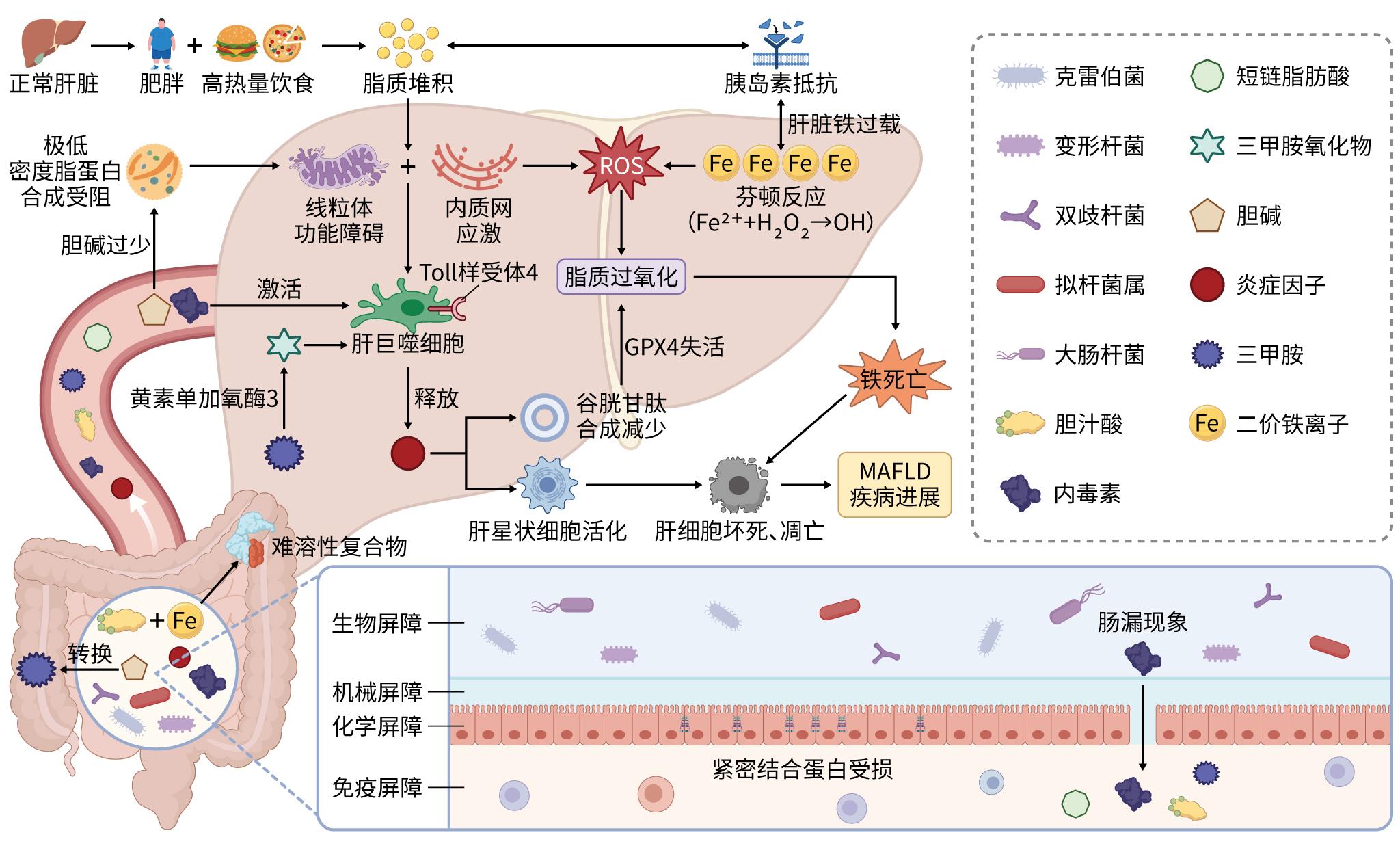
图(1)
计量
- 文章访问数: 15
- HTML全文浏览量: 7
- PDF下载量: 1
- 被引次数: 0