PDF下载 ( 20329 KB)

基于数据独立采集定量蛋白质组学的代谢相关脂肪性肝病诊断标志物的筛选及验证
DOI: 10.12449/JCH260912
Screening and validation of diagnostic biomarkers in metabolic dysfunction-associated fatty liver disease based on data-independent acquisition quantitative proteomics
-
摘要:
目的 筛选代谢相关脂肪性肝病(MAFLD)患者血清关键差异蛋白并进行验证,为该病的诊断标志物筛选及干预策略提供依据。 方法 选取2024年12月—2025年1月在成都中医药大学附属第三医院确诊的20例MAFLD患者(MAFLD组)为研究对象,同时纳入20例健康受试者(HC组),收集两组的人口统计学及血液生化指标。采用数据独立采集定量蛋白质组学检测两组血清蛋白的相对表达水平,通过基因本体功能富集分析、京都基因与基因组百科全书通路富集分析、蛋白质互作网络、Hub节点互作及加权基因共表达网络分析筛选显著差异蛋白。采用高脂饮食诱导建立MAFLD大鼠模型,随机分为对照组和模型组,检测血清生化指标、炎症因子及肝组织病理改变,并验证肝组织MER原癌基因酪氨酸激酶(MERTK)、基质金属蛋白酶9(MMP-9)的蛋白表达水平。计量资料组间比较采用成组t检验或Mann-Whitney U检验。计数资料组间比较采用χ2检验或Fisher精确检验。 结果 与HC组相比,MAFLD组血清天冬氨酸氨基转移酶(t=-7.348)、丙氨酸氨基转移酶(Z=5.088)、碱性磷酸酶(t=-3.943)、甘油三酯(Z=2.070)和高密度脂蛋白胆固醇(t=3.867)水平均显著升高(P值均<0.05)。数据独立采集血清蛋白质组学共鉴定3 559种蛋白,其中160个差异表达蛋白主要富集于代谢紊乱、免疫炎症反应及细胞外基质重塑等通路。蛋白质互作网络及Hub节点分析提示,丝氨酸蛋白酶抑制酶家族E成员1、MMP-9、细胞间黏附分子1等为核心调控蛋白。加权基因共表达网络分析筛选出与MAFLD表型呈显著正相关的MEyellow模块(cor=0.55,P<0.001),其功能富集于胞葬通路。该模块中MERTK(P<0.001)和MMP-9(P<0.001)表达最为显著。经高脂饮食诱导成功构建MAFLD大鼠模型,与对照组相比,模型组大鼠肝脏体积增大、颜色变黄、表面油腻;苏木精-伊红染色结果显示肝小叶结构紊乱、肝细胞脂肪变性及炎细胞浸润;油红O染色可见大量脂滴沉积;MASSON染色未见明显纤维条索。与对照组相比,模型组血清天冬氨酸氨基转移酶(t=-5.148)、丙氨酸氨基转移酶(t=-5.663)、总胆红素(t=-13.061)、甘油三酯(Z=-2.722)、总胆固醇(t=3.588)及肿瘤坏死因子α(t=-16.741)水平均显著升高(P值均<0.05),肝组织MERTK(t=-11.448,P<0.001)和MMP-9(t=-10.875,P<0.001)蛋白表达显著增加。 结论 MMP-9与MERTK在MAFLD中呈异常表达,可能成为该病潜在的诊断标志物。 Abstract:Objective To screen for and validate key differentially expressed proteins in serum of patients with metabolic dysfunction‑associated fatty liver disease (MAFLD), and to provide a basis for identifying diagnostic biomarkers and developing intervention strategies for this disease. Methods A total of 20 patients who were diagnosed with MAFLD in the third affiliated hospital of Chengdu University of Traditional Chinese Medicine from December 2024 to January 2025 were enrolled as MAFLD group, and 20 healthy controls were enrolled as HC group. Demographic data and serum biochemical parameters were collected from both groups. Data-independent acquisition (DIA) quantitative proteomics was used to measure the relative expression levels of serum proteins, and related methods were used to obtain significantly differentially expressed proteins, including the gene ontology functional enrichment analysis, the Kyoto Encyclopedia of Genes and Genomes pathway enrichment analysis, protein-protein interaction (PPI) network, Hub node interaction, and weighted gene co-expression network analysis (WGCNA). Rats were given a high-fat diet to establish a model of MAFLD, and randomly assigned to the control group and model group. Serum biochemical parameters and inflammatory factors were measured, liver histopathological changes were observed, and the protein expression levels of MER proto-oncogene tyrosine kinase (MERTK) and matrix metalloproteinase-9 (MMP-9) in liver tissue were verified. The independent-samples t test was used for comparison of normally distributed continuous data between groups, while the Mann-Whitney U test was used for comparison of non-normally distributed continuous data or data with heterogeneity of variance between groups; the chi-square test or the Fisher’s exact test was used for comparison of categorical data between groups. Results Compared with the HC group, the MAFLD group had significant increases in the serum levels of aspartate aminotransferase (t=-7.348, P<0.05), alanine aminotransferase (Z=5.088, P<0.05), alkaline phosphatase (t=-3.943, P<0.05), triglyceride (Z=2.070, P<0.05), and high-density lipoprotein cholesterol (t=3.867, P<0.05). A total of 3 559 proteins were identified by DIA serum proteomics, among which 160 differentially expressed proteins were mainly enriched in pathways including metabolic disorders, immune inflammatory response, and extracellular matrix remodeling. The PPI network and the Hub node analysis showed that serpin family E member 1, MMP-9, and intercellular cell adhesion molecule-1 were core regulatory proteins. WGCNA showed that the MEyellow module was significantly positively correlated with MAFLD phenotype (cor=0.55, P<0.001), which was functionally enriched in the efferocytosis pathway. MERTK (P<0.001) and MMP-9 (P<0.001) showed the most significant differential expression in this module. A rat model of MAFLD was successfully established using a high-fat diet, and compared with the control group, the model group had enlarged, yellowish, and greasy liver and disordered structure of hepatic lobules, hepatocyte fatty degeneration, and inflammatory cell infiltration based on HE staining, as well as massive lipid droplet deposition based on oil red O staining, while no obvious fibrous cords were observed in Masson staining. Compared with the control group, the model group had significant increases in the serum levels of aspartate aminotransferase (t=-5.148, P<0.05), alanine aminotransferase (t=-5.663, P<0.05), total bilirubin (t=-13.061, P<0.05), triglyceride (Z=-2.722, P<0.05), total cholesterol (t=3.588, P<0.05), and tumor necrosis factor-α (t=-16.741, P<0.05), as well as significant increases in the protein expression levels of MERTK (t=-11.448, P<0.001) and MMP-9 (t=-10.875, P<0.001) in liver tissue. Conclusion MMP-9 and MERTK are abnormally expressed in MAFLD and may be used as potential diagnostic biomarkers for this disease. -

注: a,每组样本鉴定到的蛋白质数量;b,血清蛋白质组学主成分分析;c,蛋白表达量层次聚类热图;d,差异蛋白数目统计图;e,差异蛋白质火山图。MAFLD,代谢相关脂肪性肝病;HC,健康对照。
图 1 MAFLD组与HC组的血清蛋白质组学特征
Figure 1. Serum proteomic characteristics between the MAFLD group and the HC group
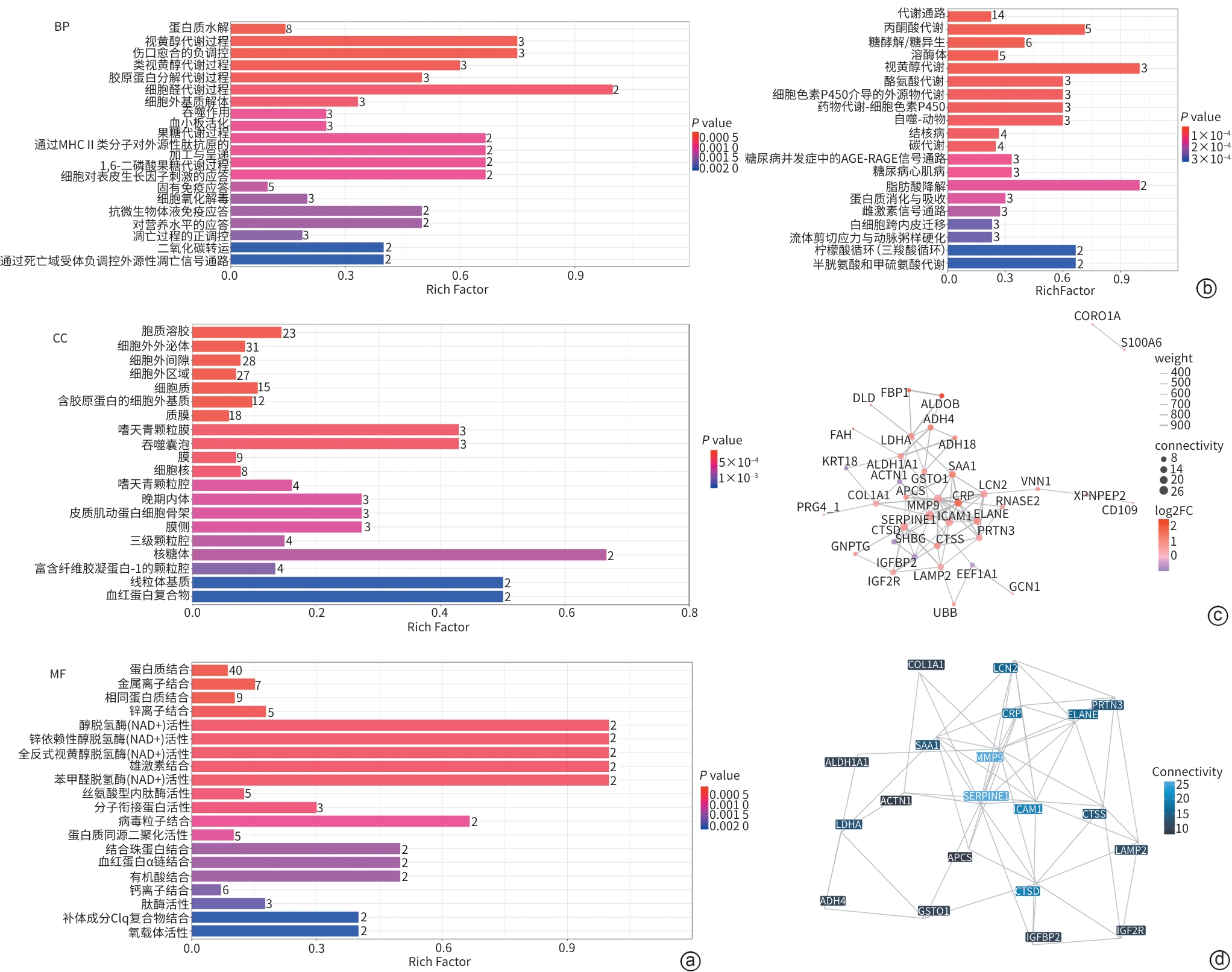
注: a,差异表达蛋白质BP、CC、MF;b,KEGG富集条形图;c,差异蛋白互作网络图;d,Hub节点互作网络图。BP,生物学过程;CC,细胞组分;MF,分子功能;KEGG,京都基因与基因组百科全书;MAFLD,代谢相关脂肪性肝病;HC,健康对照。
图 2 MAFLD组与HC组差异蛋白富集分析
Figure 2. Enrichment analysis of differentially expressed proteins between the MAFLD group and the HC group

注: a,模块-表型关联热图;b,特征蛋白邻接热图+模块聚类树;c,GO功能富集条形图;d,KEGG通路富集图;e,MAFLD组与HC组黄色模块成员度-蛋白显著性散点图;f,MERTK、MMP-9蛋白在MAFLD组与HC组中的丰度。GO,基因本体;KEGG,京都基因与基因组百科全书;MERTK,MER原癌基因酪氨酸激酶;MMP-9,基质金属蛋白酶9;MAFLD,代谢相关脂肪性肝病;HC,健康对照;WGCNA,加权基因共表达网络分析。
图 3 MAFLD组与HC组WGCNA结果
Figure 3. WGCNA of the MAFLD group and the HC group
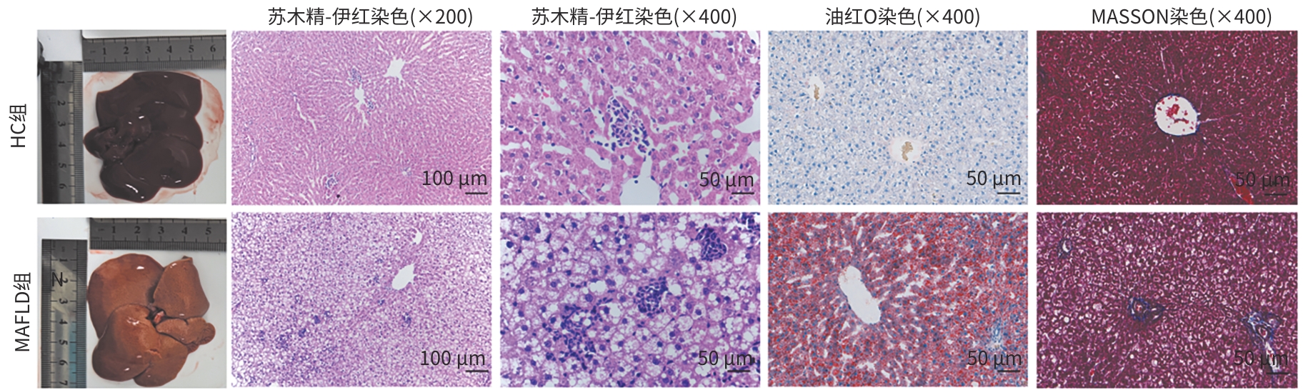
注: MAFLD,代谢相关脂肪性肝病;HC,健康对照。
图 4 各组大鼠肝脏肉眼观及不同染色情况
Figure 4. Gross liver morphology and different staining of rat liver tissues in each group
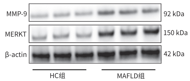
注: MERTK,MER原癌基因酪氨酸激酶;MMP-9,基质金属蛋白酶9;MAFLD,代谢相关脂肪性肝病;HC,健康对照。
图 5 各组大鼠肝组织MERTK、MMP-9蛋白条带
Figure 5. Protein bands of MERTK and MMP-9 in rat liver tissues
表 1 受试者人口统计学及血液生化指标
Table 1. Demographics and blood biochemical indicators of subjects
指标 HC组(n=20) MAFLD组(n=20) 统计值 P值 年龄(岁) 37.00(29.00~41.75) 35.00(30.25~38.00) Z=-0.732 0.464 男性[例(%)] 6(30) 12(60) χ2=3.636 0.057 FBG(mmol/L) 5.16(4.87~5.52) 5.27(4.94~6.07) Z=1.042 0.298 ALT(U/L) 16.50(12.00~23.50) 92.00(49.25~113.00) Z=5.088 <0.001 AST(U/L) 18.80±3.65 45.40±15.77 t=-7.348 <0.001 ALP(U/L) 59.90±10.32 83.60±24.82 t=-3.943 0.001 TC(mmol/L) 4.82±0.88 4.48±0.96 t=1.169 0.250 TG(mmol/L) 1.27(0.78~1.67) 1.51(1.23~3.06) Z=2.070 0.038 HDL-C(mmol/L) 1.64±0.40 1.22±0.28 t=3.867 <0.001 注:FBG,纤维蛋白原;ALT,丙氨酸氨基转移酶;AST,天冬氨酸氨基转移酶;ALP,碱性磷酸酶;TC,总胆固醇;TG,甘油三酯;HDL-C,高密度脂蛋白胆固醇;MAFLD,代谢相关脂肪性肝病;HC,健康对照。
 下载: 导出CSV
表 2 各组大鼠血清AST、ALT、TBil、TG、TC及TNF-α水平
Table 2. Serum levels of AST, ALT, TBil, TG, TC, and TNF-α
组别 动物数(只) AST(U/L) ALT(U/L) TBil(μmol/L) TG(mmol/L) TC(mmol/L) TNF-α(ng/mg) HC组 6 89.51±5.41 90.70±3.07 0.64±0.27 0.86(0.75~0.97) 1.84±0.20 0.27±0.04 MAFLD组 6 110.20±8.22 108.67±7.14 6.28±1.02 1.34(1.17~1.89) 1.44±0.18 0.74±0.06 统计值 t=-5.148 t=-5.663 t=-13.061 Z=-2.722 t=3.588 t=-16.741 P值 <0.001 <0.001 <0.001 0.004 0.005 <0.001 注:AST,天冬氨酸氨基转移酶;ALT,丙氨酸氨基转移酶;TBil,总胆红素;TG,甘油三酯;TC,总胆固醇;TNF-α,肿瘤坏死因子α;MAFLD,代谢相关脂肪性肝病;HC,健康对照。
下载: 导出CSV
表 3 各组大鼠肝组织MERTK、MMP-9蛋白表达水平
Table 3. Protein expression levels of MERTK and MMP-9 in rat liver tissues
组别 MERTK/β-actin MMP-9/β-actin HC组 0.37±0.04 0.46±0.03 MAFLD组 0.84±0.06 0.83±0.05 t值 -11.448 -10.875 P值 <0.001 <0.001 注:MERTK,MER原癌基因酪氨酸激酶;MMP-9,基质金属蛋白酶9;MAFLD,代谢相关脂肪性肝病;HC,健康对照。
下载: 导出CSV
-
[1] Teng M L, Ng C H, Huang D Q, et al. Global incidence and prevalence of nonalcoholic fatty liver disease[J]. Clin Mol Hepatol, 2023, 29( Suppl): S32- S42. DOI: 10.3350/cmh.2022.0365. [2] Riazi K, Azhari H, Charette J H, et al. The prevalence and incidence of NAFLD worldwide: A systematic review and meta-analysis[J]. Lancet Gastroenterol Hepatol, 2022, 7( 9): 851- 861. DOI: 10.1016/S2468-1253(22)00165-0. [3] Chinese Society of Hepatology, Chinese Medical Association. Guidelines for the prevention and treatment of metabolic dysfunction-associated(non-alcoholic) fatty liver disease(version 2024)[J]. J Pract Hepatol, 2024, 27( 4): 494- 510. DOI: 10.3760/cma.j.cn501113-20240327-00163.中华医学会肝病学分会. 代谢相关(非酒精性)脂肪性肝病防治指南(2024年版)[J]. 实用肝脏病杂志, 2024, 27( 4): 494- 510. DOI: 10.3760/cma.j.cn501113-20240327-00163. [4] Nan Y M, An J H, Bao J F, et al. The Chinese Society of Hepatology position statement on the redefinition of fatty liver disease[J]. J Hepatol, 2021, 75( 2): 454- 461. DOI: 10.1016/j.jhep.2021.05.003. [5] Febbraio M A, Reibe S, Shalapour S, et al. Preclinical models for studying NASH-driven HCC: How useful are they?[J]. Cell Metab, 2019, 29( 1): 18- 26. DOI: 10.1016/j.cmet.2018.10.012. [6] Stevanović J, Beleza J, Coxito P, et al. Physical exercise and liver“fitness”: Role of mitochondrial function and epigenetics-related mechanisms in non-alcoholic fatty liver disease[J]. Mol Metab, 2020, 32: 1- 14. DOI: 10.1016/j.molmet.2019.11.015. [7] Fröhlich K, Fahrner M, Brombacher E, et al. Data-independent acquisition: A milestone and prospect in clinical mass spectrometry–based proteomics[J]. Mol Cell Proteom, 2024, 23( 8): 100800. DOI: 10.1016/j.mcpro.2024.100800. [8] Lei N, Song H F, Zeng L, et al. Persistent lipid accumulation leads to persistent exacerbation of endoplasmic reticulum stress and inflammation in progressive NASH via the IRE1α/TRAF2 complex[J]. Molecules, 2023, 28( 7): 3185. DOI: 10.3390/molecules28073185. [9] Dan Lijuan, Song Hongfei, Li Xiuyan, et al. Mechanism of Sini Power combined with Linggui Zhugan Decoction on NAFLD based on transcriptomics and metabolomics[J]. China J Chin Materia Med, 2025, 50( 23): 6685- 6693. DOI: 10.19540/j.cnki.cjcmm.20250708.706.淡丽娟, 宋虹霏, 李岫滟, 等. 基于转录组学与代谢组学探讨四逆散联合苓桂术甘汤对NAFLD的作用机制[J]. 中国中药杂志, 2025, 50( 23): 6685- 6693. DOI: 10.19540/j.cnki.cjcmm.20250708.706. [10] Guo Z W, Wu D J, Mao R H, et al. Global burden of MAFLD, MAFLD related cirrhosis and MASH related liver cancer from 1990 to 2021[J]. Sci Rep, 2025, 15( 1): 7083. DOI: 10.1038/s41598-025-91312-5. [11] Quek J, Chan K E, Wong Z Y, et al. Global prevalence of non-alcoholic fatty liver disease and non-alcoholic steatohepatitis in the overweight and obese population: A systematic review and meta-analysis[J]. Lancet Gastroenterol Hepatol, 2023, 8( 1): 20- 30. DOI: 10.1016/S2468-1253(22)00317-X. [12] Horn P, Tacke F. Metabolic reprogramming in liver fibrosis[J]. Cell Metab, 2024, 36( 7): 1439- 1455. DOI: 10.1016/j.cmet.2024.05.003. [13] Li Xiuyan, Lei Na, Song Hongfei, et al. The role of endoplasmic reticulum stress in non-alcoholic fatty liver disease and related targeted therapies[J]. J Clin Hepatol, 2024, 40( 11): 2300- 2305. DOI: 10.12449/JCH241126.李岫滟, 雷娜, 宋虹霏, 等. 内质网应激在非酒精性脂肪性肝病中的作用及相关靶向治疗[J]. 临床肝胆病杂志, 2024, 40( 11): 2300- 2305. DOI: 10.12449/JCH241126. [14] Wang Dan, Zhang Jinxue, Su Lining, et al. Mechanism of action of mitochondrial dysfunction in metabolic associated fatty liver disease and the current status of research on traditional Chinese medicine[J]. J Clin Hepatol, 2025, 41( 12): 2615- 2621. DOI: 10.12449/JCH251224.王丹, 张金雪, 苏李宁, 等. 线粒体功能紊乱在代谢相关脂肪性肝病中的作用机制及中医药干预研究现状[J]. 临床肝胆病杂志, 2025, 41( 12): 2615- 2621. DOI: 10.12449/JCH251224. [15] Fromenty B, Roden M. Mitochondrial alterations in fatty liver diseases[J]. J Hepatol, 2023, 78( 2): 415- 429. DOI: 10.1016/j.jhep.2022.09.020. [16] Xu S J, Lu F, Gao J H, et al. Inflammation-mediated metabolic regulation in adipose tissue[J]. Obes Rev, 2024, 25( 6): e13724. DOI: 10.1111/obr.13724. [17] Lou R H, Shui W Q. Acquisition and analysis of DIA-based proteomic data: A comprehensive survey in 2023[J]. Mol Cell Proteom, 2024, 23( 2): 100712. DOI: 10.1016/j.mcpro.2024.100712. [18] Tian Z L, He W X, Tang J N, et al. Identification of important modules and biomarkers in breast cancer based on WGCNA[J]. Onco Targets Ther, 2020, 13: 6805- 6817. DOI: 10.2147/OTT.S258439. [19] Poon I K H, Ravichandran K S. Targeting efferocytosis in inflammaging[J]. Annu Rev Pharmacol Toxicol, 2024, 64: 339- 357. DOI: 10.1146/annurev-pharmtox-032723-110507. [20] Shi H X, Moore M P, Wang X B, et al. Efferocytosis in liver disease[J]. JHEP Rep, 2024, 6( 1): 100960. DOI: 10.1016/j.jhepr.2023.100960. [21] Wang X C, He Q F, Zhou C L, et al. Prolonged hypernutrition impairs TREM2-dependent efferocytosis to license chronic liver inflammation and NASH development[J]. Immunity, 2023, 56( 1): 58- 77.e11. DOI: 10.1016/j.immuni.2022.11.013. [22] Lee Y S, Olefsky J. Eating macrophages for a healthy anti-NASH meal[J]. Immunity, 2023, 56( 1): 3- 5. DOI: 10.1016/j.immuni.2022.12.011. [23] Petta S, Valenti L, Marra F, et al. MERTK rs4374383 polymorphism affects the severity of fibrosis in non-alcoholic fatty liver disease[J]. J Hepatol, 2016, 64( 3): 682- 690. DOI: 10.1016/j.jhep.2015.10.016. [24] Cai B S, Dongiovanni P, Corey K E, et al. Macrophage MerTK promotes liver fibrosis in nonalcoholic steatohepatitis[J]. Cell Metab, 2020, 31( 2): 406- 421. e 7. DOI: 10.1016/j.cmet.2019.11.013. [25] Bartneck M, Warzecha K T, Tacke F. Therapeutic targeting of liver inflammation and fibrosis by nanomedicine[J]. Hepatobiliary Surg Nutr, 2014, 3( 6): 364- 376. DOI: 10.3978/j.issn.2304-3881.2014.11.02. [26] Giannitrapani L, Soresi M, Bondì M L, et al. Nanotechnology applications for the therapy of liver fibrosis[J]. World J Gastroenterol, 2014, 20( 23): 7242- 7251. DOI: 10.3748/wjg.v20.i23.7242. [27] Verma R P, Hansch C. Matrix metalloproteinases(MMPs): Chemical-biological functions and(Q)SARs[J]. Bioorg Med Chem, 2007, 15( 6): 2223- 2268. DOI: 10.1016/j.bmc.2007.01.011. [28] Kurbatova I V, Topchieva L V, Dudanova O P, et al. Role of MMP-2 and MMP-9 in the relationship between inflammation, fibrosis, and apoptosis during progression of non-alcoholic fatty liver disease and diagnostic significance of plasma levels of their active forms[J]. Biochem Mosc, 2024, 89( 11): 1998- 2022. DOI: 10.1134/S0006297924110130. [29] Livzan M A, Akhmedov V A, Krolevets T S, et al. The informative value of non-invasive liver fibrosis markers in patients with nonalcoholic fatty liver disease[J]. Ter Arkh, 2016, 88( 12): 62- 68. DOI: 10.17116/terarkh2016881262-68. [30] Goyale A, Jain A, Smith C, et al. Assessment of non-alcoholic fatty liver disease(NAFLD) severity with novel serum-based markers: A pilot study[J]. PLoS One, 2021, 16( 11): e0260313. DOI: 10.1371/journal.pone.0260313. -

 本文二维码
计量
- 文章访问数: 12
- HTML全文浏览量: 4
- PDF下载量: 0
- 被引次数: 0