

 PDF下载 ( 1278 KB)

线粒体功能障碍与细胞间通讯在代谢相关脂肪性肝炎中的作用机制
DOI: 10.12449/JCH260833
利益冲突声明:本文不存在任何利益冲突。
作者贡献声明:罗婉艺负责撰写论文;张津铭负责修改论文;罗婉艺、曾国城和马骁天负责归纳文献;于牧鑫和郑楚伟负责拟定写作思路;季霞和张津铭负责指导撰写文章并最后定稿。
Mechanism of action of mitochondrial dysfunction and intercellular communication in metabolic dysfunction-associated steatohepatitis
-
摘要: 代谢相关脂肪性肝病(MAFLD)在脂质毒性、炎症浸润等因素作用下,肝细胞线粒体稳态失衡并出现功能障碍,进而进展为代谢相关脂肪性肝炎(MASH)。近年研究证实,线粒体损伤与慢性炎症密切相关,而细胞外囊泡是连接二者的核心媒介。本文系统综述了MAFLD/MASH中肝细胞线粒体损伤的分子机制,重点阐明肝细胞来源细胞外囊泡对肝内炎症的调控作用,并探讨线粒体靶向治疗及细胞间通讯干预在MAFLD/MASH中的潜在应用前景,旨在为开发此类疾病的新型治疗策略提供理论依据。Abstract: Metabolic dysfunction-associated fatty liver disease (MAFLD) progresses to metabolic dysfunction-associated steatohepatitis (MASH) due to mitochondrial homeostasis imbalance and dysfunction in hepatocytes caused by various factors such as lipotoxicity and inflammatory infiltration. Recent studies have confirmed that mitochondrial injury is closely associated with chronic inflammation, with extracellular vesicle (EV) acting as a key mediator linking the two processes. This article systematically reviews the molecular mechanisms of hepatocyte mitochondrial dysfunction in MAFLD/MASH, highlights the regulatory role of hepatocyte-derived EV in intrahepatic inflammation, and discusses the potential application prospects of mitochondria-targeted therapies and intercellular communication interventions in MAFLD/MASH, in order to provide a theoretical basis for developing novel therapeutic strategies for these diseases.
-
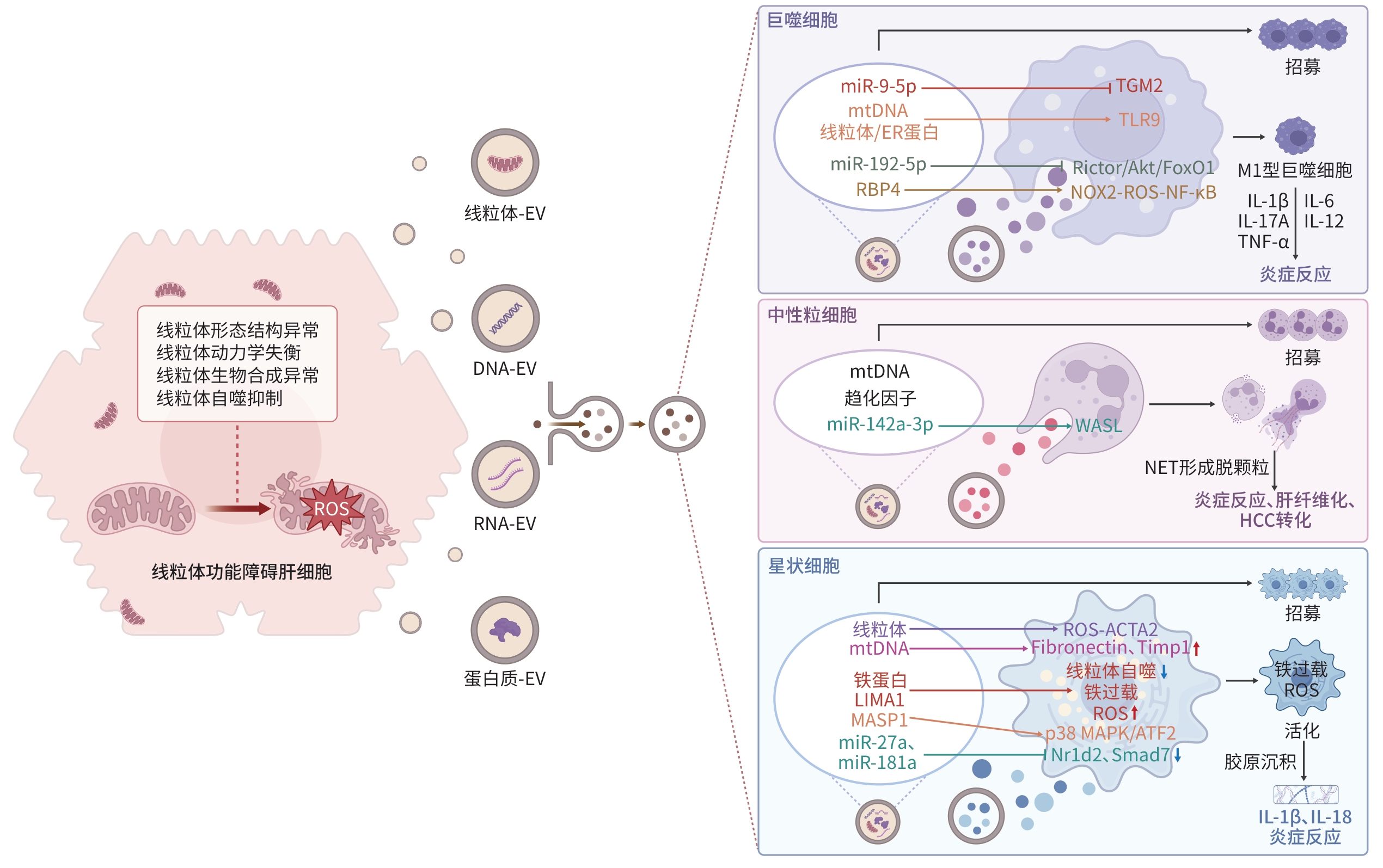
注: EV,细胞外囊泡;mtDNA,线粒体DNA;miR,微RNA;ER,内质网;RBP4,视黄醇结合蛋白4;TGM2,转谷氨酰胺酶2;TLR9,Toll样受体9;Rictor,雷帕霉素不敏感伴侣蛋白;Akt,蛋白激酶B;FoxO1,叉头框蛋白O1;NOX2,还原型烟酰胺腺嘌呤二核苷酸磷酸氧化酶2;ROS,活性氧;NF-κB,核因子κB;IL,白细胞介素;TNF-α,肿瘤坏死因子α;M1,巨噬细胞M1型(促炎型);WASL,威斯科特-奥尔德里奇综合征蛋白样肌动蛋白成核促进因子;NET,中性粒细胞胞外陷阱;HCC,肝细胞癌;LIMA1,LIM结构域与肌动蛋白结合蛋白1;MASP1,甘露聚糖结合凝集素相关丝氨酸蛋白酶1;ACTA2,α-平滑肌肌动蛋白2;Fibronectin,纤维连接蛋白;Timp1,金属蛋白酶组织抑制剂1;p38 MAPK,p38丝裂原活化蛋白激酶;ATF2,活化转录因子2;Nr1d2,核受体亚家族1D组成员2;Smad7,母系抗十五表态蛋白同源物7;MASH,代谢相关脂肪性肝炎。
图 1 肝细胞来源EV在MASH慢性炎症网络中的调控作用
Figure 1. Regulation of hepatocyte-derived extracellular vesicle in the chronic inflammatory network of MASH
-
[1] Younossi Z M, Kalligeros M, Henry L. Epidemiology of metabolic dysfunction-associated steatotic liver disease[J]. Clin Mol Hepatol, 2025, 31: S32- S50. DOI: 10.3350/cmh.2024.0431. [2] Schneider C V, Schneider K M, Raptis A, et al. Prevalence of at-risk MASH, MetALD and alcohol-associated steatotic liver disease in the general population[J]. Aliment Pharmacol Ther, 2024, 59( 10): 1271- 1281. DOI: 10.1111/apt.17958. [3] Fromenty B, Roden M. Mitochondrial alterations in fatty liver diseases[J]. J Hepatol, 2023, 78( 2): 415- 429. DOI: 10.1016/j.jhep.2022.09.020. [4] Shami G J, Samarska I V, Koek G H, et al. Giant mitochondria in human liver disease[J]. Liver Int, 2023, 43( 11): 2365- 2378. DOI: 10.1111/liv.15711. [5] Takeichi Y, Miyazawa T, Sakamoto S, et al. Non-alcoholic fatty liver disease in mice with hepatocyte-specific deletion of mitochondrial fission factor[J]. Diabetologia, 2021, 64( 9): 2092- 2107. DOI: 10.1007/s00125-021-05488-2. [6] Yamada T, Murata D, Kleiner D E, et al. Prevention and regression of megamitochondria and steatosis by blocking mitochondrial fusion in the liver[J]. iScience, 2022, 25( 4): 103996. DOI: 10.1016/j.isci.2022.103996. [7] Popov L D. Mitochondrial biogenesis: An update[J]. J Cell Mol Med, 2020, 24( 9): 4892- 4899. DOI: 10.1111/jcmm.15194. [8] Zhang L Z, Zhang Y H, Chang X X, et al. Imbalance in mitochondrial dynamics induced by low PGC-1α expression contributes to hepatocyte EMT and liver fibrosis[J]. Cell Death Dis, 2020, 11( 4): 226. DOI: 10.1038/s41419-020-2429-9. [9] Yang J W, Zou Y, Chen J, et al. Didymin alleviates metabolic dysfunction-associated fatty liver disease(MAFLD) via the stimulation of Sirt1-mediated lipophagy and mitochondrial biogenesis[J]. J Transl Med, 2023, 21( 1): 921. DOI: 10.1186/s12967-023-04790-4. [10] Zeng C F, Chen M K. Progress in nonalcoholic fatty liver disease: SIRT family regulates mitochondrial biogenesis[J]. Biomolecules, 2022, 12( 8): 1079. DOI: 10.3390/biom12081079. [11] Dong Y J, Hu M J, Tan K W, et al. ZNF143 inhibits hepatocyte mitophagy and promotes non-alcoholic fatty liver disease by targeting increased lncRNA NEAT1 expression to activate ROCK2 pathway[J]. Epigenetics, 2023, 18( 1): 2239592. DOI: 10.1080/15592294.2023.2239592. [12] Chen Y F, Yang F J, Shi Y J, et al. RNF31 alleviates liver steatosis by promoting p53/BNIP3-related mitophagy in hepatocytes[J]. Free Radic Biol Med, 2024, 219: 163- 179. DOI: 10.1016/j.freeradbiomed.2024.04.214. [13] Jin K P, Shi Y Z, Zhang H T, et al. A TNFα/Miz1-positive feedback loop inhibits mitophagy in hepatocytes and propagates non-alcoholic steatohepatitis[J]. J Hepatol, 2023, 79( 2): 403- 416. DOI: 10.1016/j.jhep.2023.03.039. [14] Mohammadipoor A, Hershfield M R, Linsenbardt H R, et al. Biological function of Extracellular Vesicles(EVs): A review of the field[J]. Mol Biol Rep, 2023, 50( 10): 8639- 8651. DOI: 10.1007/s11033-023-08624-w. [15] Gao Y H, Mi N N, Wu W X, et al. Transfer of inflammatory mitochondria via extracellular vesicles from M1 macrophages induces ferroptosis of pancreatic beta cells in acute pancreatitis[J]. J Extracell Vesicles, 2024, 13( 2): e12410. DOI: 10.1002/jev2.12410. [16] Konaka H, Kato Y, Hirano T, et al. Secretion of mitochondrial DNA via exosomes promotes inflammation in Behçet’s syndrome[J]. EMBO J, 2023, 42( 20): e112573. DOI: 10.15252/embj.2022112573. [17] Su X, Brassard A, Bartolomucci A, et al. Tumour extracellular vesicles induce neutrophil extracellular traps to promote lymph node metastasis[J]. J Extracell Vesicles, 2023, 12( 8): e12341. DOI: 10.1002/jev2.12341. [18] Zhang L, Zheng B, Bai Y, et al. Exosomes-transferred LINC00668 contributes to thrombosis by promoting NETs formation in inflammatory bowel disease[J]. Adv Sci(Weinh), 2023, 10( 28): e2300560. DOI: 10.1002/advs.202300560. [19] Khaksari S, Abnous K, Hadizadeh F, et al. Signal amplification strategies in biosensing of extracellular vesicles(EVs)[J]. Talanta, 2023, 256: 124244. DOI: 10.1016/j.talanta.2022.124244. [20] Garcia-Martinez I, Alen R, Pereira L, et al. Saturated fatty acid-enriched small extracellular vesicles mediate a crosstalk inducing liver inflammation and hepatocyte insulin resistance[J]. JHEP Rep, 2023, 5( 8): 100756. DOI: 10.1016/j.jhepr.2023.100756. [21] Hernández A, Reyes D, Geng Y N, et al. Extracellular vesicles derived from fat-laden hepatocytes undergoing chemical hypoxia promote a pro-fibrotic phenotype in hepatic stellate cells[J]. Biochim Biophys Acta Mol Basis Dis, 2020, 1866( 10): 165857. DOI: 10.1016/j.bbadis.2020.165857. [22] Rosina M, Ceci V, Turchi R, et al. Ejection of damaged mitochondria and their removal by macrophages ensure efficient thermogenesis in brown adipose tissue[J]. Cell Metab, 2022, 34( 4): 533- 548. DOI: 10.1016/j.cmet.2022.02.016. [23] Dasgupta D, Nakao Y, Mauer A S, et al. IRE1A stimulates hepatocyte-derived extracellular vesicles that promote inflammation in mice with steatohepatitis[J]. Gastroenterology, 2020, 159( 4): 1487- 1503. DOI: 10.1053/j.gastro.2020.06.031. [24] Liu H Y, Niu Q H, Wang T, et al. Lipotoxic hepatocytes promote nonalcoholic fatty liver disease progression by delivering microRNA-9-5p and activating macrophages[J]. Int J Biol Sci, 2021, 17( 14): 3745- 3759. DOI: 10.7150/ijbs.57610. [25] Liu X L, Pan Q, Cao H X, et al. Lipotoxic hepatocyte-derived exosomal microRNA 192-5p activates macrophages through rictor/Akt/forkhead box transcription factor O1 signaling in nonalcoholic fatty liver disease[J]. Hepatology, 2020, 72( 2): 454- 469. DOI: 10.1002/hep.31050. [26] Eguchi A, Yan R, Pan S Q, et al. Comprehensive characterization of hepatocyte-derived extracellular vesicles identifies direct miRNA-based regulation of hepatic stellate cells and DAMP-based hepatic macrophage IL-1β and IL-17 upregulation in alcoholic hepatitis mice[J]. J Mol Med(Berl), 2020, 98( 7): 1021- 1034. DOI: 10.1007/s00109-020-01926-7. [27] Yao J M, Ying H Z, Zhang H H, et al. Exosomal RBP4 potentiated hepatic lipid accumulation and inflammation in high-fat-diet-fed mice by promoting M1 polarization of Kupffer cells[J]. Free Radic Biol Med, 2023, 195: 58- 73. DOI: 10.1016/j.freeradbiomed.2022.12.085. [28] Hwang S, Yun H, Moon S, et al. Role of neutrophils in the pathogenesis of nonalcoholic steatohepatitis[J]. Front Endocrinol, 2021, 12: 751802. DOI: 10.3389/fendo.2021.751802. [29] Huby T, Gautier E L. Immune cell-mediated features of non-alcoholic steatohepatitis[J]. Nat Rev Immunol, 2022, 22( 7): 429- 443. DOI: 10.1038/s41577-021-00639-3. [30] Cao Z M, Zhao M, Sun H, et al. Roles of mitochondria in neutrophils[J]. Front Immunol, 2022, 13: 934444. DOI: 10.3389/fimmu.2022.934444. [31] Xu M, Xu H, Ling Y W, et al. Neutrophil extracellular traps-triggered hepatocellular senescence exacerbates lipotoxicity in non-alcoholic steatohepatitis[J]. J Adv Res, 2026, 79: 521- 534. DOI: 10.1016/j.jare.2025.03.015. [32] Yu M X, Li X W, Xu L, et al. Neutrophil extracellular traps induce intrahepatic thrombotic tendency and liver damage in cholestatic liver disease[J]. Hepatol Commun, 2024, 8( 8): e0513. DOI: 10.1097/HC9.0000000000000513. [33] Xia Y J, Wang Y, Xiong Q, et al. Neutrophil extracellular traps promote MASH fibrosis by metabolic reprogramming of HSC[J]. Hepatology, 2025, 81( 3): 947- 961. DOI: 10.1097/HEP.0000000000000762. [34] Wang H, Zhang H J, Wang Y, et al. Regulatory T-cell and neutrophil extracellular trap interaction contributes to carcinogenesis in non-alcoholic steatohepatitis[J]. J Hepatol, 2021, 75( 6): 1271- 1283. DOI: 10.1016/j.jhep.2021.07.032. [35] He Y, Rodrigues R M, Wang X L, et al. Neutrophil-to-hepatocyte communication via LDLR-dependent miR-223-enriched extracellular vesicle transfer ameliorates nonalcoholic steatohepatitis[J]. J Clin Invest, 2021, 131( 3): e141513. DOI: 10.1172/JCI141513. [36] Ayilam Ramachandran R, Lemoff A, Robertson D M. Extracellular vesicles released by host epithelial cells during Pseudomonas aeruginosa infection function as homing beacons for neutrophils[J]. Cell Commun Signal, 2024, 22( 1): 341. DOI: 10.1186/s12964-024-01609-7. [37] Ma J, Cao H X, Rodrigues R M, et al. Chronic-plus-binge alcohol intake induces production of proinflammatory mtDNA-enriched extracellular vesicles and steatohepatitis via ASK1/p38MAPKα-dependent mechanisms[J]. JCI Insight, 2020, 5( 14): e136496. DOI: 10.1172/jci.insight.136496. [38] Wang L F, Zhu Z F, Liao Y, et al. Host liver-derived extracellular vesicles deliver miR-142a-3p induces neutrophil extracellular traps via targeting WASL to block the development of Schistosoma japonicum[J]. Mol Ther, 2022, 30( 5): 2092- 2107. DOI: 10.1016/j.ymthe.2022.03.016. [39] Yang A T, Kim Y O, Yan X Z, et al. Fibroblast activation protein activates macrophages and promotes parenchymal liver inflammation and fibrosis[J]. Cell Mol Gastroenterol Hepatol, 2023, 15( 4): 841- 867. DOI: 10.1016/j.jcmgh.2022.12.005. [40] Yamaguchi M, Kanazawa T, Morino S, et al. Increased tropism of extracellular vesicles derived from palmitic acid-treated hepatocytes to activated hepatic stellate cells[J]. Membranes, 2022, 12( 10): 1023. DOI: 10.3390/membranes12101023. [41] Koenen M T, Brandt E F, Kaczor D M, et al. Extracellular vesicles from steatotic hepatocytes provoke pro-fibrotic responses in cultured stellate cells[J]. Biomolecules, 2022, 12( 5): 698. DOI: 10.3390/biom12050698. [42] Yang L, Gao Y L, Jiang S, et al. Aflatoxin B1-exposed hepatocyte-derived extracellular vesicles: Initiating hepatic stellate cell-mediated liver fibrosis through a p53-Parkin-dependent mitophagy pathway[J]. Ecotoxicol Environ Saf, 2024, 277: 116363. DOI: 10.1016/j.ecoenv.2024.116363. [43] Li Y J, Liu R P, Ding M N, et al. Tetramethylpyrazine prevents liver fibrotic injury in mice by targeting hepatocyte-derived and mitochondrial DNA-enriched extracellular vesicles[J]. Acta Pharmacol Sin, 2022, 43( 8): 2026- 2041. DOI: 10.1038/s41401-021-00843-w. [44] Sun M X, Tang M, Qian Y T, et al. Extracellular vesicles-derived ferritin from lipid-induced hepatocytes regulates activation of hepatic stellate cells[J]. Heliyon, 2024, 10( 13): e33741. DOI: 10.1016/j.heliyon.2024.e33741. [45] Li S H, Yang F J, Cheng F, et al. Lipotoxic hepatocyte derived LIMA1 enriched small extracellular vesicles promote hepatic stellate cells activation via inhibiting mitophagy[J]. Cell Mol Biol Lett, 2024, 29( 1): 82. DOI: 10.1186/s11658-024-00596-4. [46] Liu X Z, Tan S W, Liu H L, et al. Hepatocyte-derived MASP1-enriched small extracellular vesicles activate HSCs to promote liver fibrosis[J]. Hepatology, 2023, 77( 4): 1181- 1197. DOI: 10.1002/hep.32662. [47] Safran M, Masoud R, Sultan M, et al. Extracellular vesicular transmission of miR-423-5p from HepG2 cells inhibits the differentiation of hepatic stellate cells[J]. Cells, 2022, 11( 10): 1715. DOI: 10.3390/cells11101715. [48] Pugliese N, Plaz Torres M C, Petta S, et al. Is there an‘ideal’ diet for patients with NAFLD?[J]. Eur J Clin Invest, 2022, 52( 3): e13659. DOI: 10.1111/eci.13659. [49] Lin B X, Wu T, Nasb M, et al. Regular exercise alleviates metabolic dysfunction-associated steatohepatitis through rescuing mitochondrial oxidative stress and dysfunction in liver[J]. Free Radic Biol Med, 2025, 230: 163- 176. DOI: 10.1016/j.freeradbiomed.2025.02.017. [50] Petta S, Targher G, Romeo S, et al. The first MASH drug therapy on the horizon: Current perspectives of resmetirom[J]. Liver Int, 2024, 44( 7): 1526- 1536. DOI: 10.1111/liv.15930. [51] Dall M, Hassing A S, Treebak J T. NAD+ and NAFLD-caution, causality and careful optimism[J]. J Physiol, 2022, 600( 5): 1135- 1154. DOI: 10.1113/JP280908. [52] Yang J H, Liu L, Oda Y, et al. Extracellular vesicles and Cx43-gap junction channels are the main routes for mitochondrial transfer from ultra-purified mesenchymal stem cells, RECs[J]. Int J Mol Sci, 2023, 24( 12): 10294. DOI: 10.3390/ijms241210294. [53] Zhao M, Liu S Y, Wang C S, et al. Mesenchymal stem cell-derived extracellular vesicles attenuate mitochondrial damage and inflammation by stabilizing mitochondrial DNA[J]. ACS Nano, 2021, 15( 1): 1519- 1538. DOI: 10.1021/acsnano.0c08947. [54] Chen Y F, Yang F J, Wang Y J, et al. Mesenchymal stem cell-derived small extracellular vesicles reduced hepatic lipid accumulation in MASLD by suppressing mitochondrial fission[J]. Stem Cell Res Ther, 2025, 16( 1): 116. DOI: 10.1186/s13287-025-04228-2. [55] Chen Y, Xie K J, Chen C Y, et al. BMDM-derived ORP8 suppresses lipotoxicity and inflammation by relieving endoplasmic reticulum stress in mice with MASH[J]. Mol Med, 2025, 31( 1): 213. DOI: 10.1186/s10020-025-01275-6. [56] Xia L J, Zhang C L, Lv N Y, et al. AdMSC-derived exosomes alleviate acute lung injury via transferring mitochondrial component to improve homeostasis of alveolar macrophages[J]. Theranostics, 2022, 12( 6): 2928- 2947. DOI: 10.7150/thno.69533. [57] Lu W, Li X Y, Wang Z Y, et al. Mesenchymal stem cell-derived extracellular vesicles accelerate diabetic wound healing by inhibiting NET-induced ferroptosis of endothelial cells[J]. Int J Biol Sci, 2024, 20( 9): 3515- 3529. DOI: 10.7150/ijbs.97150. -

 下载:
下载:
 本文二维码
本文二维码
图(1)
计量
- 文章访问数: 151
- HTML全文浏览量: 66
- PDF下载量: 17
- 被引次数: 0




