

 PDF下载 ( 802 KB)

线粒体DNA释放触发的先天免疫机制在代谢相关脂肪性肝病中的作用及靶向干预策略
DOI: 10.12449/JCH260832
利益冲突声明:本文不存在任何利益冲突。
作者贡献声明:周禹负责选题及撰写论文;李开楊负责拟定思路;杨梅负责审核及定稿;赵琦负责指导撰写文章;赵一鸣、张飞和汪倩负责查找文献及修改论文。
Role of innate immune mechanisms triggered by mitochondrial DNA release in metabolic dysfunction-associated fatty liver disease and targeted intervention strategies
-
摘要: 代谢相关脂肪性肝病(MAFLD)是全球发病率最高的慢性肝病,其发病机制复杂。线粒体在该疾病进程中发挥关键作用,研究证实线粒体功能障碍可加剧代谢紊乱、诱发先天免疫失衡,而线粒体DNA(mtDNA)是介导这两大病理效应的核心分子。mtDNA异常释放后,可被细胞内模式识别受体识别,进而触发先天免疫反应并引发组织损伤,形成“mtDNA释放-免疫激活-组织损伤”的病理通路。目前,该通路在MAFLD疾病不同阶段的作用机制以及靶向该通路的干预策略,尚缺乏系统性的综述总结。本文系统综述该通路的核心分子机制、在MAFLD发生发展中的病理作用及当前靶向该机制的干预策略,旨在为MAFLD的病理机制解析与新型治疗策略的开发提供理论依据。Abstract: Metabolic dysfunction-associated fatty liver disease (MAFLD) is the most prevalent chronic liver disease worldwide, with a complex pathogenesis. Mitochondria play a pivotal role in this disease process, and studies have confirmed that mitochondrial dysfunction can exacerbate metabolic disorders and induce innate immune imbalance, while mitochondrial DNA (mtDNA) is the core molecule mediating these two pathological effects. After the abnormal release of mtDNA, it can be recognized by intracellular pattern recognition receptors, which in turn triggers innate immune responses and causes tissue damage, forming a pathological pathway of “mtDNA release-immune activation-tissue damage”. Currently, there is a lack of systematic reviews summarizing the mechanism of action of this pathway in different stages of MAFLD and the intervention strategies targeting this pathway. This article systematically reviews the core molecular mechanism of this pathway, its pathological role in the development and progression of MAFLD, and the current intervention strategies targeting this mechanism, in order to provide a theoretical basis for analyzing the pathogenesis of MAFLD and developing novel therapeutic strategies.
-
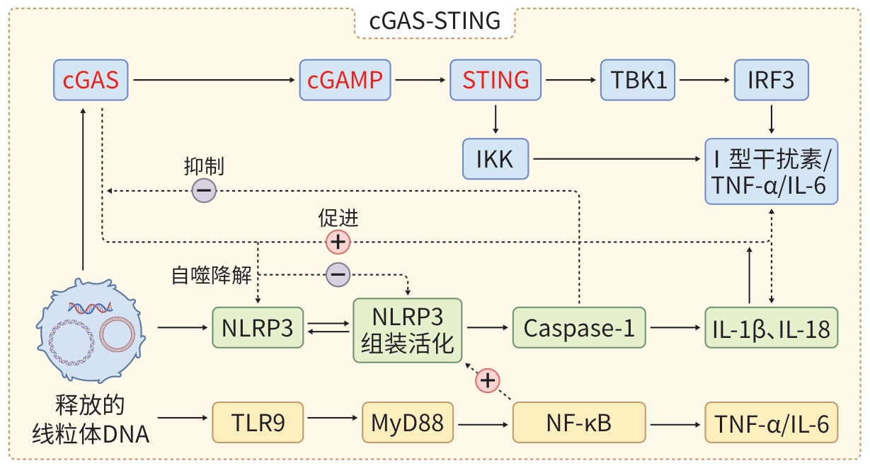
注: cGAS,环鸟苷酸-腺苷酸合酶;cGAMP,环鸟苷酸-腺苷酸;STING,干扰素基因刺激因子;TBK1,TANK结合激酶1;IRF3,干扰素调节因子3;IKK,IκB激酶;TNF-α,肿瘤坏死因子α;IL-6,白细胞介素6;NLRP3,核苷酸结合寡聚结构域样受体家族热蛋白结构域相关蛋白3;Caspase-1,胱天蛋白酶1;IL-1β,白细胞介素1β;IL-18,白细胞介素18;TLR9,Toll样受体9;MyD88,髓系分化初级反应蛋白质88;NF-κB,核因子κB。
图 1 线粒体DNA触发先天免疫机制的信号通路
Figure 1. Signal pathway of innate immune mechanisms triggered by mitochondrial DNA
-
[1] Guo Z W, Wu D J, Mao R H, et al. Global burden of MAFLD, MAFLD related cirrhosis and MASH related liver cancer from 1990 to 2021[J]. Sci Rep, 2025, 15( 1): 7083. DOI: 10.1038/s41598-025-91312-5. [2] Carvalho-Gontijo R, Han C J, Zhang L, et al. Metabolic injury of hepatocytes promotes progression of NAFLD and AALD[J]. Semin Liver Dis, 2022, 42( 3): 233- 249. DOI: 10.1055/s-0042-1755316. [3] Chen P, Yao L C, Yuan M Q, et al. Mitochondrial dysfunction: A promising therapeutic target for liver diseases[J]. Genes Dis, 2024, 11( 3): 101115. DOI: 10.1016/j.gendis.2023.101115. [4] Shi T T, Yu L P, Zhuang R X, et al. Regulation of mitochondrial function by natural products for the treatment of metabolic associated fatty liver disease[J]. Can J Gastroenterol Hepatol, 2021, 2021: 5527315. DOI: 10.1155/2021/5527315. [5] Yoon J, Kim S, Lee M, et al. Mitochondrial nucleic acids in innate immunity and beyond[J]. Exp Mol Med, 2023, 55( 12): 2508- 2518. DOI: 10.1038/s12276-023-01121-x. [6] Boyapati R K, Tamborska A, Dorward D A, et al. Advances in the understanding of mitochondrial DNA as a pathogenic factor in inflammatory diseases[J]. F1000Res, 2017, 6: 169. DOI: 10.12688/f1000research.10397.1. [7] Kausar S, Yang L Q, Abbas M N, et al. Mitochondrial DNA: A key regulator of anti-microbial innate immunity[J]. Genes, 2020, 11( 1): 86. DOI: 10.3390/genes11010086. [8] Zhao Y X, Liu B H, Xu L, et al. ROS-induced mtDNA release: The emerging messenger for communication between neurons and innate immune cells during neurodegenerative disorder progression[J]. Antioxidants(Basel), 2021, 10( 12): 1917. DOI: 10.3390/antiox10121917. [9] Gardner J J, Cushen S C, Oliveira da Silva R N, et al. Oxidative stress induces release of mitochondrial DNA into the extracellular space in human placental villous trophoblast BeWo cells[J]. Am J Physiol Cell Physiol, 2024, 326( 6): C1776- C1788. DOI: 10.1152/ajpcell.00091.2024. [10] Carvalho G, Repolês B M, Mendes I, et al. Mitochondrial DNA instability in mammalian cells[J]. Antioxid Redox Signal, 2022, 36( 13-15): 885- 905. DOI: 10.1089/ars.2021.0091. [11] Newman L E, Shadel G S. Mitochondrial DNA release in innate immune signaling[J]. Annu Rev Biochem, 2023, 92: 299- 332. DOI: 10.1146/annurev-biochem-032620-104401. [12] Riley J S, Tait S W. Mitochondrial DNA in inflammation and immunity[J]. EMBO Rep, 2020, 21( 4): e49799. DOI: 10.15252/embr.201949799. [13] Barber G N. STING: Infection, inflammation and cancer[J]. Nat Rev Immunol, 2015, 15( 12): 760- 770. DOI: 10.1038/nri3921. [14] Zhang X W, Bai X C, Chen Z J. Structures and mechanisms in the cGAS-STING innate immunity pathway[J]. Immunity, 2020, 53( 1): 43- 53. DOI: 10.1016/j.immuni.2020.05.013. [15] Jo E K, Kim J K, Shin D M, et al. Molecular mechanisms regulating NLRP3 inflammasome activation[J]. Cell Mol Immunol, 2016, 13( 2): 148- 159. DOI: 10.1038/cmi.2015.95. [16] Zhong Z Y, Liang S, Sanchez-Lopez E, et al. New mitochondrial DNA synthesis enables NLRP3 inflammasome activation[J]. Nature, 2018, 560( 7717): 198- 203. DOI: 10.1038/s41586-018-0372-z. [17] Handa P, Vemulakonda A, Kowdley K V, et al. Mitochondrial DNA from hepatocytes as a ligand for TLR9: Drivers of nonalcoholic steatohepatitis?[J]. World J Gastroenterol, 2016, 22( 31): 6965- 6971. DOI: 10.3748/wjg.v22.i31.6965. [18] Rutz M, Metzger J, Gellert T, et al. Toll-like receptor 9 binds single-stranded CpG-DNA in a sequence- and pH-dependent manner[J]. Eur J Immunol, 2004, 34( 9): 2541- 2550. DOI: 10.1002/eji.200425218. [19] Tao G W, Liao W Y, Hou J F, et al. Advances in crosstalk among innate immune pathways activated by mitochondrial DNA[J]. Heliyon, 2024, 10( 1): e24029. DOI: 10.1016/j.heliyon.2024.e24029. [20] Luo X, Zhao Y, Luo Y P, et al. Cytosolic mtDNA-cGAS-STING axis contributes to sepsis-induced acute kidney injury via activating the NLRP3 inflammasome[J]. Clin Exp Nephrol, 2024, 28( 5): 375- 390. DOI: 10.1007/s10157-023-02448-5. [21] Aarreberg L D, Esser-Nobis K, Driscoll C, et al. Interleukin-1β induces mtDNA release to activate innate immune signaling via cGAS-STING[J]. Mol Cell, 2019, 74( 4): 801- 815. DOI: 10.1016/j.molcel.2019.02.038. [22] Zhang R X, Kang R, Tang D L. The STING1 network regulates autophagy and cell death[J]. Sig Transduct Target Ther, 2021, 6( 1): 208. DOI: 10.1038/s41392-021-00613-4. [23] Wang Y T, Ning X H, Gao P F, et al. Inflammasome activation triggers caspase-1-mediated cleavage of cGAS to regulate responses to DNA virus infection[J]. Immunity, 2017, 46( 3): 393- 404. DOI: 10.1016/j.immuni.2017.02.011. [24] Kim S K, Park K Y, Choe J Y. Toll-like receptor 9 is involved in NLRP3 inflammasome activation and IL-1β production through monosodium urate-induced mitochondrial DNA[J]. Inflammation, 2020, 43( 6): 2301- 2311. DOI: 10.1007/s10753-020-01299-6. [25] Xu D W, Tian Y Z, Xia Q, et al. The cGAS-STING pathway: Novel perspectives in liver diseases[J]. Front Immunol, 2021, 12: 682736. DOI: 10.3389/fimmu.2021.682736. [26] Gong J H, Gao X L, Ge S Y, et al. The role of cGAS-STING signalling in metabolic diseases: From signalling networks to targeted intervention[J]. Int J Biol Sci, 2024, 20( 1): 152- 174. DOI: 10.7150/ijbs.84890. [27] Garcia-Martinez I, Santoro N, Chen Y L, et al. Hepatocyte mitochondrial DNA drives nonalcoholic steatohepatitis by activation of TLR9[J]. J Clin Invest, 2016, 126( 3): 859- 864. DOI: 10.1172/JCI83885. [28] Zhao Y, Zhou Y N, Wang D, et al. Mitochondrial dysfunction in metabolic dysfunction fatty liver disease(MAFLD)[J]. Int J Mol Sci, 2023, 24( 24): 17514. DOI: 10.3390/ijms242417514. [29] Clare K, Dillon J F, Brennan P N. Reactive oxygen species and oxidative stress in the pathogenesis of MAFLD[J]. J Clin Transl Hepatol, 2022, 10( 5): 939- 946. DOI: 10.14218/JCTH.2022.00067. [30] Schlatter R, Schmich K, Lutz A, et al. Modeling the TNFα-induced apoptosis pathway in hepatocytes[J]. PLoS One, 2011, 6( 4): e18646. DOI: 10.1371/journal.pone.0018646. [31] An P, Wei L L, Zhao S S, et al. Hepatocyte mitochondria-derived danger signals directly activate hepatic stellate cells and drive progression of liver fibrosis[J]. Nat Commun, 2020, 11: 2362. DOI: 10.1038/s41467-020-16092-0. [32] Arumugam S, Li B H, Boodapati S L T, et al. Mitochondrial DNA and the STING pathway are required for hepatic stellate cell activation[J]. Hepatology, 2023, 78( 5): 1448- 1461. DOI: 10.1097/HEP.0000000000000388. [33] Smith-Cortinez N, van Eunen K, Heegsma J, et al. Simultaneous induction of glycolysis and oxidative phosphorylation during activation of hepatic stellate cells reveals novel mitochondrial targets to treat liver fibrosis[J]. Cells, 2020, 9( 11): 2456. DOI: 10.3390/cells9112456. [34] Ran J Q, Yin S X, Issa R, et al. Key role of macrophages in the progression of hepatic fibrosis[J]. Hepatol Commun, 2025, 9( 1): e0602. DOI: 10.1097/HC9.0000000000000602. [35] Li H O, Zhou Y J, Wang H Z, et al. Crosstalk between liver macrophages and surrounding cells in nonalcoholic steatohepatitis[J]. Front Immunol, 2020, 11: 1169. DOI: 10.3389/fimmu.2020.01169. [36] Babuta M, Morel C, de Carvalho Ribeiro M, et al. Neutrophil extracellular traps activate hepatic stellate cells and monocytes via NLRP3 sensing in alcohol-induced acceleration of MASH fibrosis[J]. Gut, 2024, 73( 11): 1854- 1869. DOI: 10.1136/gutjnl-2023-331447. [37] Xiao Y, Zhao C, Tai Y, et al. STING mediates hepatocyte pyroptosis in liver fibrosis by Epigenetically activating the NLRP3 inflammasome[J]. Redox Biol, 2023, 62: 102691. DOI: 10.1016/j.redox.2023.102691. [38] Litwiniuk A, Bik W, Kalisz M, et al. Inflammasome NLRP3 potentially links obesity-associated low-grade systemic inflammation and insulin resistance with Alzheimer’s disease[J]. Int J Mol Sci, 2021, 22( 11): 5603. DOI: 10.3390/ijms22115603. [39] Tanti J F, Jager J. Cellular mechanisms of insulin resistance: Role of stress-regulated serine kinases and insulin receptor substrates(IRS) serine phosphorylation[J]. Curr Opin Pharmacol, 2009, 9( 6): 753- 762. DOI: 10.1016/j.coph.2009.07.004. [40] Guo Y W, Zhang X Y, Zhao Z Y, et al. NF-κB/HDAC1/SREBP1c pathway mediates the inflammation signal in progression of hepatic steatosis[J]. Acta Pharm Sin B, 2020, 10( 5): 825- 836. DOI: 10.1016/j.apsb.2020.02.005. [41] Jin Y, Tan Y J, Chen L P, et al. Reactive oxygen species induces lipid droplet accumulation in HepG2 cells by increasing perilipin 2 expression[J]. Int J Mol Sci, 2018, 19( 11): 3445. DOI: 10.3390/ijms19113445. [42] Xu J Q, Shen J Y, Yuan R L, et al. Mitochondrial targeting therapeutics: Promising role of natural products in non-alcoholic fatty liver disease[J]. Front Pharmacol, 2021, 12: 796207. DOI: 10.3389/fphar.2021.796207. [43] Izdebska M, Herbet M, Gawrońska-Grzywacz M, et al. Resveratrol limits lipogenesis and enhance mitochondrial activity in HepG2 cells[J]. J Pharm Pharm Sci, 2018, 21( 1): 504- 515. DOI: 10.18433/jpps29994. [44] Rafiei H, Omidian K, Bandy B. Dietary polyphenols protect against oleic acid-induced steatosis in an in vitro model of NAFLD by modulating lipid metabolism and improving mitochondrial function[J]. Nutrients, 2019, 11( 3): 541. DOI: 10.3390/nu11030541. [45] Chen X F, Yi L, Song S Y, et al. Puerarin attenuates palmitate-induced mitochondrial dysfunction, impaired mitophagy and inflammation in L6 myotubes[J]. Life Sci, 2018, 206: 84- 92. DOI: 10.1016/j.lfs.2018.05.041. [46] Wang S, Yang F J, Shang L C, et al. Puerarin protects against high-fat high-sucrose diet-induced non-alcoholic fatty liver disease by modulating PARP-1/PI3K/AKT signaling pathway and facilitating mitochondrial homeostasis[J]. Phytother Res, 2019, 33( 9): 2347- 2359. DOI: 10.1002/ptr.6417. [47] Zhang W, Hong R T, Tian T L. Silymarin’s protective effects and possible mechanisms on alcoholic fatty liver for rats[J]. Biomol Ther, 2013, 21( 4): 264- 269. DOI: 10.4062/biomolther.2013.020. [48] Liu M Q, Wu Y, Li Y, et al. Alleviation of metabolic dysfunction-associated steatotic liver disease by silymarin is associated with maintaining mitochondrial homeostasis via regulation of optic atrophy 1[J]. J Pharmacol Exp Ther, 2025, 392( 7): 103611. DOI: 10.1016/j.jpet.2025.103611. [49] Alves I, Araújo E M Q, Dalgaard L T, et al. Protective effects of sulforaphane preventing inflammation and oxidative stress to enhance metabolic health: A narrative review[J]. Nutrients, 2025, 17( 3): 428. DOI: 10.3390/nu17030428. [50] Stefano J T, Duarte S M B, Ribeiro Leite Altikes R G, et al. Non-pharmacological management options for MAFLD: A practical guide[J]. Ther Adv Endocrinol Metab, 2023, 14: 20420188231160394. DOI: 10.1177/20420188231160394. [51] Davidson M H. Mechanisms for the hypotriglyceridemic effect of marine omega-3 fatty acids[J]. Am J Cardiol, 2006, 98( 4): 27- 33. DOI: 10.1016/j.amjcard.2005.12.024. [52] Pérez-Monter C, Álvarez-Arce A, Nuño-Lambarri N, et al. Inulin improves diet-induced hepatic steatosis and increases intestinal Akkermansia genus level[J]. Int J Mol Sci, 2022, 23( 2): 991. DOI: 10.3390/ijms23020991. [53] Kilicarslan You D, Fuwad A, Lee K H, et al. Evaluation of the protective role of vitamin E against ROS-driven lipid oxidation in model cell membranes[J]. Antioxidants(Basel), 2024, 13( 9): 1135. DOI: 10.3390/antiox13091135. [54] Wen X, Fan J C, Duan X M, et al. Mitochondrial DNA in exercise-mediated innate immune responses[J]. Int J Mol Sci, 2025, 26( 7): 3069. DOI: 10.3390/ijms26073069. [55] Stevanović J, Beleza J, Coxito P, et al. Physical exercise and liver“fitness”: Role of mitochondrial function and epigenetics-related mechanisms in non-alcoholic fatty liver disease[J]. Mol Metab, 2020, 32: 1- 14. DOI: 10.1016/j.molmet.2019.11.015. [56] Farzanegi P, Dana A, Ebrahimpoor Z, et al. Mechanisms of beneficial effects of exercise training on non-alcoholic fatty liver disease(NAFLD): Roles of oxidative stress and inflammation[J]. Eur J Sport Sci, 2019, 19( 7): 994- 1003. DOI: 10.1080/17461391.2019.1571114. -

 下载:
下载:
 本文二维码
本文二维码
图(2)
计量
- 文章访问数: 6
- HTML全文浏览量: 1
- PDF下载量: 0
- 被引次数: 0




