

 PDF下载 ( 3007 KB)

高尔基体蛋白73对可切除肝细胞癌患者肝纤维化程度及预后的评估价值
DOI: 10.12449/JCH260820
Value of Golgi protein 73 in assessing liver fibrosis degree and prognosis of patients with resectable hepatocellular carcinoma
-
摘要:
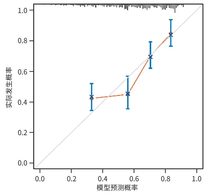
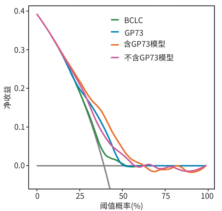
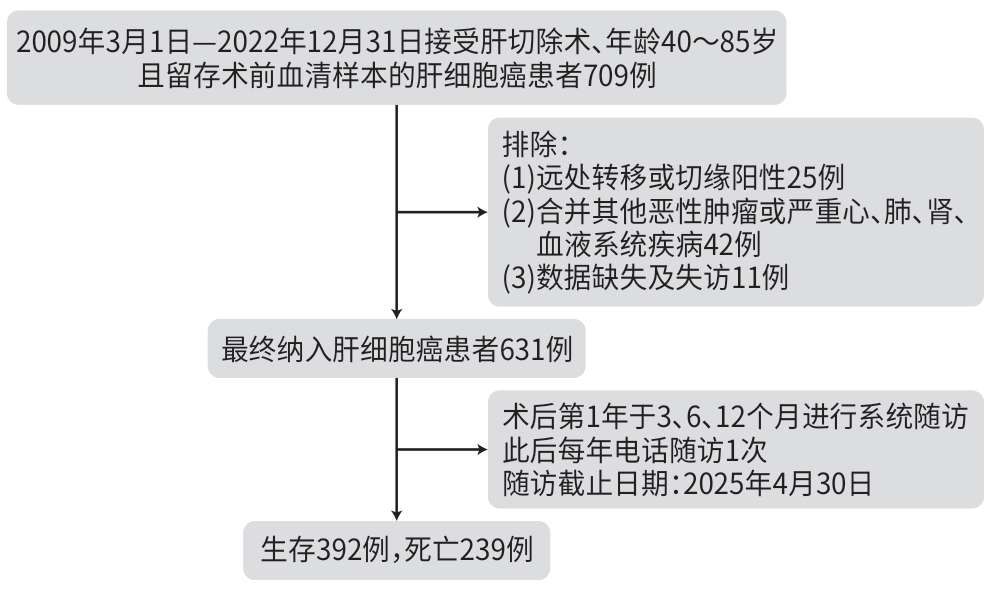
目的 评估术前血清高尔基体蛋白73(GP73)在可切除肝细胞癌(HCC)患者中诊断肝纤维化程度的效能,并探讨其对术后生存的预测作用,以期为该人群的风险分层与个体化决策提供依据。 方法 纳入2009年3月1日—2022年12月31日于吉林大学第一医院接受肝切除术的HCC患者631例。收集患者的人口学、病理学及血清学特征,并检测术前血清GP73水平。以病理Scheuer评分为肝纤维化诊断的金标准,采用Spearman相关分析评估GP73与肝纤维化的相关性,应用受试者操作特征曲线及曲线下面积(AUC)评估血清GP73与血清学复合标志物对肝纤维化的诊断价值,AUC的比较采用Delong检验。以全因死亡为预后结局,通过X-Tile 3.6.1软件确定GP73的最佳截断值,采用单因素和多因素Cox回归分析确定预后独立影响因素,并构建列线图模型。采用10折交叉验证评估模型的内部一致性,计算一致性指数(C-index)、综合判别改善指数(IDI)、净重分类改善指数(NRI),并绘制校准曲线及决策曲线分析评估模型的预测效能。 结果 血清GP73水平与肝纤维化分期呈正相关(rs =0.255,P<0.001)。血清GP73诊断显著肝纤维化的AUC为0.704,显著优于γ-谷氨酰转移酶/血小板比值(AUC=0.634,Z=2.00,P=0.046)和S指数(AUC=0.637,Z=1.98,P=0.048);与天冬氨酸氨基转移酶/血小板比值指数(AUC=0.722)及纤维化-4指数(AUC=0.695)相比,差异均无统计学意义(P值均>0.05)。HCC患者术后中位随访时间为48.66个月,累计死亡239例(37.9%)。GP73预测可切除HCC患者术后死亡的最佳截断值为65 ng/mL,GP73高表达(≥65 ng/mL)单独预测HCC患者预后的C-index为0.617,优于临床常用的巴塞罗那肝癌临床分期(BCLC分期)(C-index=0.551,P<0.001)。多因素Cox回归分析显示,血清GP73[风险比(hazard ratio,HR)=2.56,95%置信区间(confidence interval,CI):1.86~3.52,P<0.001]、性别(HR=1.58,95%CI:1.10~2.28,P=0.014)、脉管浸润(HR=1.69,95%CI:1.30~2.21,P<0.001)、总胆红素(HR=1.49,95%CI:1.16~1.93,P=0.002)及肿瘤最大径(HR=1.07,95%CI:1.04~1.11,P<0.001)是HCC患者肝切除术后全因死亡的独立危险因素。为评价在其余4个预测变量基础上加入GP73能否改善模型性能,将纳入GP73、性别、脉管浸润、总胆红素和肿瘤最大径的模型定义为含GP73模型;此外,在保留性别、脉管浸润、总胆红素和肿瘤最大径4个变量的基础上剔除GP73,构建不含GP73模型。结果显示,含GP73模型预测HCC患者术后5年生存率的AUC为0.704(95%CI:0.656~0.753),10折交叉验证平均C-index为0.716(95%CI:0.682~0.750),高于不含GP73模型(C-index=0.682,95%CI:0.645~0.719,P=0.008)和BCLC分期(C-index=0.551,95%CI:0.523~0.580,P<0.001)。与不含GP73模型相比,含GP73模型的IDI为0.039(95%CI:0.016~0.069,P<0.001),NRI为0.259(95%CI:0.179~0.335,P<0.001);与BCLC分期相比,含GP73模型的IDI为0.112(95%CI:0.069~0.163,P<0.001),NRI为0.272(95%CI:0.169~0.380,P<0.001)。校准曲线和决策曲线分析显示,含GP73模型具有良好的校准度和临床净获益。 结论 GP73能够作为可切除HCC患者肝纤维化诊断的血清学标志物,其术前水平对HCC患者肝切除术后生存具有一定预测价值。基于GP73构建的综合预后模型有助于优化术后风险分层,为HCC精准医疗提供决策依据。 Abstract:Objective To assess the performance of preoperative serum Golgi protein 73 (GP73) in diagnosing liver fibrosis degree in patients with resectable hepatocellular carcinoma (HCC), to investigate its value in predicting postoperative survival, and to provide a basis for risk stratification and individualized decision-making in this population. Methods A total of 631 patients with HCC who underwent liver resection at The First Hospital of Jilin University from March 1, 2009 to December 31, 2022 were enrolled. The demographic, pathological, and serological features of the patients were collected, and the serum level of GP73 was measured before surgery. Scheuer score determined by liver biopsy was used as the gold standard for diagnosing liver fibrosis; the Spearman correlation analysis was used to investigate the correlation between GP73 and liver fibrosis; the receiver operating characteristic (ROC) curve and the area under the ROC curve (AUC) were used to assess the value of serum GP73 and compound serological markers in the diagnosis of liver fibrosis, and the Delong test was used for comparison of AUC. With all-cause mortality as the outcome, X-Tile 3.6.1 software was used to determine the optimal cut-off value of GP73; the univariate and multivariate Cox regression analyses were used to identify independent influencing factors for prognosis, and a nomogram model was constructed. Ten-fold cross-validation was used for internal validation; index of concordance (C-index), integrated discrimination improvement (IDI) index, and net reclassification improvement (NRI) index were calculated; calibration curve and decision curve analysis were used to assess the predictive performance of the model. Results The serum level of GP73 was positively correlated with Scheuer liver fibrosis stage (rs =0.255, P<0.001). Serum GP73 had a significantly higher AUC than gamma-glutamyl transferase-to-platelet ratio (0.704 vs 0.634, Z=2.00, P=0.046) and S index (0.704 vs 0.637, Z=1.98, P=0.048) in the diagnosis of significant liver fibrosis, with no significant difference compared with aspartate aminotransferase-to-platelet ratio index (AUC=0.722) and fibrosis-4 index (AUC=0.695) (both P>0.05). During a median follow-up time of 48.66 months, 239 patients (37.9%) died. GP73 had an optimal cut-off value of 65 ng/mL in predicting postoperative mortality in patients with resectable HCC, and high GP73 expression (≥65 ng/mL) alone achieved a C-index of 0.617 for predicting the prognosis of HCC patients, which was better than the C-index of 0.551 for Barcelona Clinic Liver Cancer (BCLC) stage (P<0.001). The multivariate Cox regression analysis showed that serum GP73 (hazard ratio [HR]=2.56, 95% confidence interval [CI]: 1.86 — 3.52, P<0.001), sex (HR=1.58, 95%CI: 1.10 — 2.28, P=0.014), vascular invasion (HR=1.69, 95%CI: 1.30 — 2.21, P<0.001), total bilirubin (HR=1.49, 95%CI: 1.16 — 1.93, P=0.002), and maximum tumor diameter (HR=1.07, 95%CI: 1.04 — 1.11, P<0.001) were independent risk factors for all-cause mortality after hepatectomy in patients with HCC. In order to assess whether the addition of GP73 to the other four variables could further improve model performance, the model based on GP73, sex, vascular invasion, total bilirubin, and maximum tumor diameter was defined as the GP73-included model, and the model based on all five variables except GP73 was defined as the GP73-excluded model. The GP73-included model achieved an AUC of 0.704 (95%CI: 0.656 — 0.753) for predicting 5-year survival rate after surgery, and ten-fold cross-validation showed that this model had a mean C-index of 0.716 (95%CI: 0.682 — 0.750), which was higher than the C-index of the GP73-excluded model (C-index=0.682, 95%CI: 0.645 — 0.719, P=0.008) and BCLC stage (C-index=0.551, 95%CI: 0.523 — 0.580, P<0.001). Compared with the GP73-excluded model, the GP73-included model yielded an IDI index of 0.039 (95%CI: 0.016 — 0.069, P<0.001) and an NRI index of 0.259 (95%CI: 0.179 — 0.335, P<0.001), and compared with BCLC stage, the GP73-included model had an IDI index of 0.112 (95%CI: 0.069 — 0.163, P<0.001) and an NRI index of 0.272 (95%CI: 0.169 — 0.380, P<0.001). Calibration curve and decision curve analyses showed that the GP73-included model had good calibration and net clinical benefit. Conclusion GP73 can be used as a serological biomarker for diagnosing liver fibrosis in patients with resectable HCC, and preoperative GP73 level has a certain value in predicting the survival of these patients after hepatectomy. The integrated prognostic model incorporating GP73 can help to optimize postoperative risk stratification, thereby providing a basis for decision-making in precise treatment of HCC. -
Key words:
- Carcinoma, Hepatocellular /
- Golgi Protein 73 /
- Hepatic Fibrosis /
- Prognosis /
- Nomograms
-
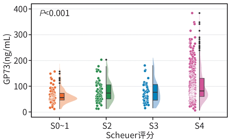
注: GP73,高尔基体蛋白73;HCC,肝细胞癌。
图 2 不同肝纤维化分期HCC患者的血清GP73水平
Figure 2. Serum GP73 in patients with hepatocellular carcinoma according to Scheuer score
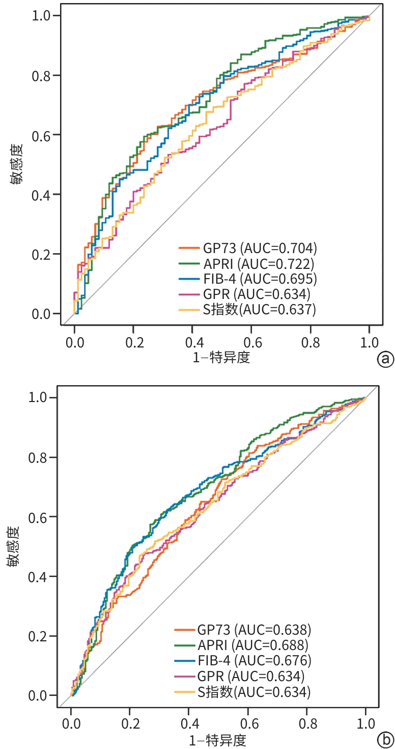
注: a,显著肝纤维化;b,肝硬化。GP73,高尔基体蛋白73;APRI,天冬氨酸氨基转移酶/血小板比值指数;FIB-4,纤维化-4指数;GPR,γ-谷氨酰转移酶/血小板比值;ROC曲线,受试者操作特征曲线;AUC,曲线下面积。
图 3 GP73、APRI、FIB-4、GPR及S指数对肝纤维化诊断的ROC曲线
Figure 3. ROC curves of GP73, APRI, FIB-4, GPR, and S-index for diagnosing liver fibrosis
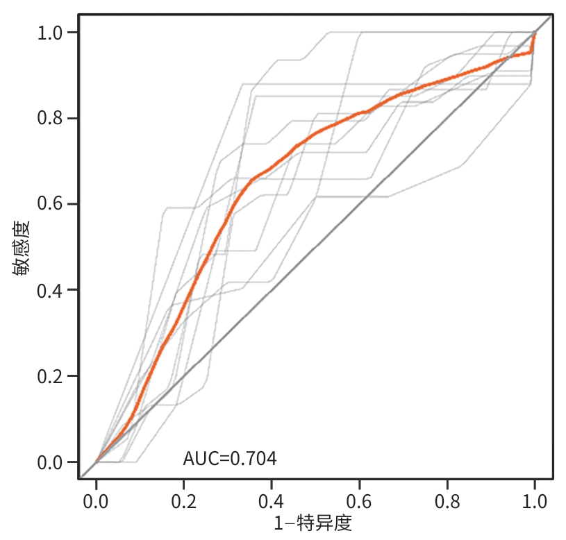
注: 灰色曲线为10折交叉验证各折时间依赖ROC曲线,橙色曲线为10折平均时间依赖ROC曲线。ROC曲线,受试者操作特征曲线;AUC,曲线下面积。
图 5 5年时间依赖ROC曲线
Figure 5. 5-year time-dependent ROC curve
表 1 HCC患者基线特征
Table 1. Subject characteristics of HCC patients
变量 数值 变量 数值 年龄[例(%)] 神经浸润[例(%)] <60岁 397(62.9) 否 627(99.4) ≥60岁 234(37.1) 是 4(0.6) 性别[例(%)] Scheuer评分[例(%)] 男 496(78.6) S0~1 88(13.9) 女 135(21.4) S2 78(12.4) Child-Pugh分级[例(%)] S3 51(8.1) A级 569(90.2) S4 414(65.6) B级 62(9.8) AFP[例(%)] 肝炎类型[例(%)] <400 ng/mL 471(74.6) 不伴病毒感染 14(2.2) ≥400 ng/mL 160(25.4) HBV感染 517(81.9) AFP-L3%[例(%)] HCV感染 52(8.2) <10% 387(61.3) 共同感染 48(7.6) ≥10% 244(38.7) BCLC分期[例(%)] DCP[例(%)] 0+A期 545(86.4) <40 ng/mL 222(35.2) B期 86(13.6) ≥40 ng/mL 409(64.8) 肿瘤最大径[例(%)] GP73(ng/mL) 77.2(54.5~115.8) <5 cm 372(59.0) Alb(g/L) 38.8(36.1~41.5) ≥5 cm 259(41.0) TBil(μmol/L) 15.1(11.2~20.7) 肿瘤数量[例(%)] AST(U/L) 32.3(24.1~48.2) 单发 510(80.8) ALT(U/L) 31.3(20.8~48.0) 多发 121(19.2) GGT(U/L) 57.0(33.4~97.3) 肿瘤组织分化[例(%)] PLT(×109/L) 150.0(108.5~190.0) 高分化 35(5.5) APRI 0.57(0.37~1.06) 中分化 426(67.5) FIB-4 2.31(1.55~3.61) 低分化 170(26.9) GPR 0.69(0.40~1.32) 脉管浸润[例(%)] S指数 0.26(0.14~0.56) 否 366(58.0) 是 265(42.0) 注:HCC,肝细胞癌;Child-Pugh分级,蔡尔德-皮尤分级;HBV,乙型肝炎病毒;HCV,丙型肝炎病毒;BCLC分期,巴塞罗那肝癌临床分期;AFP,甲胎蛋白;AFP-L3%,甲胎蛋白异质体L3百分比;DCP,异常凝血酶原;GP73,高尔基体蛋白73;Alb,白蛋白;TBil,总胆红素;AST,天冬氨酸氨基转移酶;ALT,丙氨酸氨基转移酶;GGT,γ-谷氨酰转移酶;PLT,血小板;APRI,天冬氨酸氨基转移酶/血小板比值指数;FIB-4,纤维化-4指数;GPR,γ-谷氨酰转移酶/血小板比值。
 下载: 导出CSV
表 2 不同血清GP73表达水平HCC患者的临床病理特征
Table 2. Clinicopathological characteristics between HCC patients with low and high serum GP73 levels
变量 GP73低表
达组
(n=235)GP73高表
达组
(n=396)χ2值 P值 年龄[例(%)] 0.13 0.714 <60岁 150(63.8) 247(62.4) ≥60岁 85(36.2) 149(37.6) 性别[例(%)] 1.59 0.208 男 191(81.3) 305(77.0) 女 44(18.7) 91(23.0) Child-Pugh分级[例(%)] 17.43 <0.001 A级 227(96.6) 342(86.4) B级 8(3.4) 54(13.6) 肝炎类型[例(%)] 16.99 <0.001 不伴病毒感染 1(0.4) 13(3.3) HBV感染 186(79.1) 331(83.6) HCV感染 31(13.2) 21(5.3) 共同感染 17(7.2) 31(7.8) BCLC分期[例(%)] 3.71 0.054 0+A期 211(89.8) 334(84.3) B期 24(10.2) 62(15.7) 肝硬化[例(%)] 29.22 <0.001 否 112(47.7) 105(26.5) 是 123(52.3) 291(73.5) 肿瘤最大径[例(%)] 11.73 <0.001 <5 cm 159(67.7) 213(53.8) ≥5 cm 76(32.3) 183(46.2) 肿瘤数量[例(%)] 1.12 0.290 单发 195(83.0) 315(79.5) 多发 40(17.0) 81(20.5) 肿瘤组织分化[例(%)] 10.69 0.005 高分化 16(6.8) 19(4.8) 中分化 173(73.6) 253(63.9) 低分化 46(19.6) 124(31.3) 脉管浸润[例(%)] 7.76 0.005 否 153(65.1) 213(53.8) 是 82(34.9) 183(46.2) 神经浸润[例(%)] 0.631 否 233(99.1) 394(99.5) 是 2(0.9) 2(0.5) 注:GP73,高尔基体蛋白73;HCC,肝细胞癌;Child-Pugh分级,蔡尔德-皮尤分级;HBV,乙型肝炎病毒;HCV,丙型肝炎病毒;BCLC分期,巴塞罗那肝癌临床分期。
下载: 导出CSV
表 3 HCC切除术后预后的单因素及多因素Cox回归分析
Table 3. Univariate and multivariate Cox analysis of prognosis of HCC resection
变量 单因素分析 多因素分析 HR 95%CI P值 HR 95%CI P值 年龄(≥60岁 vs <60岁) 0.99 0.76~1.30 0.934 性别(男 vs 女) 1.61 1.13~2.31 0.009 1.58 1.10~2.28 0.014 BMI(kg/m2) 0.95 0.91~1.00 0.054 肝炎类型 HBV感染 vs 不伴病毒感染 1.09 0.65~1.81 0.750 HCV感染 vs 不伴病毒感染 0.50 0.23~1.11 0.090 共同感染 vs 不伴病毒感染 0.98 0.43~2.25 0.967 Child-Pugh分级(B级 vs A级) 2.18 1.51~3.14 <0.001 BCLC分期(B期 vs 0+A期) 1.70 1.23~2.34 0.001 肝硬化(是 vs 否) 1.25 0.95~1.64 0.118 肿瘤最大径(cm) 1.12 1.08~1.16 <0.001 1.07 1.04~1.11 <0.001 肿瘤数量(多发 vs 单发) 1.57 1.17~2.11 0.003 肿瘤组织分化 中分化 vs 高分化 1.07 0.65~1.76 0.798 低分化 vs 高分化 1.60 0.96~2.67 0.074 脉管浸润(是 vs 否) 2.09 1.62~2.70 <0.001 1.69 1.30~2.21 <0.001 神经浸润(是vs 否) 1.41 0.35~5.68 0.630 Alb(<35 g/L vs ≥35 g/L) 1.50 1.09~2.07 0.013 TBil(≥17.1 μmol/L vs <17.1 μmol/L) 1.58 1.23~2.04 <0.001 1.49 1.16~1.93 0.002 AST(≥40 U/L vs <40 U/L) 1.91 1.48~2.46 <0.001 ALT(≥40 U/L vs <40 U/L) 1.35 1.04~1.75 0.022 PLT(≥125 ×109/L vs <125×109/L) 1.25 0.96~1.62 0.094 PT(≥13 s vs <13 s) 1.16 0.84~1.58 0.367 NLR(≥1.90 vs <1.90) 1.64 1.25~2.14 <0.001 MLR(≥0.27 vs <0.27) 1.47 1.13~1.90 0.004 AFP(≥400 ng/mL vs <400 ng/mL) 1.46 1.11~1.92 0.007 AFP-L3%(≥10% vs <10%) 1.44 1.11~1.86 0.006 DCP(≥40 ng/mL vs <40 ng/mL) 1.47 1.12~1.93 0.006 GP73(≥65 ng/mL vs <65 ng/mL) 2.84 2.08~3.89 <0.001 2.56 1.86~3.52 <0.001 注:HCC,肝细胞癌;HR,风险比;CI,置信区间;BMI,体重指数;HBV,乙型肝炎病毒;HCV,丙型肝炎病毒;Child-Pugh分级,蔡尔德-皮尤分级;BCLC分期,巴塞罗那肝癌临床分期;Alb,白蛋白;TBil,总胆红素;AST,天冬氨酸氨基转移酶;ALT,丙氨酸氨基转移酶;PLT,血小板;PT,凝血酶原时间;NLR,中性粒细胞/淋巴细胞比值;MLR,单核细胞/淋巴细胞比值;AFP,甲胎蛋白;AFP‑L3%,甲胎蛋白异质体L3百分比;DCP,异常凝血酶原;GP73,高尔基体蛋白73。
下载: 导出CSV
-
[1] Bray F, Laversanne M, Sung H, et al. Global cancer statistics 2022: GLOBOCAN estimates of incidence and mortality worldwide for 36 cancers in 185 countries[J]. CA Cancer J Clin, 2024, 74( 3): 229- 263. DOI: 10.3322/caac.21834. [2] Wang Z X, Qin H J, Liu S, et al. Precision diagnosis of hepatocellular carcinoma[J]. Chin Med J, 2023, 136( 10): 1155- 1165. DOI: 10.1097/CM9.0000000000002641. [3] Yao L Q, Fan H X, Chen F J, et al. Heterogeneity in prognosis for early-stage hepatocellular carcinoma after hepatectomy: A multicenter analysis of 4, 623 patients across five major staging systems[J]. Eur J Surg Oncol, 2025, 51( 10): 110383. DOI: 10.1016/j.ejso.2025.110383. [4] Sonohara F, Yamada S, Tanaka N, et al. Comparison of non-invasive liver reserve and fibrosis models: Implications for surgery and prognosis for hepatocellular carcinoma[J]. Hepatol Res, 2019, 49( 11): 1305- 1315. DOI: 10.1111/hepr.13400. [5] Zhang J W, Zhang L, Yang X B, et al. Liver fibrosis as a predictor of liver failure and outcome following ALPPS among patients with primary liver cancer[J]. Sci Rep, 2024, 14: 15827. DOI: 10.1038/s41598-024-65924-2. [6] Loomba R, Adams L A. Advances in non-invasive assessment of hepatic fibrosis[J]. Gut, 2020, 69( 7): 1343- 1352. DOI: 10.1136/gutjnl-2018-317593. [7] Smith A, Baumgartner K, Bositis C. Cirrhosis: Diagnosis and management[J]. Am Fam Physician, 2019, 100( 12): 759- 770. [8] Lan B, Luo C, Pocha C, et al. Comparison of various liver cancer staging systems in predicting prognosis after initial transcatheter arterial chemoembolization: A retrospective study from China[J]. J Gastrointest Oncol, 2024, 15( 6): 2599- 2612. DOI: 10.21037/jgo-2024-850. [9] Ke M Y, Wu X N, Zhang Y, et al. Serum GP73 predicts posthepatectomy outcomes in patients with hepatocellular carcinoma[J]. J Transl Med, 2019, 17( 1): 140. DOI: 10.1186/s12967-019-1889-0. [10] Wang Q, Cui H X, Zhu Y P, et al. Serum Golgi protein 73(GP73) is a diagnostic and prognostic marker of hepatocellular carcinoma[J]. Front Med, 2025, 12: 1571761. DOI: 10.3389/fmed.2025.1571761. [11] Feng P P, Hu X Y, Zhou S N, et al. Golgi protein 73: The driver of inflammation in the immune and tumor microenvironment[J]. Front Immunol, 2024, 15: 1508034. DOI: 10.3389/fimmu.2024.1508034. [12] An Xinyu, Qiao Jie, Hu Lingxi, et al. A study on the diagnostic value of GP73 in liver fibrosis among patients with chronic liver disease[J]. Chin J Intern Med, 2023, 62( 1): 49- 53. DOI: 10.3760/cma.j.cn112138-20220514-00371.安薪宇, 乔杰, 胡灵溪, 等. GP73对慢性肝病患者肝纤维化诊断价值的研究[J]. 中华内科杂志, 2023, 62( 1): 49- 53. DOI: 10.3760/cma.j.cn112138-20220514-00371. [13] Liver Disease Committee, Chinese Association of Integrative Medicine. Guidelines for the diagnosis and treatment of liver fibrosis in integrative medicine practice(2019)[J]. J Clin Hepatol, 2019, 35( 7): 1444- 1449. DOI: 10.3969/j.issn.1001-5256.2019.07.007.中国中西医结合学会肝病专业委员会. 肝纤维化中西医结合诊疗指南(2019年版)[J]. 临床肝胆病杂志, 2019, 35( 7): 1444- 1449. DOI: 10.3969/j.issn.1001-5256.2019.07.007. [14] Borcak D, Yesilbag Z, Ozdemir Y E, et al. Assessing liver fibrosis in chronic hepatitis B: Liver biopsy or non-invasive fibrosis markers?[J]. J Clin Med, 2025, 14( 22): 8164. DOI: 10.3390/jcm14228164. [15] Fan Haina, Ma Yangqing, Sun Xin, et al. Role of serum Golgi protein 73 in the assessment of pathological prognosis and its inflammatory influencing factors for hepatitis B virus-related liver fibrosis[J]. Chin J Hepatol, 2025, 33( 8): 772- 780. DOI: 10.3760/cma.j.cn501113-20240419-00212.范海纳, 马杨青, 孙鑫, 等. 血清高尔基体蛋白73对乙型肝炎肝纤维化病理预后的评估作用及其炎症影响因素[J]. 中华肝脏病杂志, 2025, 33( 8): 772- 780. DOI: 10.3760/cma.j.cn501113-20240419-00212. [16] Ye J Z, Gao X, Huang X, et al. Integrating single-cell and spatial transcriptomics to uncover and elucidate GP73-mediated pro-angiogenic regulatory networks in hepatocellular carcinoma[J]. Research(Wash D C), 2024, 7: 0387. DOI: 10.34133/research.0387. [17] Liu Y M, Hu X Y, Zhou S N, et al. Golgi protein 73 promotes angiogenesis in hepatocellular carcinoma[J]. Research(Wash D C), 2024, 7: 0425. DOI: 10.34133/research.0425. [18] Zhang J, Zhang M K, Ma H M, et al. A meta-analysis of the prognostic significance of Golgi protein 73 in hepatocellular carcinoma in Chinese patients[J]. Arch Med Sci, 2020, 16( 5): 1104- 1110. DOI: 10.5114/aoms.2019.83821. [19] Chen L J, Chang Y J, Chang Y J. Survival predictability between the American joint committee on cancer 8th edition staging system and the Barcelona clinic liver cancer classification in patients with hepatocellular carcinoma[J]. Oncologist, 2021, 26( 3): e445- e453. DOI: 10.1002/onco.13535. [20] Ren Meng, Yang Yi, Li Ziming, et al. Clinical features and prognosis of hepatocellular carcinoma: A single-center study of 850 cases[J]. J Clin Hepatol, 2024, 40( 10): 2019- 2026. DOI: 10.12449/JCH241015.任朦, 杨益, 李紫明, 等. 850例单中心肝细胞癌患者的临床特征与预后分析[J]. 临床肝胆病杂志, 2024, 40( 10): 2019- 2026. DOI: 10.12449/JCH241015. -

 本文二维码
本文二维码
计量
- 文章访问数: 176
- HTML全文浏览量: 50
- PDF下载量: 22
- 被引次数: 0




