
 PDF下载 ( 18708 KB)

高血糖环境下Yes相关蛋白1和人类胶原蛋白Ⅵ α1链在胰腺癌中的表达水平及其对患者预后的影响
DOI: 10.12449/JCH260519
Expression levels of Yes-associated protein 1 and human collagen type Ⅵ alpha 1 in pancreatic cancer under hyperglycemic conditions and their impact on prognosis
-
摘要:
目的 探讨高血糖环境下Yes相关蛋白1(YAP1)和人类胶原蛋白Ⅵ α1链(COL6A1)在胰腺癌中的表达变化及其与预后的关系。 方法 使用GEPIA数据库及R语言分析YAP1与COL6A1在胰腺癌中的表达水平和相关性,并评估其对胰腺癌患者预后的影响。通过cBioPortal数据库筛选胰腺癌中YAP1和COL6A1的共表达基因,并进行基因本体论(GO)和京都基因与基因组百科全书(KEGG)富集分析。实时定量荧光聚合酶链式反应(qPCR)和蛋白质免疫印迹(WB)法检测高糖环境与正常糖环境中人胰腺癌细胞系PANC-1中YAP1和COL6A1基因及蛋白的表达水平。构建2型糖尿病胰腺癌裸鼠模型,使用免疫组织化学检测裸鼠皮下肿瘤组织中YAP1和COL6A1的表达情况。收集2016年1月—2020年1月广西医科大学第一附属医院、广西医科大学附属肿瘤医院、广西医科大学附属武鸣医院共96例胰腺癌患者的组织标本和临床资料,其中48例胰腺癌合并糖尿病患者纳入研究组,48例单纯胰腺癌患者纳入对照组,使用免疫组织化学法检测两组患者肿瘤组织中YAP1和COL6A1的表达水平并分析其与临床指标、生存预后的关系。计量资料组间比较采用成组t检验,计数资料组间比较采用χ2检验或Mann-Whitney U检验,正态分布连续变量采用Pearson相关性分析,非连续变量或非正态分布采用Spearman相关性分析。采用Kaplan-Meier法绘制生存曲线,Log-rank检验比较生存率。采用Cox比例风险模型进行单因素及多因素分析。 结果 YAP1和COL6A1在胰腺癌中均呈高表达,两者具有强相关性(r=0.85,P<0.001),且与患者不良预后相关(风险比分别为1.8、1.5,P值均<0.05)。YAP1和COL6A1分别与219和122个基因显著相关(r值分别为0.83、0.86,P值均<0.001),GO和KEGG富集分析表明其通过细胞外基质-受体相互作用、Hippo信号通路和磷脂酰肌醇3-激酶/蛋白激酶B信号通路影响胰腺癌的发生和发展。WB和qPCR结果显示,高糖环境下人胰腺癌细胞系PANC-1中YAP1和COL6A1的基因(t值分别为4.726、6.197,P值均<0.05)与蛋白(t值分别为6.826、5.254,P值均<0.05)表达均显著升高。免疫组织化学结果显示,模型组裸鼠皮下肿瘤组织中YAP1和COL6A1的表达显著升高,两者呈正相关(r=0.985,P<0.001)。临床胰腺癌组织标本免疫组织化学显示,YAP1、COL6A1蛋白在研究组中阳性表达上升,两者呈正相关(r=0.882,P<0.001)。临床数据多因素分析显示,淋巴结转移(风险比=0.083,95%置信区间:0.030~0.231)、远处器官转移(风险比=0.166,95%置信区间:0.065~0.420)、YAP1表达量(风险比=2.027,95%置信区间:1.065~3.857)和COL6A1表达量(风险比=2.044,95%置信区间:1.019~4.099)是胰腺癌患者预后的独立影响因素(P值均<0.05)。生存分析显示,YAP1高表达组的2年生存率显著低于YAP1低表达组(6.82% vs 25.00%,χ2=16.382,P<0.001);COL6A1高表达组的2年生存率显著低于COL6A1低表达组(14.58% vs 18.75%,χ2=4.579,P=0.032)。 结论 高血糖环境促进YAP1和COL6A1在胰腺癌中高表达,且与患者的不良预后相关。 -
关键词:
- 胰腺肿瘤 /
- 高血糖症 /
- Yes相关蛋白1 /
- 人类胶原蛋白Ⅵ α1链
Abstract:Objective To investigate the changes in the expression levels of Yes-associated protein 1 (YAP1) and human collagen type Ⅵ alpha 1 (COL6A1) in pancreatic cancer under hyperglycemic conditions and their association with prognosis. Methods GEPIA database and R software were used to analyze the expression levels of YAP1 and COL6A1 and their correlation in pancreatic cancer, as well as their impact on the prognosis of patients with pancreatic cancer. The cBioPortal database was used to identify the co-expressed genes of YAP1 and COL6A1 in pancreatic cancer, and the gene ontology (GO) and Kyoto Encyclopedia of Genes and Genomes (KEGG) enrichment analyses were performed. Quantitative real-time PCR and Western blot were used to measure the mRNA and protein expression levels of YAP1 and COL6A1 in human pancreatic cancer PANC-1 cells cultured under hyperglycemic or normoglycemic conditions. A nude mouse model of pancreatic cancer with type 2 diabetes was established, and immunohistochemistry was used to measure the expression of YAP1 and COL6A1 in subcutaneous tumor tissue. Clinical specimens and data of 96 patients with pancreatic cancer were collected from January 2016 to January 2020 in the First Affiliated Hospital of Guangxi Medical University, Guangxi Medical University Cancer Hospital and Wuming Hospital Affiliated to Guangxi Medical University. Among them, 48 patients with pancreatic cancer and diabetes were enrolled as study group, and 48 patients with pancreatic cancer alone were enrolled as control group; immunohistochemistry was used to measure the expression levels of YAP1 and COL6A1 in tissue samples from both groups, and their association with clinical indicators and survival outcomes was analyzed. The t-test was used for comparison of continuous data between groups, and the chi-square test or the Mann-Whitney U test was used for comparison of categorical data between groups; the Pearson correlation analysis was used for normally distributed continuous variables, while the Spearman correlation analysis was used for discontinuous variables or non-normally distributed variables. The Kaplan-Meier method was used to plot survival curves, and the Log-rank test was used for comparison of survival rates. The Cox proportional hazards model was used to perform univariate and multivariate analyses. Results Both YAP1 and COL6A1 were highly expressed in pancreatic cancer, with a strong correlation between them (r=0.85, P<0.001), and they were associated with the poor prognosis of patients (hazard ratio [HR]=1.8 and 1.5, both P<0.05). YAP1 and COL6A1 were significantly correlated with 219 and 122 genes, respectively (r=0.83 and 0.86, both P<0.05), and the GO and KEGG enrichment analyses showed that these genes affected the development and progression of pancreatic cancer through extracellular matrix-receptor interaction, the Hippo signaling pathway, and the phosphatidylinositol 3-kinase/protein kinase B signaling pathway. Quantitative real-time PCR and Western blot showed that there were significant increases in the mRNA and protein expression levels of YAP1 and COL6A1 in human pancreatic cancer PANC-1 cells under hyperglycemic conditions (mRNA expression: t=4.726 and 6.197, both P<0.05; protein expression: t=6.826 and 5.254, both P<0.05). Immunohistochemistry showed that there were significant increases in the expression levels of YAP1 and COL6A1 in subcutaneous tumor tissue of nude mice, with a positive correlation between them (r=0.985, P<0.001). For the clinical specimens of pancreatic cancer tissue, immunohistochemistry showed that there were significant increases in the positive expression levels of YAP1 and COL6A1 proteins in the study group, with a positive correlation between them (r=0.882, P<0.001). The multivariate analysis of clinical data showed that lymph node metastasis (HR=0.083, 95% confidence interval [CI]: 0.030 — 0.231, P<0.05), distant organ metastasis (HR=0.166, 95%CI: 0.065 — 0.420, P<0.05), YAP1 expression level (HR=2.027, 95%CI: 1.065 — 3.857, P<0.05), and COL6A1 expression level (HR=2.044, 95%CI: 1.019 — 4.099, P<0.05) were independent influencing factors for the prognosis of patients with pancreatic cancer. The survival analysis showed that the high YAP1 expression group had a significantly lower 2-year survival rate than the low YAP1 expression group (6.82% vs 25.00%, χ2=16.382, P<0.001), and the high COL6A1 expression group had a significantly lower 2-year survival rate than the low COL6A1 expression group (14.58% vs 18.75%, χ2=4.579, P=0.032). Conclusion The hyperglycemic environment promotes the high expression of YAP1 and COL6A1 in pancreatic cancer, which is associated with poor prognosis in patients. -
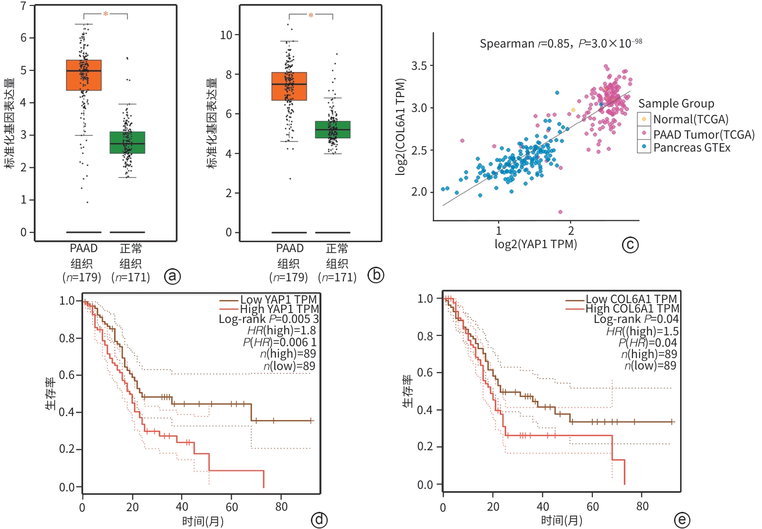
注: a,YAP1基因在PAAD及正常胰腺组织中的表达,*P<0.05;b,COL6A1基因在PAAD及正常胰腺组织中的表达,*P<0.05;c,YAP1和COL6A1基因的表达在PAAD中的相关性;d,YAP1基因表达水平对患者总生存期的影响;e,COL6A1基因表达水平对患者总生存期的影响。YAP1,Yes相关蛋白1;COL6A1,人类胶原蛋白Ⅵ α1链;PAAD,胰腺导管腺癌;HR,风险比。
图 1 YAP1和COL6A1基因在PAAD中的表达、相关性及预后
Figure 1. Expression, correlation, and prognostic value of YAP1 and COL6A1 genes in PAAD
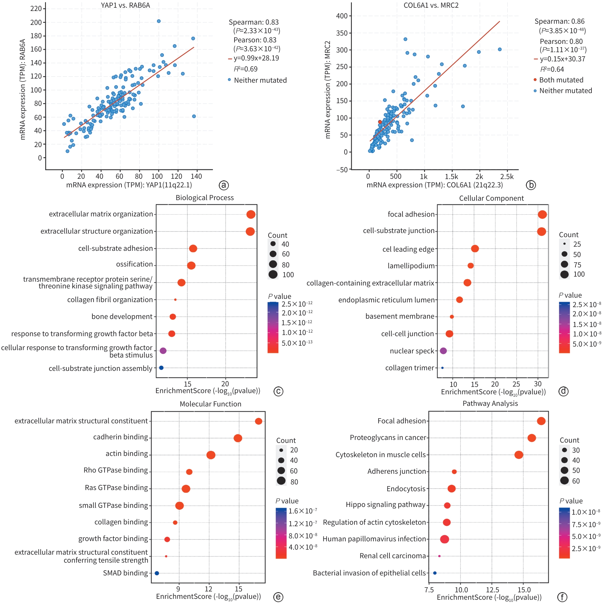
注: a,PAAD中YAP1与RAB6A表达显著相关(r=0.83,P<0.001),横纵坐标分别表示YAP1与RAB6A的基因表达量;b,PAAD中COL6A1与MRC2表达显著相关(r=0.86,P<0.001),横纵坐标分别表示COL6A1与MRC2的基因表达量;c,YAP1和COL6A1在PAAD中共表达基因的生物过程分析;d,YAP1和COL6A1在PAAD中共表达基因的细胞组分分析;e,YAP1和COL6A1在PAAD中共表达基因的分子功能分析;f,YAP1和COL6A1在PAAD中共表达基因的KEGG通路分析。YAP1,Yes相关蛋白1;COL6A1,人类胶原蛋白Ⅵ α1链;PAAD,胰腺导管腺癌。
图 2 YAP1和COL6A1在PAAD中的共表达基因及富集分析
Figure 2. Co-expression genes and enrichment analysis of YAP1 and COL6A1 in PAAD
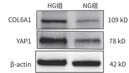
注: YAP1,Yes相关蛋白1;COL6A1,人类胶原蛋白Ⅵ α1链。
图 3 蛋白质免疫印迹检测胰腺癌细胞YAP1和COL6A1蛋白的表达
Figure 3. Western blot experiment detects the expression of YAP1 and COL6A1 proteins in pancreatic cancer cells
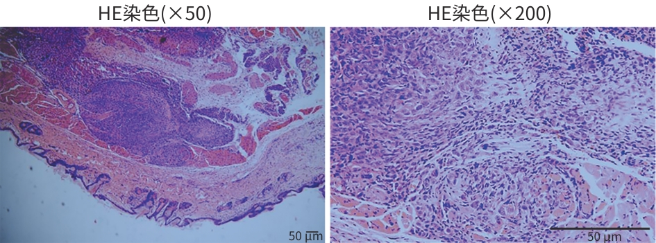
图 4 裸鼠皮下肿瘤组织HE染色
Figure 4. Hematoxylin and eosin staining of subcutaneous tumor tissue in nude mice
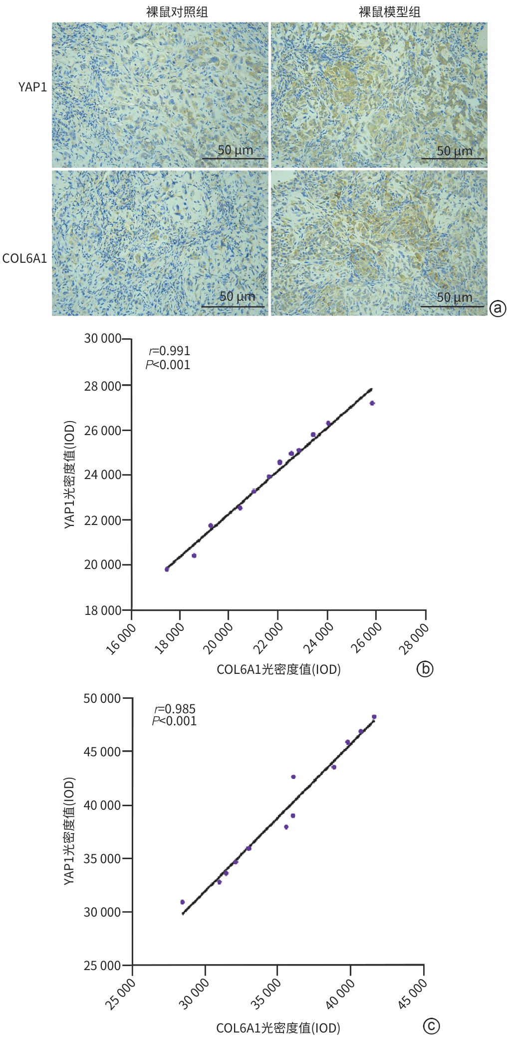
注: a,裸鼠模型组和裸鼠对照组YAP1、COL6A1蛋白表达结果(×200);b,裸鼠对照组YAP1、COL6A1蛋白相关性分析;c,裸鼠模型组YAP1、COL6A1蛋白相关性分析。YAP1,Yes相关蛋白1;COL6A1,人类胶原蛋白Ⅵ α1链;IOD,累计光密度值。
图 5 免疫组织化学检测两组YAP1、COL6A1蛋白表达
Figure 5. Immunohistochemical detection of YAP1 and COL6A1 protein expression in each group
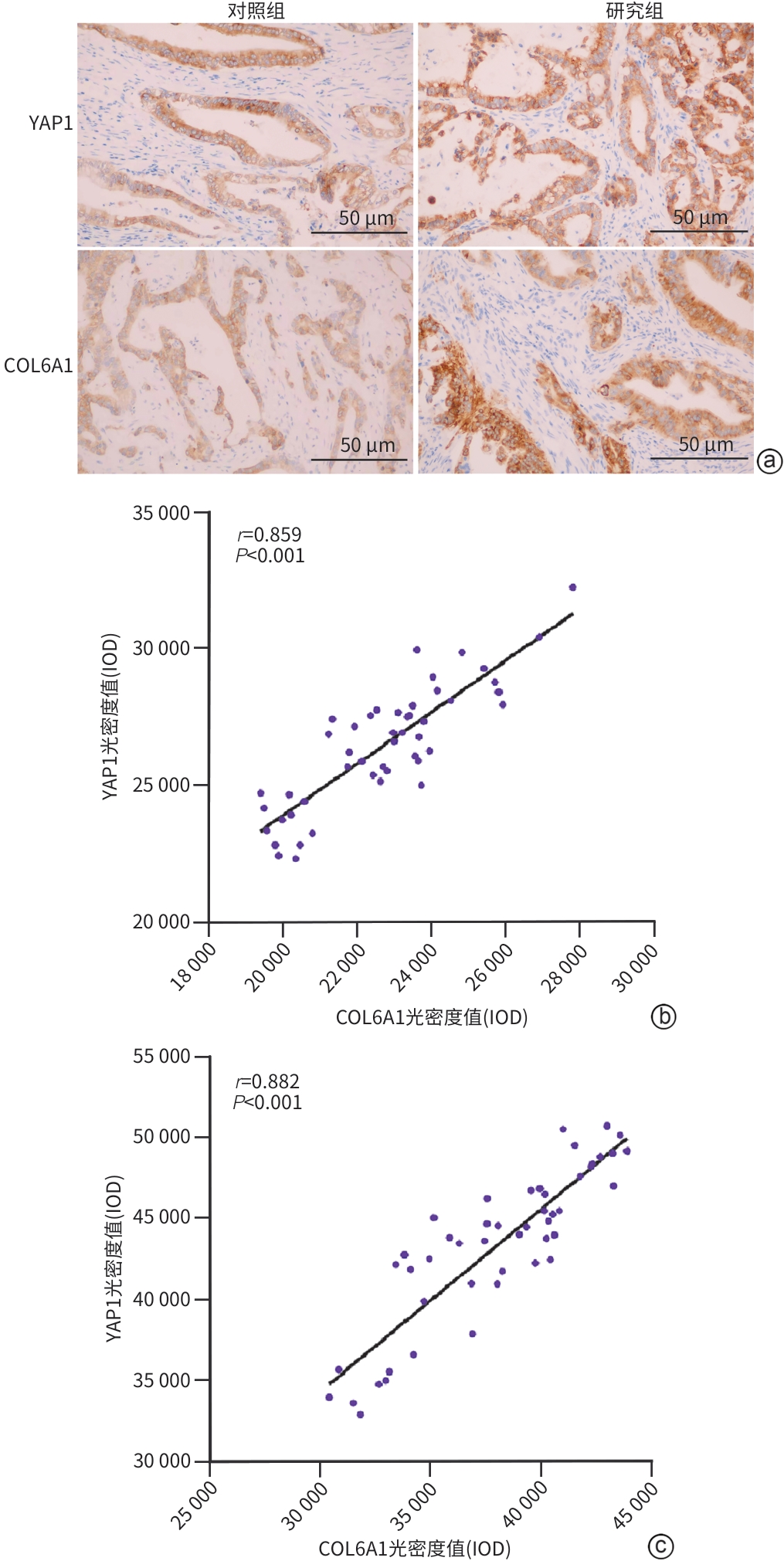
注: a,对照组和研究组YAP1、COL6A1蛋白表达结果(×200);b,对照组YAP1、COL6A1蛋白相关性分析;c,研究组YAP1、COL6A1蛋白相关性分析。YAP1,Yes相关蛋白1;COL6A1,人类胶原蛋白Ⅵ α1链;IOD,累计光密度值。
图 6 免疫组织化学检测各组YAP1、COL6A1蛋白表达
Figure 6. Immunohistochemical detection of YAP1 and COL6A1 protein expression in each group
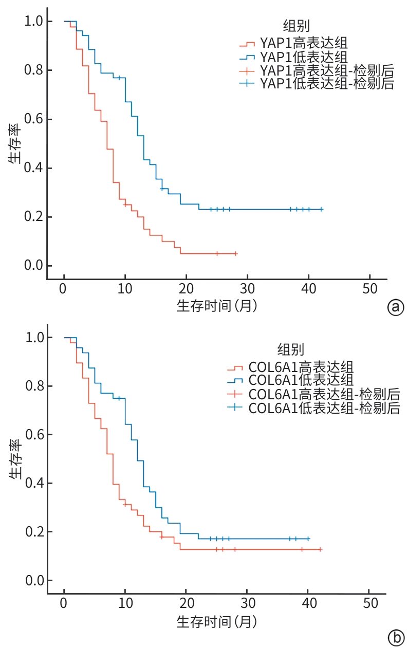
注: a,YAP1生存曲线;b,COL6A1生存曲线。YAP1,Yes相关蛋白1;COL6A1,人类胶原蛋白Ⅵ α1链。
图 7 YAP1和COL6A1高/低表达组胰腺癌患者的生存曲线
Figure 7. Survival curve of pancreatic cancer patients with high/low YAP1 and COL6A1 expression group
表 1 不同处理组YAP1、COL6A1蛋白相对表达量
Table 1. The relative expression levels of YAP1 and COL6A1 proteins in different treatment groups
组别 YAP1 COL6A1 NG组 0.490±0.013 0.246±0.112 HG组 0.696±0.067 0.743±0.058 t值 6.826 5.254 P值 0.006 0.002 注:YAP1,Yes相关蛋白1;COL6A1,人类胶原蛋白Ⅵ α1链。
 下载: 导出CSV
表 2 不同处理组YAP1和COL6A1基因相对表达量
Table 2. Gene expression of YAP1 and COL6A1 in different treatment groups
组别 YAP1 COL6A1 NG组 1.035±0.302 1.013±0.174 HG组 2.788±0.782 3.140±0.751 t值 4.726 6.197 P值 0.005 <0.001 注:YAP1,Yes相关蛋白1;COL6A1,人类胶原蛋白Ⅵ α1链。
下载: 导出CSV
表 3 两组YAP1、COL6A1累计光密度值比较
Table 3. Comparison of YAP1 and COL6A1 optical density values between the groups
组别 YAP1 COL6A1 裸鼠对照组 23 815.066±2 399.870 21 575.033±2 399.870 裸鼠模型组 39 396.598±5 938.120 35 394.846±4 254.425 t值 8.472 9.801 P值 <0.001 <0.001 注:YAP1,Yes相关蛋白1;COL6A1,人类胶原蛋白Ⅵ α1链。
下载: 导出CSV
表 4 两组YAP1、COL6A1蛋白相对表达量比较
Table 4. Comparison of YAP1 and COL6A1 protein expression levels between the pancreatic cancer-only group and the pancreatic cancer group with comorbid diabetes
组别 YAP1 COL6A1 对照组 26 471.573±2 241.120 22 705.187±2 034.836 研究组 43 476.033±4 824.713 38 073.580±3 788.025 t值 22.150 24.760 P值 <0.001 <0.001 注:YAP1,Yes相关蛋白1;COL6A1,人类胶原蛋白Ⅵ α1链。
下载: 导出CSV
表 5 临床资料基线表
Table 5. Baseline table of clinical characteristics
指标 对照组(n=48) 研究组(n=48) 统计值 P值 性别[例(%)] χ2=0.043 0.837 男 27(56.25) 28(58.33) 女 21(43.75) 20(41.67) 年龄(岁) 62.042±8.108 61.330±8.931 t=0.407 0.685 分化程度[例(%)] Z=1.298 0.194 高分化导管腺癌 10(20.83) 7(14.58) 中分化导管腺癌 23(47.92) 20(41.67) 低分化导管腺癌 15(31.25) 21(43.75) AJCC分期[例(%)] Z=3.716 0.001 Ⅰ期 12(25.00) 6(12.50) Ⅱ期 25(52.08) 12(25.00) Ⅲ期 9(18.75) 19(39.58) Ⅳ期 2(4.17) 11(22.92) 神经侵犯[例(%)] χ²=1.042 0.307 有 21(43.75) 26(54.17) 无 27(56.25) 22(45.83) 血管侵犯[例(%)] χ²=0.042 0.838 有 22(45.83) 23(47.92) 无 26(54.17) 25(52.08) 淋巴结转移[例(%)] χ²=10.741 0.001 有 14(29.17) 30(62.50) 无 34(70.83) 18(37.50) 远处器官转移[例(%)] χ²=7.207 0.007 有 2(4.17) 11(22.92) 无 46(95.83) 37(77.08) 注:AJCC,美国癌症联合委员会。
下载: 导出CSV
表 6 影响胰腺癌患者远期生存的单因素分析
Table 6. Analysis of single factors affecting long-term survival of patients with pancreatic cancer
因素 β值 HR 95%CI P值 肿瘤大小 1.859 6.419 3.166~13.015 <0.001 血管侵犯 -1.278 0.279 0.174~0.445 <0.001 神经侵犯 -1.120 0.326 0.206~0.517 <0.001 淋巴结转移 -3.369 0.034 0.014~0.085 <0.001 远处器官转移 -2.995 0.050 0.021~0.117 <0.001 分化程度 -0.776 0.460 0.331~0.640 <0.001 YAP1 1.638 5.146 3.044~8.966 <0.001 COL6A1 1.769 5.863 3.452~9.958 <0.001 HbA1c(%) 0.540 1.717 1.105~2.667 <0.001 注:YAP1,Yes相关蛋白1;COL6A1,人类胶原蛋白Ⅵ α1链;HbA1c,糖化血红蛋白;HR,风险比;CI,置信区间。
下载: 导出CSV
表 7 影响胰腺癌患者远期生存的多因素分析
Table 7. Analysis of multiple factors affecting long-term survival of patients with pancreatic cancer
因素 HR 95%CI P值 淋巴结转移 0.083 0.030~0.231 <0.001 远处器官转移 0.166 0.065~0.420 <0.001 YAP1 2.027 1.065~3.857 0.031 COL6A1 2.044 1.019~4.099 0.044 注:YAP1,Yes相关蛋白1;COL6A1,人类胶原蛋白Ⅵ α1链;HR,风险比;CI,置信区间。
下载: 导出CSV
表 8 肿瘤组织中YAP1和COL6A1蛋白表达水平与胰腺癌患者预后的关系
Table 8. Relationship between YAP1 and COL6A1 protein expression level and prognosis of pancreatic cancer patients
组别 例数 2年生存数[例(%)] χ2值 P值 YAP1 16.382 <0.001 高表达组 44 3(6.82) 低表达组 52 13(25.00) COL6A1 4.579 0.032 高表达组 48 7(14.58) 低表达组 48 9(18.75) 注:YAP1,Yes相关蛋白1;COL6A1,人类胶原蛋白Ⅵ α1链。
下载: 导出CSV
-
[1] CHEN SM, PHUC PT, NGUYEN PA, et al. A novel prediction model of the risk of pancreatic cancer among diabetes patients using multiple clinical data and machine learning[J]. Cancer Med, 2023, 12( 19): 19987- 19999. DOI: 10.1002/cam4.6547. [2] QIN C, LI TY, LIN C, et al. The systematic role of pancreatic cancer exosomes: Distant communication, liquid biopsy and future therapy[J]. Cancer Cell Int, 2024, 24( 1): 264. DOI: 10.1186/s12935-024-03456-5. [3] WANG C, LYU J, MA YL. Analysis of the clinical characteristics and prognostic risk factors in 100 patients with pancreatic cancer[J]. J Clin Exp Med, 2025, 24( 23): 2510- 2514. DOI: 10.3969/j.issn.1671-4695.2025.23.013.王琛, 吕婧, 马于丽. 胰腺癌100例患者临床特征及预后的危险因素分析[J]. 临床和实验医学杂志, 2025, 24( 23): 2510- 2514. DOI: 10.3969/j.issn.1671-4695.2025.23.013. [4] ANDERSEN DK, KORC M, PETERSEN GM, et al. Diabetes, pancreatogenic diabetes, and pancreatic cancer[J]. Diabetes, 2017, 66( 5): 1103- 1110. DOI: 10.2337/db16-1477. [5] LEGA IC, LIPSCOMBE LL. Review: Diabetes, obesity, and cancer-pathophysiology and clinical implications[J]. Endocr Rev, 2020, 41( 1): bnz014. DOI: 10.1210/endrev/bnz014. [6] RENEHAN AG, TIPPING O, WANG MY. Diabetes and cancer: Doubts of a causal link[J]. Int J Cancer, 2024, 154( 11): 1875- 1876. DOI: 10.1002/ijc.34862. [7] COSMIN STAN M, PAUL D. Diabetes and cancer: A twisted bond[J]. Oncol Rev, 2024, 18: 1354549. DOI: 10.3389/or.2024.1354549. [8] KHAN I, KAMAL A, AKHTAR S. Diabetes driven oncogenesis and anticancer potential of repurposed antidiabetic drug: A systemic review[J]. Cell Biochem Biophys, 2024, 82( 3): 1907- 1929. DOI: 10.1007/s12013-024-01387-6. [9] PLISZKA M, SZABLEWSKI L. Associations between diabetes mellitus and selected cancers[J]. Int J Mol Sci, 2024, 25( 13): 7476. DOI: 10.3390/ijms25137476. [10] CHEN JC, WANG XY, HE Q, et al. YAP activation in renal proximal tubule cells drives diabetic renal interstitial fibrogenesis[J]. Diabetes, 2020, 69( 11): 2446- 2457. DOI: 10.2337/db20-0579. [11] LI S, ZHU H, CHEN HD, et al. Glucose promotes epithelial-mesenchymal transitions in bladder cancer by regulating the functions of YAP1 and TAZ[J]. J Cell Mol Med, 2020, 24( 18): 10391- 10401. DOI: 10.1111/jcmm.15653. [12] ZHANG Y, LIU ZY, YANG X, et al. H3K27 acetylation activated-COL6A1 promotes osteosarcoma lung metastasis by repressing STAT1 and activating pulmonary cancer-associated fibroblasts[J]. Theranostics, 2021, 11( 3): 1473- 1492. DOI: 10.7150/thno.51245. [13] ZHANG X, QIAO YX, WU Q, et al. The essential role of YAP O-GlcNAcylation in high-glucose-stimulated liver tumorigenesis[J]. Nat Commun, 2017, 8: 15280. DOI: 10.1038/ncomms15280. [14] ORTILLON J, LE BAIL JC, VILLARD E, et al. High glucose activates YAP signaling to promote vascular inflammation[J]. Front Physiol, 2021, 12: 665994. DOI: 10.3389/fphys.2021.665994. [15] YU Q, ZHANG Z, ZHANG HJ. Effect of glucose variability on pancreatic cancer through regulation of COL6A1[J]. Cancer Manag Res, 2021, 13: 1291- 1298. DOI: 10.2147/CMAR.S293473. [16] XU YN, HUANG XH, TANG ZP, et al. Establishment and in vivo imaging observation of a nude mouse model of type 2 diabetes mellitus and pancreatic cancer[J]. J Clin Hepatol, 2024, 40( 6): 1231- 1239. DOI: 10.12449/JCH240625.许永宁, 黄雪桓, 唐芷盼, 等. 2型糖尿病胰腺癌裸鼠模型的建立及活体成像观察[J]. 临床肝胆病杂志, 2024, 40( 6): 1231- 1239. DOI: 10.12449/JCH240625. [17] LIU JW, XU L, ZHAN XR. LncRNA MALAT1 regulates diabetic cardiac fibroblasts through the Hippo-YAP signaling pathway[J]. Biochem Cell Biol, 2020, 98( 5): 537- 547. DOI: 10.1139/bcb-2019-0434. [18] WANG Y, XU JM, CHENG ZF. YAP1 promotes high glucose-induced inflammation and extracellular matrix deposition in glomerular mesangial cells by modulating NF-κB/JMJD3 pathway[J]. Exp Ther Med, 2021, 22( 6): 1349. DOI: 10.3892/etm.2021.10784. [19] BERTERO T, OLDHAM WM, GRASSET EM, et al. Tumor-stroma mechanics coordinate amino acid availability to sustain tumor growth and malignancy[J]. Cell Metab, 2019, 29( 1): 124- 140.e10. DOI: 10.1016/j.cmet.2018.09.012. [20] ZHANG TC, CHEN JJ, YANG H, et al. Stromal softness confines pancreatic cancer growth through lysosomal-cathepsin mediated YAP1 degradation[J]. Cell Mol Life Sci, 2024, 81( 1): 442. DOI: 10.1007/s00018-024-05466-y. [21] BUGLIO G LO, CICERO A LO, CAMPORA S, et al. The multifaced role of collagen in cancer development and progression[J]. Int J Mol Sci, 2024, 25( 24): 13523. DOI: 10.3390/ijms252413523. [22] CHEN YZ, ZHAO XG, SUN J, et al. YAP1/Twist promotes fibroblast activation and lung fibrosis that conferred by miR-15a loss in IPF[J]. Cell Death Differ, 2019, 26( 9): 1832- 1844. DOI: 10.1038/s41418-018-0250-0. [23] ZHANG YL, LIU QP, NING J, et al. The proteasome-dependent degradation of ALKBH5 regulates ECM deposition in PM2.5 exposure-induced pulmonary fibrosis of mice[J]. J Hazard Mater, 2022, 432: 128655. DOI: 10.1016/j.jhazmat.2022.128655. [24] YE S, LIU Y, FULLER AM, et al. TGFβ and Hippo pathways cooperate to enhance sarcomagenesis and metastasis through the hyaluronan-mediated motility receptor(HMMR)[J]. Mol Cancer Res, 2020, 18( 4): 560- 573. DOI: 10.1158/1541-7786.MCR-19-0877. -

 本文二维码
本文二维码
计量
- 文章访问数: 9
- HTML全文浏览量: 1
- PDF下载量: 1
- 被引次数: 0

