
 PDF下载 ( 896 KB)

血清外纺锤体极样蛋白1(ESPL1)在HBV相关肝纤维化进程中的诊断价值
DOI: 10.12449/JCH240911
Diagnostic value of serum extra-spindle pole-like protein 1 in the progression of hepatitis B virus-related liver fibrosis
-
摘要:
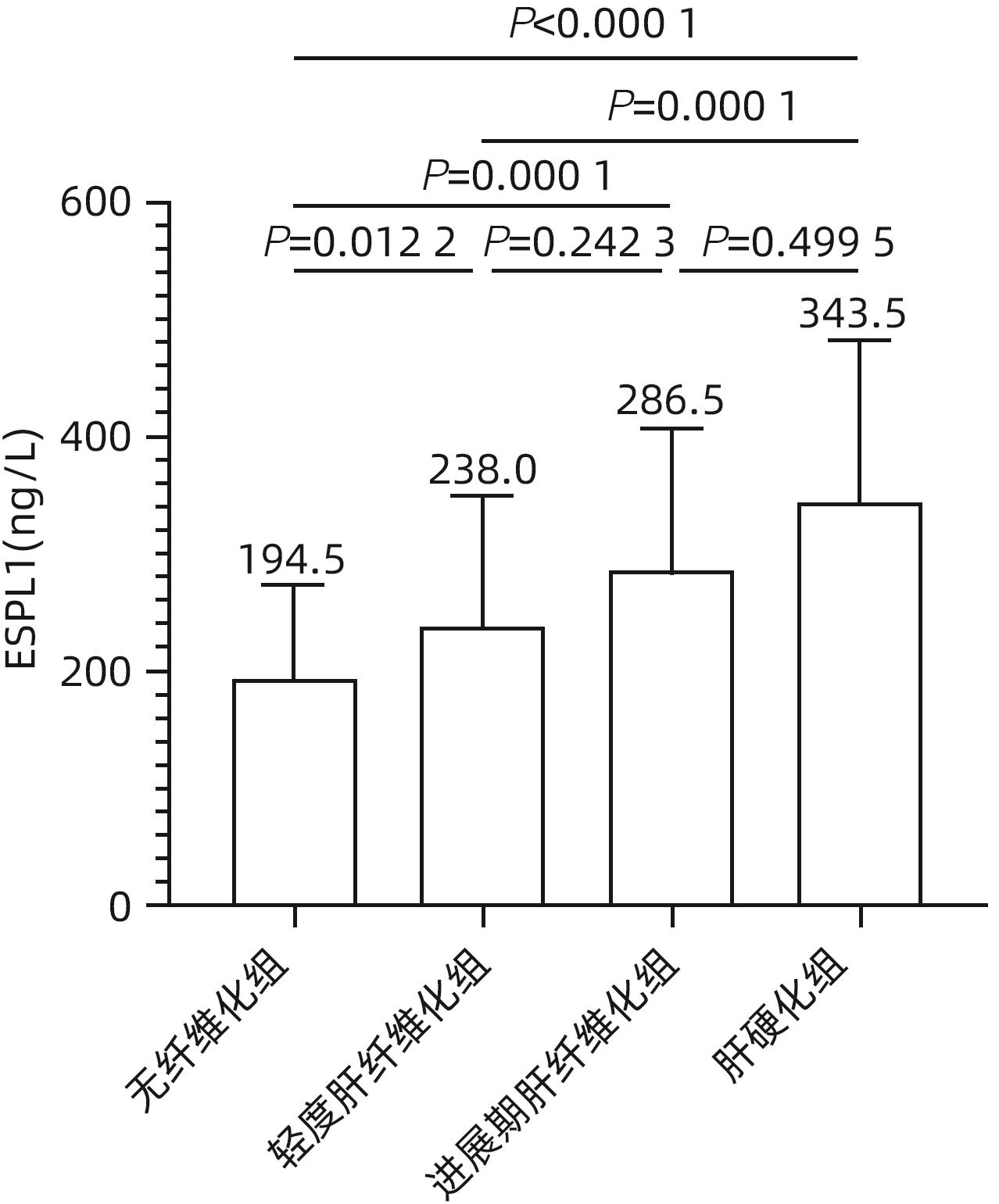
目的 探讨外纺锤体极样蛋白1(ESPL1)检测对HBV相关肝纤维化进程的临床诊断价值。 方法 选取2017年6月—2023年8月广西医科大学第一附属医院收治的HBV感染者228例,采用瞬时弹性成像系统FibroScan检测患者的肝硬度值(LSM),并根据LSM值分为无肝纤维化组(n=80)、轻度肝纤维化组(n=83)、进展期肝纤维化组(n=30)和肝硬化组(n=35)。采用酶联免疫吸附试验(ELISA)检测各组血清中的ESPL1水平。采用Kruskal-Wallis H检验比较4组患者血清ESPL1水平的差异;Spearman相关性分析方法比较ESPL1与LSM值的相关性;运用受试者工作特征(ROC)曲线分析血清ESPL1对不同肝纤维化进程的预测价值。 结果 肝硬化组血清ESPL1水平高于无肝纤维化组、轻度肝纤维化组(P值均<0.05),进展期肝纤维化组、轻度肝纤维化组血清ESPL1水平高于无肝纤维化组(P值均<0.05)。相关性分析显示:不同肝纤维化程度HBV感染者血清ESPL1与LSM值呈正相关(r=0.515,P<0.001);血清ESPL1预测肝硬化和进展期肝纤维化的曲线下面积分别为0.809和0.638,敏感度分别为87.5%和100%,特异度分别为59.7%和31.3%。 结论 血清ESPL1与HBV相关肝纤维化存在一定的相关性,血清ESPL水平越高可能提示肝纤维化程度越高;血清ESPL1可望成为辅助诊断肝硬化的血清标志物之一和动态监测HBV感染者肝纤维化进程的重要临床方法。 Abstract:Objective To investigate the clinical diagnostic value of extra-spindle pole-like protein 1 (ESPL1) in the progression of hepatitis B virus (HBV)-related liver fibrosis. Methods A total of 228 patients with HBV infection who were admitted to The First Affiliated Hospital of Guangxi Medical University from June 2017 to August 2023 were enrolled. The transient elastography system FibroScan was used to determine liver stiffness measurement (LSM) for all patients, and according to the LSM value, they were divided into non-liver fibrosis group with 80 patients, mild liver fibrosis group with 83 patients, advanced liver fibrosis group with 30 patients, and liver cirrhosis group with 35 patients. ELISA was used to measure the serum level of ESPL1. The Kruskal-Wallis H test was used for comparison of the serum level of ESPL1 between the four groups; the Spearman correlation analysis was used to investigate the correlation between ESPL1 and LSM; the receiver operating characteristic (ROC) curve was used to analyze the value of serum ESPL1 in predicting the progression of liver fibrosis. Results The liver cirrhosis group had a significantly higher serum level of ESPL1 than the non-liver fibrosis group and the mild liver fibrosis group (both P<0.05), and the advanced liver fibrosis group and the mild liver fibrosis group had a significantly higher serum level of ESPL1 than the non-liver fibrosis group (both P<0.05). The correlation analysis showed that there was a positive correlation between serum ESPL1 and LSM in the patients with HBV infection and varying degrees of liver fibrosis (r=0.515, P<0.001). Serum ESPL1 had an area under the ROC curve (AUC) of 0.809 in predicting liver cirrhosis and an AUC of 0.638 in predicting advanced liver fibrosis, with a sensitivity of 87.5% and 100%, respectively, and a specificity of 59.7% and 31.3%, respectively. Conclusion There is a certain correlation between serum ESPL1 and HBV-related liver fibrosis, and higher serum ESPL1 may indicate a higher degree of liver fibrosis. Serum ESPL1 is expected to become one of the serum markers for assisting in the diagnosis of liver cirrhosis and an important clinical method for dynamically monitoring the progression of liver fibrosis in patients with HBV infection. -
Key words:
- Hepatitis B virus /
- Hepatic Fibrosis /
- Extra Spindle Pole-like Protein 1
-
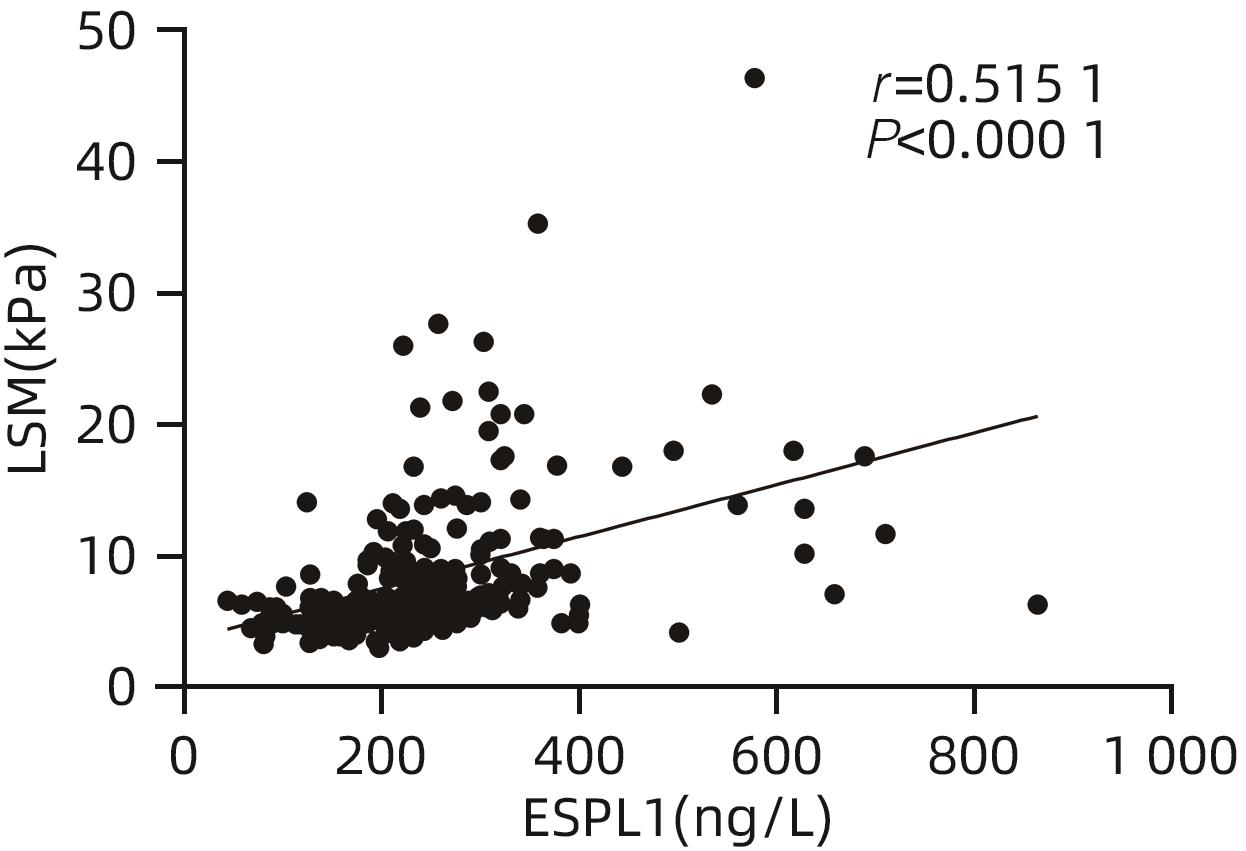
图 2 CHB患者血清ESPL1与LSM值相关性散点图
Figure 2. Scatter diagram of correlation between serum ESPL1 and LSM values in CHB patients

图 3 ESPL1诊断肝硬化效能的ROC曲线
Figure 3. Receiver operating characteristic curve of ESPL1 diagnostic efficacy for liver cirrhosis
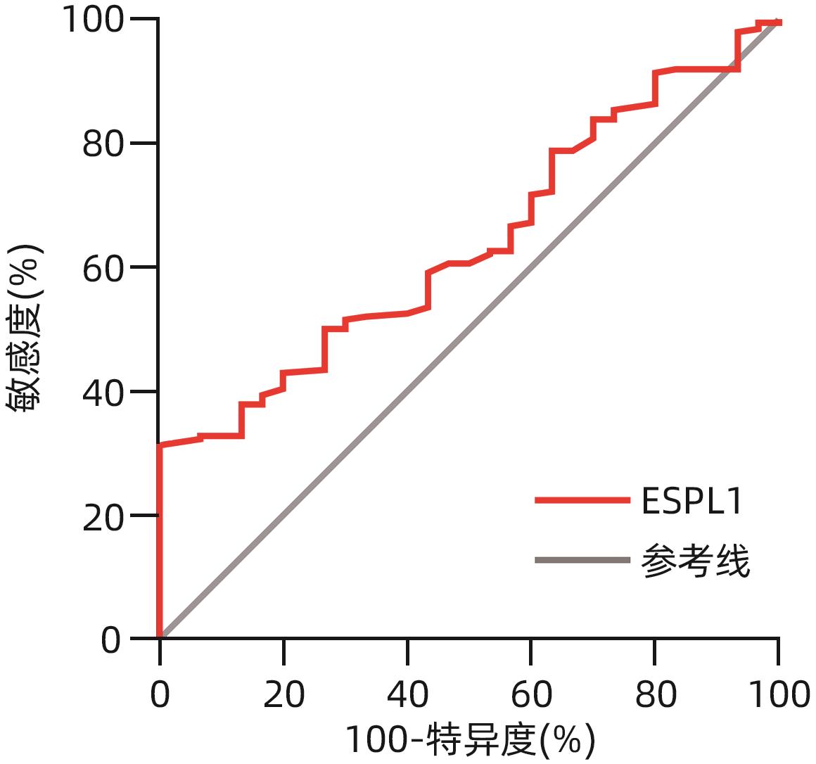
图 4 ESPL1诊断进展期肝纤维化效能的ROC曲线
Figure 4. Receiver operating characteristic curve of ESPL1 diagnostic efficacy for advanced liver fibrosis
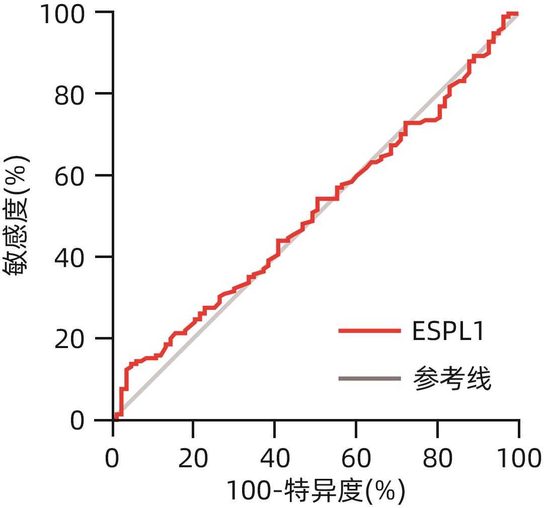
图 5 ESPL1诊断轻度肝纤维化效能的ROC曲线
Figure 5. Receiver operating characteristic curve of ESPL1 diagnostic efficacy for mild liver fibrosis
表 1 患者一般资料表
Table 1. General information table of patients
项目 无肝纤维化组(n=80) 轻度肝纤维化组(n=83) 进展期肝纤维化组(n=30) 肝硬化组(n=35) χ²值 P值 年龄(岁) 45±10 46±111) 49±8 53±102) 17.729 <0.05 男[例(%)] 46(57.5) 69(83.1)2) 25(83.3)2) 25(71.4) 15.405 <0.05 TBil(μmol/L) 14.7±5.8 17.1±7.4 17.2±9.3 23.3±15.22) 11.556 <0.05 ALT(U/L) 19.8±7.0 23.9±8.72) 25.6±8.02) 25.1±8.12) 20.057 <0.05 注:与肝硬化组比较,1)P<0.05;与无肝纤维化组比较,2)P<0.05。
 下载: 导出CSV
-
[1] FATTOVICH G, BORTOLOTTI F, DONATO F. Natural history of chronic hepatitis B: Special emphasis on disease progression and prognostic factors[J]. J Hepatol, 2008, 48( 2): 335- 352. DOI: 10.1016/j.jhep.2007.11.011. [2] CHEN YC, CHU CM, LIAW YF. Age-specific prognosis following spontaneous hepatitis B e antigen seroconversion in chronic hepatitis B[J]. Hepatology, 2010, 51( 2): 435- 444. DOI: 10.1002/hep.23348. [3] DUBERG AS, LYBECK C, FÄLT A, et al. Chronic hepatitis B virus infection and the risk of hepatocellular carcinoma by age and country of origin in people living in Sweden: A national register study[J]. Hepatol Commun, 2022, 6( 9): 2418- 2430. DOI: 10.1002/hep4.1974. [4] Chinese Society of Hepatology, Chinese Medical Association; Chinese Society of Gastroenterology, Chinese Medical Association; Chinese Society of Infectious Diseases, Chinese Medical Association. Consensus on the diagnosis and therapy of hepatic fibrosis(2019)[J]. J Clin Hepatol, 2019, 35( 10): 2163- 2172. DOI: 10.3969/j. issn.1001-5256.2019.10.007.中华医学会肝病学分会, 中华医学会消化病学分会, 中华医学会感染病学分会. 肝纤维化诊断及治疗共识(2019年)[J]. 临床肝胆病杂志, 2019, 35( 10): 2163- 2172. DOI: 10.3969/j. issn.1001-5256.2019.10.007. [5] Chinese Foundation for Hepatitis Prevention and Control; Chinese Society of Infectious Diseases, Chinese Medical Association; Chinese Society of Hepatology, Chinese Medical Association. Consensus on clinical application of transient elastography detecting liver fibrosis: A 2018 update[J]. Chin J Hepatol, 2019, 27( 3): 182- 191. DOI: 10.3760/cma.j.issn.1007-3418.2019.03.004.中国肝炎防治基金会, 中华医学会感染病学分会, 中华医学会肝病学分会, 等. 瞬时弹性成像技术诊断肝纤维化专家共识(2018年更新版)[J]. 中华肝脏病杂志, 2019, 27( 3): 182- 191. DOI: 10.3760/cma.j.issn.1007-3418.2019.03.004. [6] AGBIM U, ASRANI SK. Non-invasive assessment of liver fibrosis and prognosis: An update on serum and elastography markers[J]. Expert Rev Gastroenterol Hepatol, 2019, 13( 4): 361- 374. DOI: 10.1080/17474124.2019.1579641. [7] NEWSOME PN, SASSO M, DEEKS JJ, et al. FibroScan-AST(FAST) score for the non-invasive identification of patients with non-alcoholic steatohepatitis with significant activity and fibrosis: A prospective derivation and global validation study[J]. Lancet Gastroenterol Hepatol, 2020, 5( 4): 362- 373. DOI: 10.1016/S2468-1253(19)30383-8. [8] ZHANG X, ZHU GJ, YE XH, et al. Clinical value of GPR parameter model combined with Fibroscan for evaluating the stage of liver fibrosis in patientes with chronic hepatitis B[J]. Chin Hepatol, 2022, 27( 8): 877- 880. DOI: 10.14000/j.cnki.issn.1008-1704.2022.08.010.张鑫, 朱桂娟, 叶晓航, 等. GPR参数模型联合Fibroscan对慢性乙型肝炎肝纤维化的诊断效能[J]. 肝脏, 2022, 27( 8): 877- 880. DOI: 10.14000/j.cnki.issn.1008-1704.2022.08.010. [9] BACAC M, FUSCO C, PLANCHE A, et al. Securin and separase modulate membrane traffic by affecting endosomal acidification[J]. Traffic, 2011, 12( 5): 615- 626. DOI: 10.1111/j.1600-0854.2011.01169.x. [10] WANG RM, ZANG WW, HU BB, et al. Serum ESPL1 can be used as a biomarker for patients with hepatitis B virus-related liver cancer: A Chinese case-control study[J]. Technol Cancer Res Treat, 2020, 19: 1533033820980785. DOI: 10.1177/1533033820980785. [11] Chinese Society of Hepatology, Chinese Medical Association; Chinese Society of Infectious Diseases, Chinese Medical Association. Guidelines for the prevention and treatment of chronic hepatitis B(2022 version)[J]. Chin J Infect Dis, 2023, 41( 1): 3- 28. DOI: 10.3760/cma.j.cn311365-20230220-00050.中华医学会肝病学分会, 中华医学会感染病学分会. 慢性乙型肝炎防治指南(2022年版)[J]. 中华传染病杂志, 2023, 41( 1): 3- 28. DOI: 10.3760/cma.j.cn311365-20230220-00050. [12] KANDA T, GOTO T, HIROTSU Y, et al. Molecular mechanisms driving progression of liver cirrhosis towards hepatocellular carcinoma in chronic hepatitis B and C infections: A review[J]. Int J Mol Sci, 2019, 20( 6): 1358. DOI: 10.3390/ijms20061358. [13] LAMPROYE A, BELAICHE J, DELWAIDE J. Le FibroScan: une nouvelle méthode d’évaluation non invasive de la fibrose hépatique[The FibroScan: a new non invasive method of liver fibrosis evaluation][J]. Rev Med Liege, 2007, 62 Spec No: 68- 72. [14] AKIMA T, TAMANO M, HIRAISHI H. Liver stiffness measured by transient elastography is a predictor of hepatocellular carcinoma development in viral hepatitis[J]. Hepatol Res, 2011, 41( 10): 965- 970. DOI: 10.1111/j.1872-034X.2011.00846.x. [15] JEONG PY, KUMAR A, JOSHI PM, et al. Intertwined functions of separase and caspase in cell division and programmed cell death[J]. Sci Rep, 2020, 10( 1): 6159. DOI: 10.1038/s41598-020-63081-w. [16] SINGLETON MR, UHLMANN F. Separase-securin complex: A cunning way to control chromosome segregation[J]. Nat Struct Mol Biol, 2017, 24( 4): 337- 339. DOI: 10.1038/nsmb.3393. [17] WIRTH KG, WUTZ G, KUDO NR, et al. Separase: A universal trigger for sister chromatid disjunction but not chromosome cycle progression[J]. J Cell Biol, 2006, 172( 6): 847- 860. DOI: 10.1083/jcb.200506119. [18] RUPPENTHAL S, KLEINER H, NOLTE F, et al. Increased separase activity and occurrence of centrosome aberrations concur with transformation of MDS[J]. PLoS One, 2018, 13( 1): e0191734. DOI: 10.1371/journal.pone.0191734. [19] MUKHERJEE M, GE GQ, ZHANG NG, et al. Separase loss of function cooperates with the loss of p53 in the initiation and progression of T- and B-cell lymphoma, leukemia and aneuploidy in mice[J]. PLoS One, 2011, 6( 7): e22167. DOI: 10.1371/journal.pone.0022167. [20] JUNG KS, KIM SU, AHN SH, et al. Risk assessment of hepatitis B virus-related hepatocellular carcinoma development using liver stiffness measurement(FibroScan)[J]. Hepatology, 2011, 53( 3): 885- 894. DOI: 10.1002/hep.24121. [21] HU BB, WANG RM, LIANG HK, et al. Expression of ESPL1 gene in hepatocellular carcinoma tissue and its role in the prognosis assessment[J]. Chin Hepatol, 2023, 28( 3): 285- 289. DOI: 10.14000/j.cnki.issn.1008-1704.2023.03.008.胡伯斌, 王荣明, 梁蘅恺, 等. ESPL1基因在肝细胞癌组织中的表达及其在预后评估中的作用[J]. 肝脏, 2023, 28( 3): 285- 289. DOI: 10.14000/j.cnki.issn.1008-1704.2023.03.008. [22] DENG DL. Objective to evaluate the value of peripheral blood in the diagnosis of hepatocellular carcinoma and prediction of recurrence[D]. Nanning: Guangxi Medical University, 2021.邓德丽. 评估血清ESPL1对HBV相关肝细胞癌的诊断及预测复发的价值[D]. 南宁: 广西医科大学, 2021. -

 本文二维码
本文二维码
计量
- 文章访问数: 950
- HTML全文浏览量: 1228
- PDF下载量: 76
- 被引次数: 0

