| [1] |
MEZZANO G, JUANOLA A, CARDENAS A, et al. Global burden of disease: acute-on-chronic liver failure, a systematic review and meta-analysis[J]. Gut, 2022, 71(1): 148-155. DOI: 10.1136/gutjnl-2020-322161. |
| [2] |
GIANNELLI V, DI GREGORIO V, IEBBA V, et al. Microbiota and the gut-liver axis: bacterial translocation, inflammation and infection in cirrhosis[J]. World J Gastroenterol, 2014, 20(45): 16795-16810. DOI: 10.3748/wjg.v20.i45.16795. |
| [3] |
GOMAA EZ. Human gut microbiota/microbiome in health and diseases: a review[J]. Antonie Van Leeuwenhoek, 2020, 113(12): 2019-2040. DOI: 10.1007/s10482-020-01474-7. |
| [4] |
LAN P, YIN SM, HE Z. Research progress of gut microbiota in the prevention and treatment of colorectal cancer[J]. Chin J Dig Surg, 2022, 21(6): 730-736. DOI: 10.3760/cma.j.cn115610-20220402-00176. |
| [5] |
O'HARA AM, SHANAHAN F. The gut flora as a forgotten organ[J]. EMBO Rep, 2006, 7(7): 688-693. DOI: 10.1038/sj.embor.7400731. |
| [6] |
FAN Y, PEDERSEN O. Gut microbiota in human metabolic health and disease[J]. Nat Rev Microbiol, 2021, 19(1): 55-71. DOI: 10.1038/s41579-020-0433-9. |
| [7] |
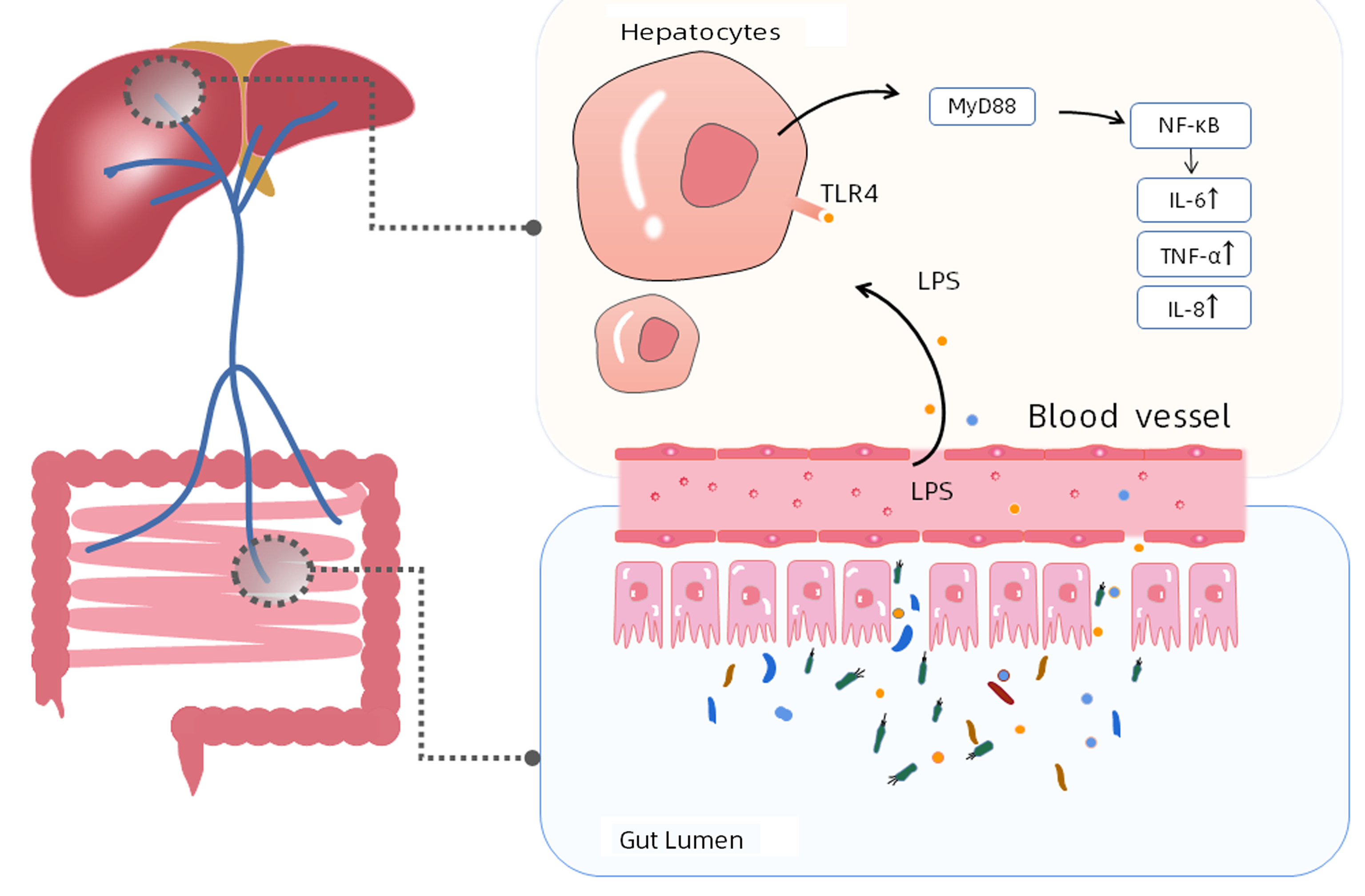
CHOPYK DM, GRAKOUI A. Contribution of the Intestinal microbiome and gut barrier to hepatic disorders[J]. Gastroenterology, 2020, 159(3): 849-863. DOI: 10.1053/j.gastro.2020.04.077. |
| [8] |
HIIPPALA K, JOUHTEN H, RONKAINEN A, et al. The potential of gut commensals in reinforcing intestinal barrier function and alleviating inflammation[J]. Nutrients, 2018, 10(8): 988. DOI: 10.3390/nu10080988. |
| [9] |
TREBICKA J, BORK P, KRAG A, et al. Utilizing the gut microbiome in decompensated cirrhosis and acute-on-chronic liver failure[J]. Nat Rev Gastroenterol Hepatol, 2021, 18(3): 167-180. DOI: 10.1038/s41575-020-00376-3. |
| [10] |
ZHANG Y, ZHANG J, XU T, et al. Allicin ameliorates intraintestinal bacterial translocation after trauma/hemorrhagic shock in rats: The role of mesenteric lymph node dendritic cell[J]. Surgery, 2017, 161(2): 546-555. DOI: 10.1016/j.surg.2016.08.029. |
| [11] |
|
| [12] |
MEIR M, FLEMMING S, BURKARD N, et al. The glial cell-line derived neurotrophic factor: a novel regulator of intestinal barrier function in health and disease[J]. Am J Physiol Gastrointest Liver Physiol, 2016, 310(11): G1118-G1123. DOI: 10.1152/ajpgi.00125.2016. |
| [13] |
MACIA L, TAN J, VIEIRA AT, et al. Metabolite-sensing receptors GPR43 and GPR109A facilitate dietary fibre-induced gut homeostasis through regulation of the inflammasome[J]. Nat Commun, 2015, 6: 6734. DOI: 10.1038/ncomms7734. |
| [14] |
KIM SE, PARK JW, KIM HS, et al. The role of gut dysbiosis in acute-on-chronic liver failure[J]. Int J Mol Sci, 2021, 22(21): 11680. DOI: 10.3390/ijms222111680. |
| [15] |
BAJAJ JS, VARGAS HE, REDDY KR, et al. Association between intestinal microbiota collected at hospital admission and outcomes of patients with cirrhosis[J]. Clin Gastroenterol Hepatol, 2019, 17(4): 756-765.e3. DOI: 10.1016/j.cgh.2018.07.022. |
| [16] |
BAJAJ JS, HEUMAN DM, HYLEMON PB, et al. Altered profile of human gut microbiome is associated with cirrhosis and its complications[J]. J Hepatol, 2014, 60(5): 940-947. DOI: 10.1016/j.jhep.2013.12.019. |
| [17] |
KAKIYAMA G, PANDAK WM, GILLEVET PM, et al. Modulation of the fecal bile acid profile by gut microbiota in cirrhosis[J]. J Hepatol, 2013, 58(5): 949-955. DOI: 10.1016/j.jhep.2013.01.003. |
| [18] |
FERNÁNDEZ J, ACEVEDO J, WIEST R, et al. European Foundation for the Study of Chronic Liver Failure. Bacterial and fungal infections in acute-on-chronic liver failure: prevalence, characteristics and impact on prognosis[J]. Gut, 2018, 67(10): 1870-1880. DOI: 10.1136/gutjnl-2017-314240. |
| [19] |
JENNE CN, KUBES P. Immune surveillance by the liver[J]. Nat Immunol, 2013, 14(10): 996-1006. DOI: 10.1038/ni.2691. |
| [20] |
ARVANITI V, D'AMICO G, FEDE G, et al. Infections in patients with cirrhosis increase mortality four-fold and should be used in determining prognosis[J]. Gastroenterology, 2010, 139(4): 1246-1256, 1256.e1-5. DOI: 10.1053/j.gastro.2010.06.019. |
| [21] |
MÜCKE MM, RUMYANTSEVA T, MÜCKE VT, et al. Bacterial infection-triggered acute-on-chronic liver failure is associated with increased mortality[J]. Liver Int, 2018, 38(4): 645-653. DOI: 10.1111/liv.13568. |
| [22] |
BRUNS T, REUKEN PA, STENGEL S, et al. The prognostic significance of bacterial DNA in patients with decompensated cirrhosis and suspected infection[J]. Liver Int, 2016, 36(8): 1133-1142. DOI: 10.1111/liv.13095. |
| [23] |
LI CQ, GUO JC, XUN YH, et al. Characterization of intestinal flora in chronic plus acute liver failure combined with spontaneous peritonitis[J]. Zhejiang J Integr Tradit Chin West Med, 2021, 31(11): 1012-1015. DOI: 10.3969/j.issn.1005-4561.2021.11.007. |
| [24] |
ALEXOPOULOU A, VASILIEVA L, AGIASOTELLI D, et al. Extensively drug-resistant bacteria are an independent predictive factor of mortality in 130 patients with spontaneous bacterial peritonitis or spontaneous bacteremia[J]. World J Gastroenterol, 2016, 22(15): 4049-4056. DOI: 10.3748/wjg.v22.i15.4049. |
| [25] |
ZHANG W, WANG GC, ZHANG T, et al. Distribution characteristics and drug resistance analysis of clinically isolated pathogens in patients complicated with decompensated hepatitis B cirrhosis and spontaneous bacterial peritonitis[J]. Int J Lab Med, 2022, 43(19): 2346-2351. DOI: 10.3969/j.issn.1673-4130.2022.19.009. |
| [26] |
ENGELMANN C, SHEIKH M, SHARMA S, et al. Toll-like receptor 4 is a therapeutic target for prevention and treatment of liver failure[J]. J Hepatol, 2020, 73(1): 102-112. DOI: 10.1016/j.jhep.2020.01.011. |
| [27] |
WANG Y, CHEN H, CHEN Q, et al. The protective mechanism of CAY10683 on intestinal mucosal barrier in acute liver failure through LPS/TLR4/MyD88 pathway[J]. Mediators Inflamm, 2018, 2018: 7859601. DOI: 10.1155/2018/7859601. |
| [28] |
BIAGIOLI M, CARINO A. Signaling from intestine to the host: how bile acids regulate intestinal and liver immunity[J]. Handb Exp Pharmacol, 2019, 256: 95-108. DOI: 10.1007/164_2019_225. |
| [29] |
GRÜNER N, MATTNER J. Bile acids and microbiota: multifaceted and versatile regulators of the liver-gut axis[J]. Int J Mol Sci, 2021, 22(3): 1397. DOI: 10.3390/ijms22031397. |
| [30] |
PÉAN N, DOIGNON I, GARCIN I, et al. The receptor TGR5 protects the liver from bile acid overload during liver regeneration in mice[J]. Hepatology, 2013, 58(4): 1451-1460. DOI: 10.1002/hep.26463. |
| [31] |
CICHOŻ-LACH H, MICHALAK A. Oxidative stress as a crucial factor in liver diseases[J]. World J Gastroenterol, 2014, 20(25): 8082-8091. DOI: 10.3748/wjg.v20.i25.8082. |
| [32] |
ZHONG W, QIAN K, XIONG J, et al. Curcumin alleviates lipopolysaccharide induced sepsis and liver failure by suppression of oxidative stress-related inflammation via PI3K/AKT and NF-κB related signaling[J]. Biomed Pharmacother, 2016, 83: 302-313. DOI: 10.1016/j.biopha.2016.06.036. |
| [33] |
|
| [34] |
GUSTOT T, LEMMERS A, MORENO C, et al. Differential liver sensitization to toll-like receptor pathways in mice with alcoholic fatty liver[J]. Hepatology, 2006, 43(5): 989-1000. DOI: 10.1002/hep.21138. |
| [35] |
BEAM A, CLINGER E, HAO L. Effect of diet and dietary components on the composition of the gut microbiota[J]. Nutrients, 2021, 13(8): 2795. DOI: 10.3390/nu13082795. |
| [36] |
SO D, WHELAN K, ROSSI M, et al. Dietary fiber intervention on gut microbiota composition in healthy adults: a systematic review and meta-analysis[J]. Am J Clin Nutr, 2018, 107(6): 965-983. DOI: 10.1093/ajcn/nqy041. |
| [37] |
KOH A, de VADDER F, KOVATCHEVA-DATCHARY P, et al. From dietary fiber to host physiology: short-chain fatty acids as key bacterial metabolites[J]. Cell, 2016, 165(6): 1332-1345. DOI: 10.1016/j.cell.2016.05.041. |
| [38] |
KOCOT AM, JAROCKA-CYRTA E, DRABIŃSKA N. Overview of the importance of biotics in gut barrier integrity[J]. Int J Mol Sci, 2022, 23(5): 2896. DOI: 10.3390/ijms23052896. |
| [39] |
CONG J, ZHOU P, ZHANG R. Intestinal microbiota-derived short chain fatty acids in host health and disease[J]. Nutrients, 2022, 14(9): 1977. DOI: 10.3390/nu14091977. |
| [40] |
HORVATH A, DURDEVIC M, LEBER B, et al. Changes in the intestinal microbiome during a multispecies probiotic intervention in compensated cirrhosis[J]. Nutrients, 2020, 12(6): 1874. DOI: 10.3390/nu12061874. |
| [41] |
PATEL VC, LEE S, MCPHAIL M, et al. Rifaximin-α reduces gut-derived inflammation and mucin degradation in cirrhosis and encephalopathy: RIFSYS randomised controlled trial[J]. J Hepatol, 2022, 76(2): 332-342. DOI: 10.1016/j.jhep.2021.09.010. |
| [42] |
BECATTINI S, TAUR Y, PAMER EG. Antibiotic-induced changes in the intestinal microbiota and disease[J]. Trends Mol Med, 2016, 22(6): 458-478. DOI: 10.1016/j.molmed.2016.04.003. |


 DownLoad:
