中文English
ISSN 1001-5256 (Print)
ISSN 2097-3497 (Online)
CN 22-1108/R
Volume 38 Issue 8
Aug.  2022
Turn off MathJax
Article Contents

Risk factors for rebleeding after emergency esophageal variceal ligation in patients with liver cirrhosis

DOI: 10.3969/j.issn.1001-5256.2022.08.016
Research funding:

National Science and Technology Major Project during the 13th Five-Year Plan Period (2017ZX10203202-003)

More Information
  • Corresponding author: LI Zhongbin, 302lizhongbin@sina.com(ORCID: 0000-0003-4878-7634)
  • Received Date: 2021-12-10
  • Accepted Date: 2022-02-03
  • Published Date: 2022-08-20
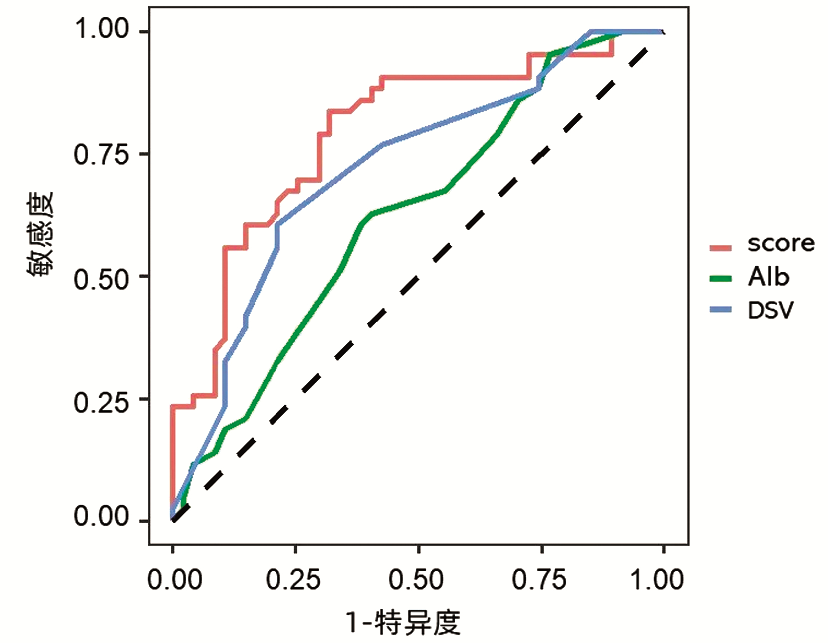
  •   Objective  To investigate the risk factors for rebleeding after emergency esophageal variceal ligation (EVL) in patients with liver cirrhosis.  Methods  A retrospective analysis was performed for the clinical and laboratory data of 290 patients with liver cirrhosis who underwent emergency EVL in The Fifth Medical Center of Chinese PLA General Hospital from January 2016 to December 2019, and according to the presence or absence of rebleeding within 1-year follow-up, they were divided into rebleeding group and non-rebleeding group. The t-test was used for comparison of normally distributed continuous data between two groups, and the Wilcoxon rank sum test was used for comparison of non-normally distributed continuous data between two groups; the chi-square test was used for comparison of categorical data between two groups. A multivariate logistic regression analysis was performed with the statistically significant factors as independent variables to screen out the independent risk factors for rebleeding after emergency EVL, and the receiver operating characteristic (ROC) curve was plotted to obtain the indices for predicting the probability of rebleeding and establish a predictive model.  Results  The univariate analysis showed that there were significant differences between the two groups in platelet count (t=-1.888, P=0.047), Child-Pugh score (χ2=5.975, P=0.049), albumin level (t=-2.229, P=0.029), and splenic vein diameter (t=3.808, P=0.001). The multivariate logistic regression analysis showed that Child-Pugh score (odds ratio [OR]=0.280, 95% confidence interval [CI]: 0.108-0.729, P=0.009), splenic vein diameter (OR=1.549, 95%CI: 1.197-2.005, P=0.001) and albumin level (OR=0.832, 95%CI: 0.729-0.949, P=0.006) were independent influencing factors for rebleeding after EVL. The predictive model based on these three factors had an area under the ROC curve of 0.796, with a sensitivity of 83.7% and a specificity of 74.5% at the cut-off value of -0.086.  Conclusion  Child-Pugh score, albumin level, and splenic vein diameter are independent risk factors for rebleeding after emergency EVL, and the combination of the three indices has the highest sensitivity and specificity in predicting rebleeding.


  • loading
  • [1]
    KRAJA B, MONE I, AKSHIJA I, et al. Predictors of esophageal varices and first variceal bleeding in liver cirrhosis patients[J]. World J Gastroenterol, 2017, 23(26): 4806-4814. DOI: 10.3748/wjg.v23.i26.4806.
    [2]
    BARI K, GARCIA-TSAO G. Treatment of portal hypertension[J]. World J Gatroenterol, 2012, 18(11): 1166-75. DOI: 10.3748/wjg.v18.i11.1166.
    [3]
    Chinese Society of Hepatology, Chinese Medical Association. Chinese guidelines on the management of liver cirrhosis[J]. J Clin Hepatol, 2019, 35(11): 2408-2425. DOI: 10.3969/j.issn.1001-5256.2019.11.006.

    中华医学会肝病学分会. 肝硬化诊治指南[J]. 临床肝胆病杂志, 2019, 35(11): 2408-2425. DOI: 10.3969/j.issn.1001-5256.2019.11.006.
    [4]
    Chinese Society of Hepatology, Chinese Medical Association, Chinese Society of Gastroenterology, Chinese Medical Association, Chinese Society of Endoscopy, Chinese Medical Association. Guidelines for the diagnosis and treatment of esophageal and gastric variceal bleeding in cirrhotic portal hypertension[J]. J Clin Hepatol, 2016, 32(2): 203-219. DOI: 10.3969/j.issn.1001-5256.2016.02.002.

    中华医学会肝病学分会, 中华医学会消化病学分会, 中华医学会内镜学分会. 肝硬化门静脉高压食管胃静脉曲张出血的防治指南[J]. 临床肝胆病杂志, 2016, 32(2): 203-219. DOI: 10.3969/j.issn.1001-5256.2016.02.002.
    [5]
    Chinese Society of Spleen and Portal Hypertension Surgery, Chinese Society of Surgery, Chinese Medical Association. Expert consensus on diagnosis and treatment of esophagogastric variceal bleeding in cirrhotic portal hypertension (2019 edition)[J]. Chin J Surg, 2019, 57(12): 885-892. DOI: 10.3760/cma.j.issn.0529-5815.2019.12.002.

    中华医学会外科学分会脾及门静脉高压外科学组. 肝硬化门静脉高压症食管、胃底静脉曲张破裂出血诊治专家共识(2019版)[J]. 中华外科杂志, 2019, 57(12): 885-892. DOI: 10.3760/cma.j.issn.0529-5815.2019.12.002.
    [6]
    LO GH. Endoscopic treatments for portal hypertension[J]. Hepatol Int, 2018, 12(Suppl 1): 91-101. DOI: 10.1007/s12072-017-9828-8.
    [7]
    ZHANG YY, CHEN GD, WANG ZF, et al. Clinical characteristics and endoscopic findings in patients with cirrhotic upper gastrointestinal variceal bleeding[J]. Chin J General Surg, 2018, 33(2): 134-137. DOI: 10.3760/cma.j.issn.1007-631X.2018.02.010.

    张媛媛, 陈国栋, 王智峰, 等. 肝硬化患者静脉曲张出血的临床和内镜特点分析[J]. 中华普通外科杂志, 2018, 33(2): 134-137. DOI: 10.3760/cma.j.issn.1007-631X.2018.02.010.
    [8]
    CHEN SY. Problems and countermeasures of endoscopic treatment of esophageal and gastric varices in cirrhotic portal hypertension[J]. Chin J Dig, 2019, 39(6): 373-375. DOI: 10.3760/cma.j.issn.0254-1432.2019.06.005.

    陈世耀. 肝硬化门静脉高压食管胃静脉曲张内镜治疗的问题与对策[J]. 中华消化杂志, 2019, 39(6): 373-375. DOI: 10.3760/cma.j.issn.0254-1432.2019.06.005.
    [9]
    de FRANCHIS R, Baveno Ⅵ Faculty. Expanding consensus in portal hypertension: Report of the Baveno Ⅵ Consensus Workshop: Stratifying risk and individualizing care for portal hypertension[J]. J Hepatol, 2015, 63(3): 743-752. DOI: 10.1016/j.jhep.2015.05.022.
    [10]
    ZHAO Q, HE SX, LIU YP, et al. Clinical study of endoscopic ligation, sclerotherapy and tissue glue embolization in the treatment of esophageal and gastric varicose vein bleeding[J]. J Clin Exp Med, 2021, 20(3): 279-282. DOI: 10.3969/j.issn.1671-4695.2021.03.016.

    赵倩, 和水祥, 刘亚萍, 等. 内镜下套扎、硬化剂及组织胶栓塞联合治疗食管胃底静脉曲张出血的临床研究[J]. 临床和实验医学杂志, 2021, 20(3): 279-282. DOI: 10.3969/j.issn.1671-4695.2021.03.016.
    [11]
    DENG SM, ZHANG JX, QI Y, et al. Application of endoscopic ligation in esophageal varices and study on high risk factors of postoperative rebleeding[J]. Clin J Med Offic, 2021, 49(6): 718-720. DOI: 10.16680/j.1671-3826.2021.06.43.

    邓水苗, 张嘉星, 齐晔, 等. 内镜下套扎术在食管静脉曲张中应用及其术后再出血高危因素研究[J]. 临床军医杂志, 2021, 49(6): 718-720. DOI: 10.16680/j.1671-3826.2021.06.43.
    [12]
    LI BM, ZHU X, SHU X, et al. Endoscopic variceal ligation plus sclerotherapy for esophageal variceal bleeding[J]. Chin J Dig Endosc, 2013, 30(2): 67-70. DOI: 10.3760/cma.j.issn.1007-5232.2013.02.003.

    李弼民, 朱萱, 舒徐, 等. 肝硬化食管静脉曲张破裂出血序贯治疗的临床应用价值[J]. 中华消化内镜杂志, 2013, 30(2): 67-70. DOI: 10.3760/cma.j.issn.1007-5232.2013.02.003.
    [13]
    CHEN J, ZENG XQ, MA LL, et al. Randomized controlled trial comparing endoscopic ligation with or without sclerotherapy for secondary prophylaxis of variceal bleeding[J]. Eur J Gastroenterol Hepatol, 2016, 28(1): 95-100. DOI: 10.1097/MEG.0000000000000499.
    [14]
    VUACHET D, CERVONI JP, VUITTON L, et al. Improved survival of cirrhotic patients with variceal bleeding over the decade 2000-2010[J]. Clin Res Hepatol Gastroenterol, 2015, 39(1): 59-67. DOI: 10.1016/j.clinre.2014.06.018.
    [15]
    CHEN XL, CHEN TW, ZHANG XM, et al. Platelet count combined with right liver volume and spleen volume measured by magnetic resonance imaging for identifying cirrhosis and esophageal varices[J]. World J Gastroenterol, 2015, 21(35): 10184-10191. DOI: 10.3748/wjg.v21.i35.10184.
    [16]
    ZHANG Y, DING HG. Risk factors for early rebleeding after endoscopic therapy for esophageal varices in cirrhotic patients[J]. J Clin Hepatol, 2021, 37(9): 2087-2091. DOI: 10.3969/j.issn.1001-5256.2021.09.017.

    张妍, 丁惠国. 肝硬化食管静脉曲张内镜治疗术后早期再出血的危险因素分析[J]. 临床肝胆病杂志, 2021, 37(9): 2087-2091. DOI: 10.3969/j.issn.1001-5256.2021.09.017.
    [17]
    TRIANTOS C, KALAFATELI M. Endoscopic treatment of esophageal varices in patients with liver cirrhosis[J]. World J Gastroenterol, 2014, 20(36): 13015-13026. DOI: 10.3748/wjg.v20.i36.13015.
    [18]
    MOSTAFA EF, MOHAMMAD AN. Incidence and predictors of rebleeding after band ligation of oesophageal varices[J]. Arab J Gastroenterol, 2014, 15(3-4): 135-141. DOI: 10.1016/j.ajg.2014.10.002.
    [19]
    JIN Y, WANG X, ZHANG LJ, et al. Risk factors for early rebleeding after esophageal variceal ligation in patients with liver cirrhosis[J]. J Clin Hepatol, 2017, 33 (11): 2147-2151. DOI: 10.3969/j.issn.1001-5256.2017.11.019.

    金燕, 王雪, 张玲娟, 等. 肝硬化患者食管静脉曲张套扎术后早期再出血的危险因素分析[J]. 临床肝胆病杂志, 2017, 33(11): 2147-2151. DOI: 10.3969/j.issn.1001-5256.2017.11.019.
    [20]
    ZHANG S, YU JH, XU GL. Advances in hemodynamics of portal hypertension[J]. J Hepatobiliary Surg, 2013, 21(3): 230-232. DOI: 10.3969/j.issn.1006-4761.2013.03.025.

    张硕, 余继海, 许戈良. 门静脉高压症血流动力学研究进展[J]. 肝胆外科杂志, 2013, 21(3): 230-232. DOI: 10.3969/j.issn.1006-4761.2013.03.025.
    [21]
    LI YF, LIU LJ, CHENG X, et al. The risk factors of esophageal variceal bleeding in patients with liver cirrhosis[J]. Pract J Clin Med, 2019, 16(1): 41-44. DOI: 10.3969/j.issn.1672-6170.2019.01.014.

    李艳芳, 刘灵军, 程星, 等. 肝硬化食管静脉曲张破裂出血危险因素分析[J]. 实用医院临床杂志, 2019, 16(1): 41-44. DOI: 10.3969/j.issn.1672-6170.2019.01.014.
    [22]
    YANG HR. Value of color Doppler ultrasound in the diagnosis of portal hypertension liver cirrhosis merged with esophageal variceal bleeding[J]. J Hainan Med Univ, 2016, 22(5): 496-498. DOI: 10.13210/j.cnki.jhmu.20151203.007.

    杨海荣. 彩色多普勒超声在肝硬化门脉高压食管静脉曲张破裂出血中的价值[J]. 海南医学院学报, 2016, 22(5): 496-498. DOI: 10.13210/j.cnki.jhmu.20151203.007.
  • 加载中

Catalog

    通讯作者: 陈斌, bchen63@163.com
    • 1. 

      沈阳化工大学材料科学与工程学院 沈阳 110142

    1. 本站搜索
    2. 百度学术搜索
    3. 万方数据库搜索
    4. CNKI搜索

    Figures(1)  / Tables(3)

    Article Metrics

    Article views (651) PDF downloads(55) Cited by()
    Proportional views
    Related

    /

    DownLoad:  Full-Size Img  PowerPoint
    Return
    Return