中文English
ISSN 1001-5256 (Print)
ISSN 2097-3497 (Online)
CN 22-1108/R
Volume 38 Issue 5
May  2022
Turn off MathJax
Article Contents

Effect of exosomes from adult human liver-derived stem cells on concanavalin A-induced acute liver injury in mice

DOI: 10.3969/j.issn.1001-5256.2022.05.024
Research funding:

National Natural Science Foundation of China (81170395);

National Natural Science Foundation of China (81570556);

Chinese Foundation for Hepatitis Prevention and Control-TianQing Liver Disease Research Fund Subject (TQGB20210013)

More Information
  • Corresponding author: HONG Feng, fenghong9508@163.com(ORCID: 0000-0003-4377-7226)
  • Received Date: 2021-09-27
  • Accepted Date: 2021-10-28
  • Published Date: 2022-05-20
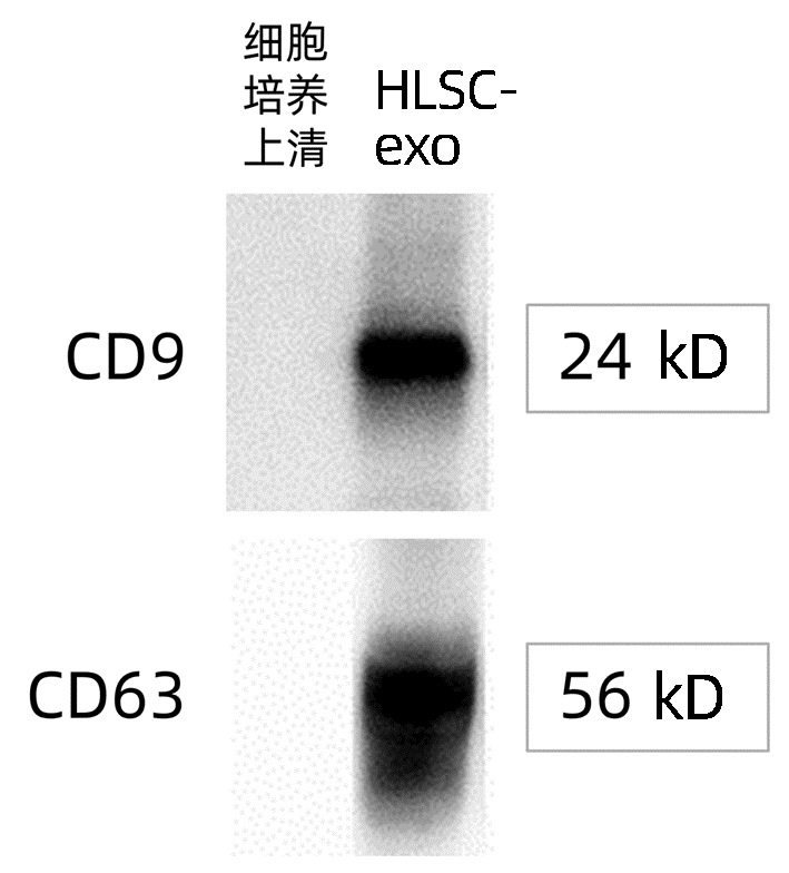
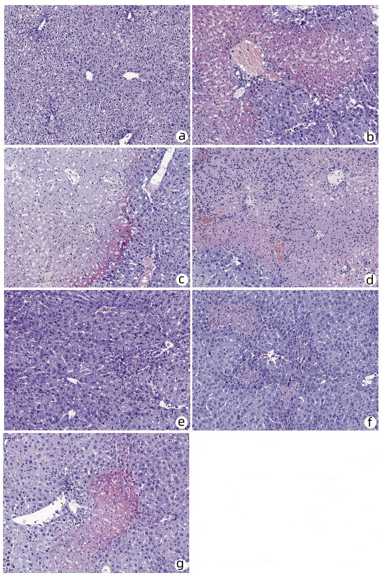
  •   Objective  To investigate the protective effect of adult human liver-derived stem cell exosomes (HLSC-exo) intravenously injected at different time points against acute liver injury induced by concanavalin A (ConA) in mice.  Methods  HLSC-exo was extracted by differential centrifugation. Western blot was used to measure the expression of the marker proteins CD9 and CD63, and nanoparticle tracking analysis was used to investigate particle size distribution. A total of 56 male C57BL/6 mice were randomly divided into blank control group, ConA model group, and HLSC-exo treatment group. The ConA model group and the HLSC-exo treatment group were further divided into 3-, 6-, and 12-hour subgroups according to the interval between phosphate buffer or HLSC-exo injection and ConA injection. The serum levels of alanine aminotransferase (ALT), aspartate aminotransferase (AST), tumor necrosis factor-α (TNF-α), and interleukin-10 (IL-10) were measured, and the gross morphology and histopathology of the liver were compared between groups. A one-way analysis of variance was used for comparison of continuous data between multiple groups, and the least significant difference t-test was used for further comparison between two groups.  Results  HLSC-exo was a membranous vesicle with a diameter of 90-110 nm, with a clear saucer-like structure under an electron microscope and marked expression of its specific marker proteins CD9 and CD63. In the blank control group, the levels of ALT and AST were 31.81±6.74 U/L and 69.75±8.30 U/L, respectively. Compared with the blank control group, the 3-, 6-, and 12-hour ConA model groups had significant increases in the levels of ALT and AST (all P < 0.001); compared with the 3-and 6-hour ConA model groups, the 3-and 6-hour HLSC-exo treatment groups had significant reductions in the levels of ALT and AST (225.58±115.59 U/L vs 1989.32±347.67 U/L, 1174.71±203.30 U/L vs 2208.33±349.96 U/L, 303.53±126.68 U/L vs 2534.27±644.72 U/L, 1340.70±262.56 U/L vs 2437.13±288.13 U/L, all P < 0.001); compared with the 6-hour HLSC-exo treatment group, the 3-hour HLSC-exo treatment group had significantly greater reductions (P < 0.001). In the blank group, the levels of IL-10 and TNF-α were 313.51±10.97 pg/ml and 476.05±7.31 pg/ml, respectively. Compared with the blank control group, the 3-, 6-, and 12-hour ConA model groups had a significant reduction in the level of IL-10 (all P < 0.001); compared with the 3-and 6-hour ConA model groups, the 3-and 6-hour HLSC-exo treatment groups had a significant increase in the level of IL-10(331.61±10.46 pg/ml vs 266.20±8.15 pg/ml, 288.13±10.74 pg/ml vs 264.41±9.12 pg/ml, both P < 0.001); compared with the 6-hour HLSC-exo treatment group, the 3-hour HLSC-exo treatment group had a significantly greater increase (P < 0.001). Compared with the blank control group, the 3-, 6-, and 12-hour ConA model groups had a significant increase in the level of TNF-α (all P < 0.001); compared with the 3-and 6-hour ConA model groups, the 3-and 6-hour HLSC-exo treatment groups had a significant reduction in the level of TNF-α (478.26±12.99 pg/ml vs 551.31±17.70 pg/ml, 515.58±7.18 pg/ml vs 556.21±11.15 pg/ml, both P < 0.001); compared with the 6-hour HLSC-exo treatment group, the 3-hour HLSC-exo treatment group had a significantly greater reduction (P < 0.001). Compared with the 3-and 6-hour ConA model groups in terms of the gross morphology and histopathology of the liver, the 3-and 6-hour HLSC-exo treatment groups had a significant reduction in the degree of hepatocyte necrosis, and the 3-hour HLSC-exo treatment group had a basically complete lobular structure, with sporadic spotty necrosis; the 12-hour HLSC-exo treatment group had no significant improvement in hepatocyte necrosis compared with the 12-hour ConA model group.  Conclusion  Intravenous injection of adult HLSC-exo can alleviate acute liver injury induced by ConA in mice, and injection at 3 hours in advance has the most significant protective effect. Regulation of cytokines is one of the important mechanisms for HLSC-exo to alleviate liver injury.


  • loading
  • [1]
    ESCORSELL À, CASTELLOTE J, SÁNCHEZ-DELGADO J, et al. Management of acute liver failure. Clinical guideline from the Catalan Society of Digestology[J]. Gastroenterol Hepatol, 2019, 42(1): 51-64. DOI: 10.1016/j.gastrohep.2018.07.013.
    [2]
    YE T, WANG T, YANG X, et al. Comparison of concanavalin a-induced murine autoimmune hepatitis models[J]. Cell Physiol Biochem, 2018, 46(3): 1241-1251. DOI: 10.1159/000489074.
    [3]
    JIANG W, TAN Y, CAI M, et al. Human umbilical cord MSC-derived exosomes suppress the development of CCl4-induced liver injury through antioxidant effect[J]. Stem Cells Int, 2018, 2018: 6079642. DOI: 10.1155/2018/6079642.
    [4]
    BI Y, LI J, YANG Y, et al. Human liver stem cells attenuate concanavalin A-induced acute liver injury by modulating myeloid-derived suppressor cells and CD4+ T cells in mice[J]. Stem Cell Res Ther, 2019, 10(1): 22. DOI: 10.1186/s13287-018-1128-2.
    [5]
    FAN Z, ZHANG K, HONG F, et al. The mechanisms of intraperitoneal transplantation of human liver-derived stem cells against concanavalin A-induced acute liver injury in mice[J]. Chin J Lab Diag, 2018, 22(6): 1070-1073. DOI: 10.3969/j.issn.1007-4287.2018.06.043.

    樊增, 张凯, 洪丰, 等. 人肝源性干细胞腹腔移植抗刀豆蛋白A诱导小鼠急性肝损伤的机制研究[J]. 中国实验诊断学, 2018, 22(6): 1070-1073. DOI: 10.3969/j.issn.1007-4287.2018.06.043.
    [6]
    HU C, ZHAO L, ZHANG L, et al. Mesenchymal stem cell-based cell-free strategies: Safe and effective treatments for liver injury[J]. Stem Cell Res Ther, 2020, 11(1): 377. DOI: 10.1186/s13287-020-01895-1.
    [7]
    LOU G, CHEN Z, ZHENG M, et al. Mesenchymal stem cell-derived exosomes as a new therapeutic strategy for liver diseases[J]. Exp Mol Med, 2017, 49(6): e346. DOI: 10.1038/emm.2017.63.
    [8]
    SHIREJINI SZ, INCI F. The Yin and Yang of exosome isolation methods: conventional practice, microfluidics, and commercial kits[J]. Biotechnol Adv, 2022, 54: 107814. DOI: 10.1016/j.biotechadv.2021.107814.
    [9]
    WANG L, YANG RN. Study on role and clinical significance of IL-9, IL-10 in immune liver damage[J]. Lab Med Clin, 2017, 14(20): 3011-3014. DOI: 10.3969/j.issn.1672-9455.2017.20.014.

    王兰, 杨瑞宁. IL-9、IL-10在免疫性肝损伤中的作用及临床意义[J]. 检验医学与临床, 2017, 14(20): 3011-3014. DOI: 10.3969/j.issn.1672-9455.2017.20.014.
    [10]
    GAO YD, TIAN Y, CHEN Y, et al. Effect of magnesium isoglycyrrhizinate on concanavalin A-induced acute liver failure in mice[J]. J Clin Hepatol, 2020, 36(7): 1571-1576. DOI: 10.3969/j.issn.1001-5256.2020.07.024.

    高钰迪, 田原, 陈煜, 等. 异甘草酸镁对刀豆蛋白A诱导的急性肝衰竭小鼠模型的影响[J]. 临床肝胆病杂志, 2020, 36(7): 1571-1576. DOI: 10.3969/j.issn.1001-5256.2020.07.024.
  • 加载中

Catalog

    通讯作者: 陈斌, bchen63@163.com
    • 1. 

      沈阳化工大学材料科学与工程学院 沈阳 110142

    1. 本站搜索
    2. 百度学术搜索
    3. 万方数据库搜索
    4. CNKI搜索

    Figures(4)  / Tables(2)

    Article Metrics

    Article views (482) PDF downloads(31) Cited by()
    Proportional views
    Related

    /

    DownLoad:  Full-Size Img  PowerPoint
    Return
    Return