| [1] |
ROEHLEN N, CROUCHET E, BAUMERT T F. Liver fibrosis: mechanistic concepts and therapeutic perspectives[J]. Cells, 2020, 9( 4): 875- 918. DOI: 10.3390/cells9040875. |
| [2] |
PAROLA M, PINZANI M. Liver fibrosis: Pathophysiology, pathogenetic targets and clinical issues[J]. Mol Aspects Med, 2019, 65: 37- 55. DOI: 10.1016/j.mam.2018.09.002. |
| [3] |
|
| [4] |
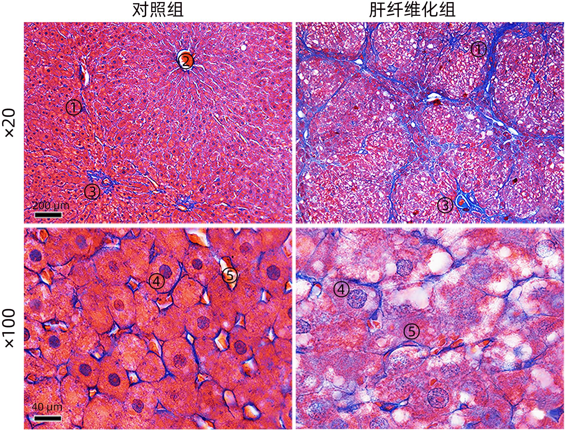
WANG CL. Effects of the changes in the number and volume of hepatic stellate cells on the volume of collagen fibers in rat fibrotic liver and the effects of magnesium isoglycetate on them——a stereological study[D]. Nanchong: North Sichuan Medical Colleage, 2019.
王川林. 肝星状细胞数量和体积变化对肝纤维化大鼠肝组织胶原纤维体积的影响及异甘草酸镁的作用——体视学研究[D]. 南充: 川北医学院, 2019.
|
| [5] |
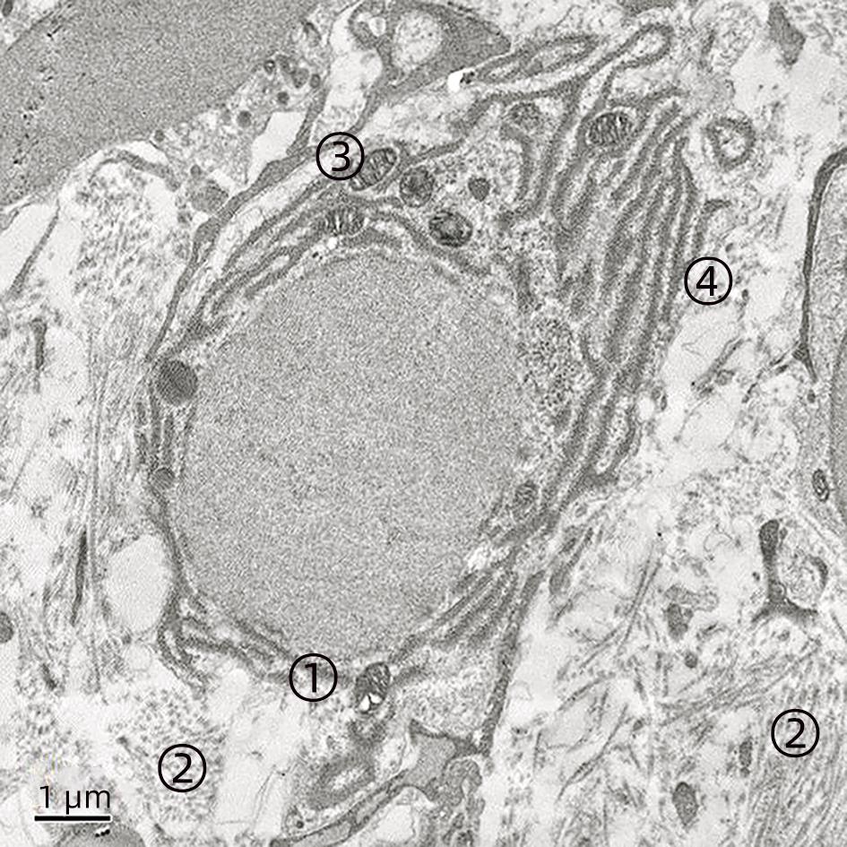
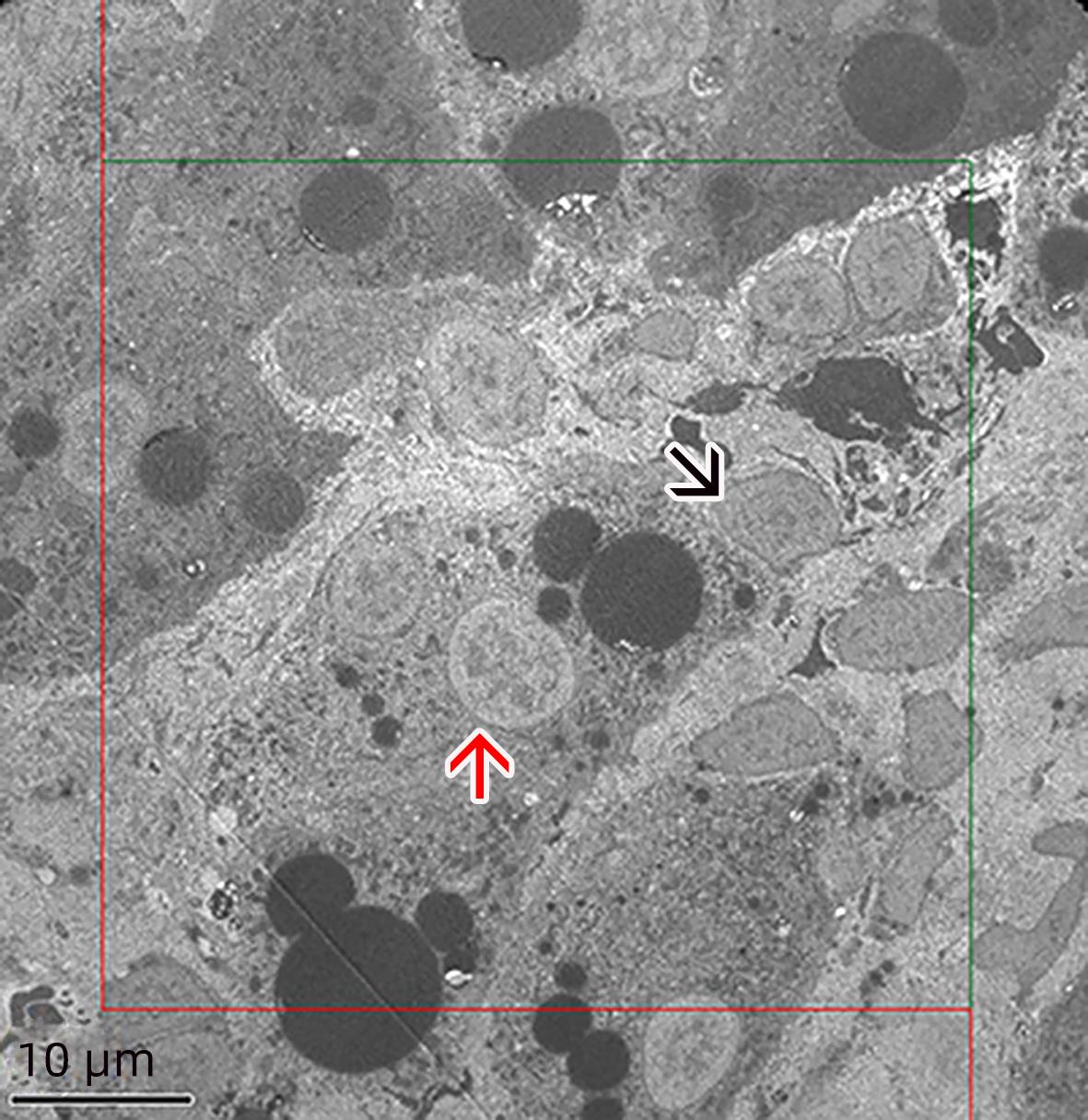
LOTOWSKA JM, SOBANIEC-LOTOWSKA ME, LEBENSZTEJN DM, et al. Ultrastructural characteristics of rat hepatic oval cells and their intercellular contacts in the model of biliary fibrosis: new insights into experimental liver fibrogenesis[J]. Gastroenterol Res Pract, 2017, 2017: 2721547. DOI: 10.1155/2017/2721547. |
| [6] |
de VOS R, DESMET V. Ultrastructural characteristics of novel epithelial cell types identified in human pathologic liver specimens with chronic ductular reaction[J]. Am J Pathol, 1992, 140( 6): 1441- 1450.
|
| [7] |
DING ZY, JIN GN, WANG W, et al. Activin a-smad signaling mediates connective tissue growth factor synthesis in liver progenitor cells[J]. Int J Mol Sci, 2016, 17( 3): 408. DOI: 10.3390/ijms17030408. |
| [8] |
MANDAL A, RAJU S, VISWANATHAN C. Cryopreserved hepatic progenitor cells derived from human embryonic stem cells can arrest progression of liver fibrosis in rats[J]. Cell Biol Int, 2016, 40( 10): 1107- 1115. DOI: 10.1002/cbin.10649. |
| [9] |
HE Z, FENG M. Activation, isolation, identification and culture of hepatic stem cells from porcine liver tissues[J]. Cell Prolif, 2011, 44( 6): 558- 566. DOI: 10.1111/j.1365-2184.2011.00781.x. |
| [10] |
DORRELL C, ERKER L, SCHUG J, et al. Prospective isolation of a bipotential clonogenic liver progenitor cell in adult mice[J]. Genes Dev, 2011, 25( 11): 1193- 1203. DOI: 10.1101/gad.2029411. |
| [11] |
WANG CL, YANG X, CUI QL, et al. Effect of magnesium isoglycyrrhizinate on the pathological changes of liver tissue in CCl 4 induced hepatic fibrosis rats[J]. J North Sichuan Med Coll, 2018, 33( 3): 360- 363. DOI: 10.3969/j.issn.1005-3697.2018.03.017. |
| [12] |
OH SH, HATCH HM, PETERSEN BE. Hepatic oval‘stem’ cell in liver regeneration[J]. Semin Cell Dev Biol, 2002, 13( 6): 405- 409. DOI: 10.1016/s1084952102001271. |
| [13] |
LIN Y, DONG MQ, LIU ZM, et al. A strategy of vascular-targeted therapy for liver fibrosis[J]. Hepatology, 2022, 76( 3): 660- 675. DOI: 10.1002/hep.32299. |
| [14] |
DHAR D, BAGLIERI J, KISSELEVA T, et al. Mechanisms of liver fibrosis and its role in liver cancer[J]. Exp Biol Med(Maywood), 2020, 245( 2): 96- 108. DOI: 10.1177/1535370219898141. |
| [15] |
AWAN SJ, BAIG MT, YAQUB F, et al. In vitro differentiated hepatic oval-like cells enhance hepatic regeneration in CCl(4)-induced hepatic injury[J]. Cell Biol Int, 2017, 41( 1): 51- 61. DOI: 10.1002/cbin.10699. |
| [16] |
YANG AT, HU DD, WANG P, et al. TGF-β1 induces the dual regulation of hepatic progenitor cells with both anti- and proliver fibrosis[J]. Stem Cells Int, 2016, 2016: 1492694. DOI: 10.1155/2016/1492694. |
| [17] |
KÖHN-GAONE J, GOGOI-TIWARI J, RAMM GA, et al. The role of liver progenitor cells during liver regeneration, fibrogenesis, and carcinogenesis[J]. Am J Physiol Gastrointest Liver Physiol, 2016, 310( 3): G143- 154. DOI: 10.1152/ajpgi.00215.2015. |
| [18] |
QIU DK, MA X, PENG YS, et al. Locational and quantitative study of hepatic oval cells in chronic liver diseases——Pathologic analysis of 29 liver samples from patients with chronic liver diseases[J]. Chin J Dig, 2000, 20( 5): 301- 303. DOI: 10.3760/j.issn:0254-1432.2000.05.004. |
| [19] |
EZHILARASAN D, SOKAL E, NAJIMI M. Hepatic fibrosis: It is time to go with hepatic stellate cell-specific therapeutic targets[J]. Hepatobiliary Pancreat Dis Int, 2018, 17( 3): 192- 197. DOI: 10.1016/j.hbpd.2018.04.003. |
| [20] |
KAUR S, SIDDIQUI H, BHAT MH. Hepatic progenitor cells in action: liver regeneration or fibrosis?[J]. Am J Pathol, 2015, 185( 9): 2342- 2350. DOI: 10.1016/j.ajpath.2015.06.004. |
| [21] |
STRAZZABOSCO M, FABRIS L. Development of the bile ducts: essentials for the clinical hepatologist[J]. J Hepatol, 2012, 56( 5): 1159- 1170. DOI: 10.1016/j.jhep.2011.09.022. |
| [22] |
WANG P, LIU T, CONG M, et al. Expression of extracellular matrix genes in cultured hepatic oval cells: an origin of hepatic stellate cells through transforming growth factor beta?[J]. Liver Int, 2009, 29( 4): 575- 584. DOI: 10.1111/j.1478-3231. |
| [23] |
FAKTOR VM, RADAEVA SA. The formation of oval-cell ducts during hepatic carcinogenesis in mice. Its relationship to the pre-existing canals of Hering[J]. Ontogenez, 1992, 23( 4): 407- 418.
|
| [24] |
BURKE ZD, SHEN CN, RALPHS KL, et al. Characterization of liver function in transdifferentiated hepatocytes[J]. J Cell Physiol, 2006, 206( 1): 147- 159. DOI: 10.1002/jcp.20438. |


 DownLoad: