中文English
ISSN 1001-5256 (Print)
ISSN 2097-3497 (Online)
CN 22-1108/R
Volume 37 Issue 12
Dec.  2021
Turn off MathJax
Article Contents

Association between serum macrophage polarization-related factors and liver fibrosis in echinococcosis multilocularis

DOI: 10.3969/j.issn.1001-5256.2021.12.017
Research funding:

National Natural Science Foundation of China (81960576);

Qinghai Province Science and Technology Plan Project (2018-ZJ-757)

  • Received Date: 2021-05-10
  • Accepted Date: 2021-07-23
  • Published Date: 2021-12-20
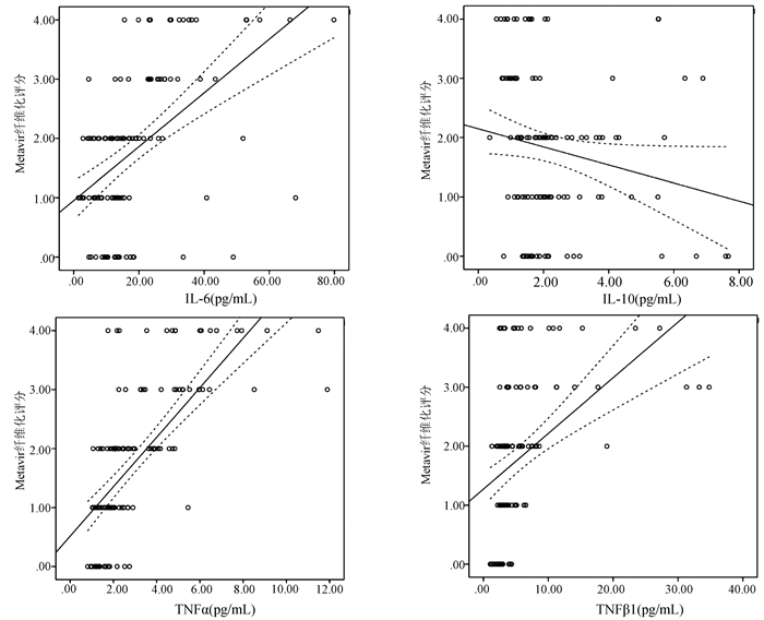
  •   Objective  To investigate the association between serum macrophage polarization-related factors and liver fibrosis in patients with alveolar echinococcosis (AE).  Methods  A total of 120 patients with AE who attended Department of Hepatobiliary and Pancreatic Surgery in The Affiliated Hospital of Qinghai University from September 2018 to October 2020 were enrolled as AE group, and 33 healthy controls were enrolled as normal control group. The two groups and the patients with varying degrees of liver fibrosis were compared in terms of the levels of interleukin-6 (IL-6), interleukin-10 (IL-10), tumor necrosis factor-α (TNF-α), and transforming growth factor-β1 (TGF-β1). Comparison of normally distributed continuous data between two groups was made by the independent samples t-test, while comparison of non-normally distributed continuous data was made by the Mann-Whitney U test or Kruskal-Wallis H test. Comparison of categorical data between groups was made by the chi-square test. Univariate and multivariate logistic regression analyses were used to investigate the association between serum macrophage polarization-related factors and liver fibrosis in patients with AE, and the receiver operating characteristic (ROC) curve was used to analyze the value of serological examination in the diagnosis of liver fibrosis in patients with AE. A Spearman correlation analysis was used to analyze the correlation of each index with HAI score and Metavir score.  Results  Compared with the normal control group, the AE group had significant increases in the serum levels of IL-6 [13.97 (9.64-23.62) pg/mL vs 1.30 (0.35-2.71) pg/mL, Z=-5.980, P < 0.001], TNF-α [2.26 (1.65-4.13) pg/mL vs 1.40 (1.04-2.10) pg/mL, Z=-3.114, P < 0.01], and TGF-β1 [3.64(2.71-5.72) pg/mL vs 2.91(2.20-3.35) pg/mL, Z=-2.594, P < 0.05], and increases in the serum levels of IL-6 (hazard ratio [HR]=2.721, 95% confidence interval [CI]: 1.730-4.280, P < 0.05) and TNF-α(HR=3.527, 95%CI: 1.158-10.747, P < 0.05) were independent risk factors for the onset of liver fibrosis in AE patients. The ROC curve analysis showed that hydatid IgG combined with the serum levels of IL-6 and TNF-α had a sensitivity of 88.4%, a specificity of 95.8%, and an area under the ROC curve of 0.951(95%CI: 0.937-0.964) in the diagnosis of liver fibrosis, which were significantly higher than those of IL-6, TNF-α, or hydatid IgG alone (Z=-3.458, -4.011, and 2.379, all P < 0.05). The Spearman analysis showed that the serum levels of IL-6, TNF-α, and TGF-β1 were positively correlated with HAI score (r=0.560, 0.644, and 0.465, all P < 0.001) and Metavir fibrosis score (r=0.530, 0.758, and 0.567, all P < 0.001), and the serum level of IL-10 was negatively correlated with HAI score (r=-0.232, P=0.011) and Metavir fibrosis score (r=-0.288, P=0.001).  Conclusion  Macrophage polarization is often observed in patients with hepatic AE, and the levels of the macrophage polarization-related factors IL-6, TNF-α, and TGF-β1 are associated with the development and progression of liver fibrosis, which can provide certain reference information for predicting the onset of liver fibrosis.


  • loading
  • [1]
    WEN H, VUITTON L, TUXUN T, et al. Echinococcosis: Advances in the 21st Century[J]. Clin Microbiol Rev, 2019, 32(2): e00075-18. DOI: 10.1128/CMR.00075-18.
    [2]
    ZHAO SY, ZHU HH, WANG XQ, et al. Present situation and progress of comprehensive treatments for hepatic alveolar echinococcosis[J]. Chin J Schisto Control, 2019, 31(6): 676-678. DOI: 10.16250/j.32.1374.2018086.

    赵顺云, 朱海宏, 王向前, 等. 肝多房棘球蚴病的综合治疗现状和进展[J]. 中国血吸虫病防治杂志, 2019, 31(6): 676-678. DOI: 10.16250/j.32.1374.2018086.
    [3]
    NONO JK, LUTZ MB, BREHM K. Expansion of host regulatory T cells by secreted products of the tapeworm echinococcus multilocularis[J]. Front Immunol, 2020, 11: 798. DOI: 10.3389/fimmu.2020.00798.
    [4]
    CAI J, HUANG L, WANG LJ, et al. The role of macrophage polarization in parasitic infections: A review[J]. Chin J Schisto Control, 2020, 32(4): 432-435. DOI: 10.16250/j.32.1374.2019252.

    蔡娟, 黄琳, 王灵军, 等. 巨噬细胞极化在寄生虫感染中的作用研究进展[J]. 中国血吸虫病防治杂志, 2020, 32(4): 432-435. DOI: 10.16250/j.32.1374.2019252.
    [5]
    International classification of ultrasound images in cystic echinococcosis for application in clinical and field epidemiological settings[J]. Acta Trop, 2003, 85(2): 253-261. DOI: 10.1016/s0001-706x(02)00223-1.
    [6]
    Chinese Doctor Association, Chinese College of Surgeons, Echinococcosis Professional Committee. Expert consensus on diagnosis and treatment of two types of hydatid disease (2015 edition)[J]. Chin J Dig Surg, 2015, 14(4): 253-264. DOI: 10.3760/cma.j.issn.1673-9752.2015.04.001.

    中国医师协会外科医师分会包虫病外科专业委员会. 两型包虫病诊断和治疗的专家共识(2015版)[J]. 中华消化外科杂志, 2015, 14(4): 253-264. DOI: 10.3760/cma.j.issn.1673-9752.2015.04.001.
    [7]
    WANG H, ZHANG CS, FANG BB, et al. Dual role of hepatic macrophages in the establishment of the echinococcus multilocularis metacestode in mice[J]. Front Immunol, 2020, 11: 600635. DOI: 10.3389/fimmu.2020.600635.
    [8]
    KASIMU AHT, ABUDUSALAMU AN, TUERGANAILI AJ, et al. Analysis of hospital expenses for patients with end-stage hepatic alveolar echinococcosis receiving ex vivo liver resection and autotransplantation[J]. Chin J Parasitol Parasit Dis, 2020, 38(1): 53-57. DOI: 10.12140/j.issn.1000-7423.2020.01.021.

    喀斯木·艾海提, 阿卜杜萨拉木·艾尼, 吐尔干艾力·阿吉, 等. 离体肝切除和自体肝移植术治疗终末期肝多房棘球蚴病的住院医疗费用分析[J]. 中国寄生虫学与寄生虫病杂志, 2020, 38(1): 53-57. DOI: 10.12140/j.issn.1000-7423.2020.01.021.
    [9]
    SUN YY, LI XF, MENG XM, et al. Macrophage phenotype in liver injury and repair[J]. Scand J Immunol, 2017, 85(3): 166-174. DOI: 10.1111/sji.12468.
    [10]
    ZHANG K, SHI Z, ZHANG M, et al. Silencing lncRNA Lfar1 alleviates the classical activation and pyoptosis of macrophage in hepatic fibrosis[J]. Cell Death Dis, 2020, 11(2): 132. DOI: 10.1038/s41419-020-2323-5.
    [11]
    LIU X, LOU JL, BAI L, et al. Phenotypic transformation of macrophages in the liver during the development of liver fibrosis in mice[J]. Labeled Immunoassays Clin Med, 2019, 26(9): 1532-1536, 1564. DOI: 10.11748/bjmy.issn.1006-1703.2019.09.020.

    刘新, 娄金丽, 白丽, 等. 小鼠肝纤维化形成过程中肝内巨噬细胞的表型转换[J]. 标记免疫分析与临床, 2019, 26(9): 1532-1536, 1564. DOI: 10.11748/bjmy.issn.1006-1703.2019.09.020.
    [12]
    JI C, ZHANG WJ, GUAN N, et al. Effect of M1 macrophages on immune status in chronic periodontitis model mice[J]. J Jilin Univ(Med Edit), 2020, 46(6): 1137-1142. DOI: 10.13481/j.1671-587x.20200605.

    籍辰, 张文娟, 关宁, 等. M1型巨噬细胞对慢性牙周炎模型小鼠免疫状态的影响[J]. 吉林大学学报(医学版), 2020, 46(6): 1137-1142. DOI: 10.13481/j.1671-587x.20200605.
    [13]
    ATRI C, GUERFALI FZ, LAOUINI D. Role of human macrophage polarization in inflammation during infectious diseases[J]. Int J Mol Sci, 2018, 19(6). DOI: 10.3390/ijms19061801.
    [14]
    GUO ZJ, ZHAI HQ, WANG NN, et al. Study of macrophage polarization on pulmonary fibrosis and signaling pathway[J]. China J Chin Mater Med, 2018, 43(22): 4370-4379. DOI: 10.19540/j.cnki.cjcmm.2018.0117.

    郭兆娟, 翟华强, 王宁宁, 等. 巨噬细胞极化在肺纤维化中的作用及信号通路研究[J]. 中国中药杂志, 2018, 43(22): 4370-4379. DOI: 10.19540/j.cnki.cjcmm.2018.0117.
    [15]
    TACKE F. Targeting hepatic macrophages to treat liver diseases[J]. J Hepatol, 2017, 66(6): 1300-1312. DOI: 10.1016/j.jhep.2017.02.026.
    [16]
    E WJ, LU YL, ZHANG LQ, et al. Characteristics and clinical value of macrophage polarization in tissues and serum of patients with hepatic alveolar echinocococosis[J]. J Pract Med, 2020, 36(9): 1198-1202. DOI: 10.3969/j.issn.1006-5725.2020.09.016.

    鄂维建, 芦永良, 张灵强, 等. 肝泡型包虫病肝组织和血清中巨噬细胞极化特点及临床意义[J]. 实用医学杂志, 2020, 36(9): 1198-1202. DOI: 10.3969/j.issn.1006-5725.2020.09.016.
    [17]
    LI X, JIN Q, YAO Q, et al. The flavonoid quercetin ameliorates liver inflammation and fibrosis by regulating hepatic macrophages activation and polarization in mice[J]. Front Pharmacol, 2018, 9: 72. DOI: 10.3389/fphar.2018.00072.
    [18]
    CHEN C, SHEN WT, LIU Y. Advances in research on the effect of macrophages in microenvironment on liver fibrosis due to schistosomiasis[J]. J Pathog Biol, 2020, 15(2): 241-245. DOI: 10.13350/j.cjpb.200225.

    陈聪, 沈文涛, 刘彦. 微环境中巨噬细胞影响血吸虫肝纤维化的研究进展[J]. 中国病原生物学杂志, 2020, 15(2): 241-245. DOI: 10.13350/j.cjpb.200225.
  • 加载中

Catalog

    通讯作者: 陈斌, bchen63@163.com
    • 1. 

      沈阳化工大学材料科学与工程学院 沈阳 110142

    1. 本站搜索
    2. 百度学术搜索
    3. 万方数据库搜索
    4. CNKI搜索

    Figures(4)  / Tables(5)

    Article Metrics

    Article views (302) PDF downloads(22) Cited by()
    Proportional views
    Related

    /

    DownLoad:  Full-Size Img  PowerPoint
    Return
    Return