中文English
ISSN 1001-5256 (Print)
ISSN 2097-3497 (Online)
CN 22-1108/R
Volume 37 Issue 2
Mar.  2021
Turn off MathJax
Article Contents

Effect of different cytopathological grading standards on the diagnosis of pancreatic cancer by endoscopic ultrasound-guided fine needle aspiration

DOI: 10.3969/j.issn.1001-5256.2021.02.028
  • Received Date: 2020-08-16
  • Accepted Date: 2020-10-16
  • Published Date: 2021-02-20
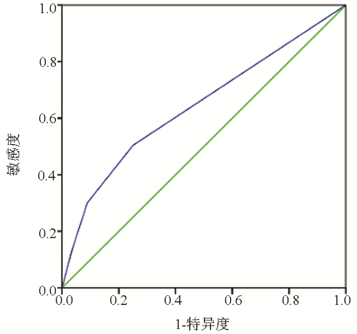
  •   Objective  To investigate the effect of different cytopathological grading standards on the efficiency of endoscopic ultrasound-guided fine needle aspiration (EUS-FNA) in the diagnosis of pancreatic cancer.  Methods  Related clinical data and pancreatic cytopathological results were collected from 256 patients with pancreatic space-occupying lesions who underwent EUS-FNA in The First Affiliated Hospital of Anhui Medical University from May 2011 to March 2019, and the influencing factors for the diagnostic efficiency of EUS-FNA were analyzed based on surgical pathology and follow-up results. The independent samples t-test or the Mann-Whitney U test was used for comparison of continuous data between two groups, and the chi-square test was used for comparison of categorical data between two groups. The receiver operating characteristic (ROC) curve was used to evaluate the value of different cytopathological grading standards in the diagnosis of pancreatic cancer.  Results  A total of 67 patients who were lost to follow-up were excluded, and a total of 189 patients were included in the study. According to the Papanicolaou cytopathological standard, there were 47 cases of heterotypic cells, 25 cases of suspected cancer cells, 20 cases of cancer cells, and 97 cases without tumor cells based on EUS-FNA. A total of 133 patients were confirmed to have pancreatic cancer by postoperative pathology and follow-up results, among whom 52 had no tumor cells, 36 had heterotypic cells, 25 had suspected cancer cells, and 20 had cancer cells based on cytopathological results. EUS-FNA had a true positive rate of 60.90% (81 patients) and a false negative rate of 39.10% (52 patients) in the diagnosis of pancreatic cancer; for the 56 patients without pancreatic cancer, EUS-FNA had a false positive rate of 19.64% (11 patients) and a true negative rate of 80.36% (45 patients). EUS-FNA had an area under the ROC curve of 0.643 (95% confidence interval: 0.561-0.724) in the diagnosis of pancreatic cancer. In combination with different cytopathological grading standards and with the diagnostic criteria of "the identification of heterotypic cells or suspected cancer cells or cancer cells was considered positive", "the identification of suspected cancer cells or cancer cells was considered positive", and "the identification of cancer cells was considered positive", the results showed that the diagnostic criteria of "the identification of heterotypic cells or suspected cancer cells or cancer cells was considered positive" improved the efficiency of EUS-FNA in the diagnosis of pancreatic cancer, with a sensitivity of 50.38% and a specificity of 75.00%. Among the 189 patients, 13 (6.88%) experienced complications after EUS-FNA, which included hyperamylasemia and abdominal pain.  Conclusion  The combination of different cytopathological grading standards can help improve the efficiency of EUS-FNA in the diagnosis of pancreatic cancer.


  • loading
  • [1]
    KANDEL P, WALLACE MB. Advanced EUS guided tissue acquisition methods for pancreatic cancer[J]. Cancers (Basel), 2018, 10(2): 54. DOI: 10.3390/cancers10020054
    [2]
    JIN KZ, HUANG QY, LIU C, et al. New advances in diagnosis and treatment of pancreatic cancer[J]. Chin J Dig Surg, 2019, 18(7): 657-661. (in Chinese) DOI: 10.3760/cma.j.issn.1673-9752.2019.07.009

    金凯舟, 黄秋依, 刘辰, 等. 胰腺癌诊断与治疗的新进展[J]. 中华消化外科杂志, 2019, 18(7): 657-661. DOI: 10.3760/cma.j.issn.1673-9752.2019.07.009
    [3]
    CAǦLAR E, HATEMI I, ATASOY D, et al. Concordance of endoscopic ultrasonography-guided fine needle aspiration diagnosis with the final diagnosis in subepithelial lesions[J]. Clin Endosc, 2013, 46(4): 379-383. DOI: 10.5946/ce.2013.46.4.379
    [4]
    YANG F, YANG F, SUN SY. Endoscopic ultrasound diagnosis of space-occupying lesions in the pancreas[J]. J Clin Hepatol, 2020, 36(8): 1704-1709. (in Chinese) DOI: 10.3969/j.issn.1001-5256.2020.08.005

    杨帆, 杨飞, 孙思予. 胰腺占位性病变的超声内镜诊断[J]. 临床肝胆病杂志, 2020, 36(8): 1704-1709. DOI: 10.3969/j.issn.1001-5256.2020.08.005
    [5]
    WANG W, SHPANER A, KRISHNA SG, et al. Use of EUS-FNA in diagnosing pancreatic neoplasm without a definitive mass on CT[J]. Gastrointest Endosc, 2013, 78(1): 73-80. DOI: 10.1016/j.gie.2013.01.040
    [6]
    ELTOUM IA, ALSTON EA, ROBERSON J. Trends in pancreatic pathology practice before and after implementation of endoscopic ultrasound-guided fine-needle aspiration: An example of disruptive innovation effect?[J]. Arch Pathol Lab Med, 2012, 136(4): 447-453. DOI: 10.5858/arpa.2011-0218-OA
    [7]
    LAYFIELD LJ, DODD L, FACTOR R, et al. Malignancy risk associated with diagnostic categories defined by the Papanicolaou Society of Cytopathology pancreaticobiliary guidelines[J]. Cancer Cytopathol, 2014, 122(6): 420-427. DOI: 10.1002/cncy.21386
    [8]
    HEWITT MJ, McPHAIL MJ, POSSAMAI L, et al. EUS-guided FNA for diagnosis of solid pancreatic neoplasms: A meta-analysis[J]. Gastrointest Endosc, 2012, 75(2): 319-331. DOI: 10.1016/j.gie.2011.08.049
    [9]
    KIM SH, WOO YS, LEE KH, et al. Preoperative EUS-guided FNA: Effects on peritoneal recurrence and survival in patients with pancreatic cancer[J]. Gastrointest Endosc, 2018, 88(6): 926-934. DOI: 10.1016/j.gie.2018.06.024
    [10]
    YAN H, JIA A, ZHANG J, et al. The diagnostic value of endoscopic ultrasonography-guided fine needle aspiration for pancreatic space-occupying diseases[J]. J Xi'an Jiaotong Univ(Med Sci), 2020, 41(3): 425-428. (in Chinese) https://www.cnki.com.cn/Article/CJFDTOTAL-XAYX202003021.htm

    严欢, 贾皑, 张娟, 等. 超声内镜引导细针穿刺活检对胰腺占位性疾病的诊断价值[J]. 西安交通大学学报(医学版), 2020, 41(3): 425-428. https://www.cnki.com.cn/Article/CJFDTOTAL-XAYX202003021.htm
    [11]
    KITANO M, YOSHIDA T, ITONAGA M, et al. Impact of endoscopic ultrasonography on diagnosis of pancreatic cancer[J]. J Gastroenterol, 2019, 54(1): 19-32. DOI: 10.1007/s00535-018-1519-2
    [12]
    CAZACU IM, LUZURIAGA CHAVEZ AA, SAFTOIU A, et al. A quarter century of EUS-FNA: Progress, milestones, and future directions[J]. Endosc Ultrasound, 2018, 7(3): 141-160. DOI: 10.4103/eus.eus_19_18
    [13]
    LEBLANC JK, EMERSON RE, DEWITT J, et al. A prospective study comparing rapid assessment of smears and ThinPrep for endoscopic ultrasound-guided fine-needle aspirates[J]. Endoscopy, 2010, 42(5): 389-394. DOI: 10.1055/s-0029-1243841
    [14]
    SHAH T, ZFASS AM. Accuracy of EUS-FNA in solid pancreatic lesions: Sometimes size does matter[J]. Dig Dis Sci, 2019, 64(7): 1734-1735. DOI: 10.1007/s10620-019-5468-2
    [15]
    ZHANG Y, ZHU Q, GONG TT, et al. Diagnostic value of EUS-FNA for pancreatic masses and its influential factors[J]. Chin J Dig Endosc, 2011, 28(9): 492-496. (in Chinese) https://www.cnki.com.cn/Article/CJFDTOTAL-YBQJ202006002.htm

    张燚, 诸琦, 龚婷婷, 等. 内镜超声引导下细针穿刺抽吸术对胰腺占位病变诊断价值及其影响因素的研究[J]. 中华消化内镜杂志, 2011, 28(9): 492-496. https://www.cnki.com.cn/Article/CJFDTOTAL-YBQJ202006002.htm
    [16]
    FAN Z, ZHANG L, ZHANG XF, et al. The diagnostic value of endoscopic ultrasonography guided fine needle aspiration for occupying pancreatic lesions[J]. Chin J Dig Endosc, 2016, 33(12): 847-850. (in Chinese) https://www.cnki.com.cn/Article/CJFDTOTAL-ZHXH200705001.htm

    范震, 张乐, 张筱凤, 等. 内镜超声引导下细针穿刺活检对胰腺占位性病变的诊断价值[J]. 中华消化内镜杂志, 2016, 33(12): 847-850. https://www.cnki.com.cn/Article/CJFDTOTAL-ZHXH200705001.htm
    [17]
    BERGERON JP, PERRY KD, HOUSER PM, et al. Endoscopic ultrasound-guided pancreatic fine-needle aspiration: Potential pitfalls in one institution's experience of 1212 procedures[J]. Cancer Cytopathol, 2015, 123(2): 98-107.
    [18]
    SIDDIQUI AA, KOWALSKI TE, SHAHID H, et al. False-positive EUS-guided FNA cytology for solid pancreatic lesions[J]. Gastrointest Endosc, 2011, 74(3): 535-540.
    [19]
    LI YQ, WANG W, SUN LQ, et al. Factors influencing the diagnostic positivity of endoscopic ultrasound guided fine needle aspiration for small pancreatic cancer and the occurrence of postoperative advent events[J]. Chin J Pancreatol, 2018, 18(4): 224-227. (in Chinese) http://med.wanfangdata.com.cn/Paper/Detail?id=PeriodicalPaper_yxbx201804003

    李玉琼, 王伟, 孙力祺, 等. 影响EUS-FNA的小胰腺癌诊断阳性率及术后并发症发生的相关因素[J]. 中华胰腺病杂志, 2018, 18(4): 224-227. http://med.wanfangdata.com.cn/Paper/Detail?id=PeriodicalPaper_yxbx201804003
    [20]
    GLEESON FC, KIPP BR, CAUDILL JL, et al. False positive endoscopic ultrasound fine needle aspiration cytology: Incidence and risk factors[J]. Gut, 2010, 59(5): 586-593.
    [21]
    YOSHINAGA S, ITOI T, YAMAO K, et al. Safety and efficacy of endoscopic ultrasound-guided fine needle aspiration for pancreatic masses: A prospective multicenter study[J]. Dig Endosc, 2020, 32(1): 114-126.
  • 加载中

Catalog

    通讯作者: 陈斌, bchen63@163.com
    • 1. 

      沈阳化工大学材料科学与工程学院 沈阳 110142

    1. 本站搜索
    2. 百度学术搜索
    3. 万方数据库搜索
    4. CNKI搜索

    Figures(1)  / Tables(3)

    Article Metrics

    Article views (441) PDF downloads(31) Cited by()
    Proportional views
    Related

    /

    DownLoad:  Full-Size Img  PowerPoint
    Return
    Return