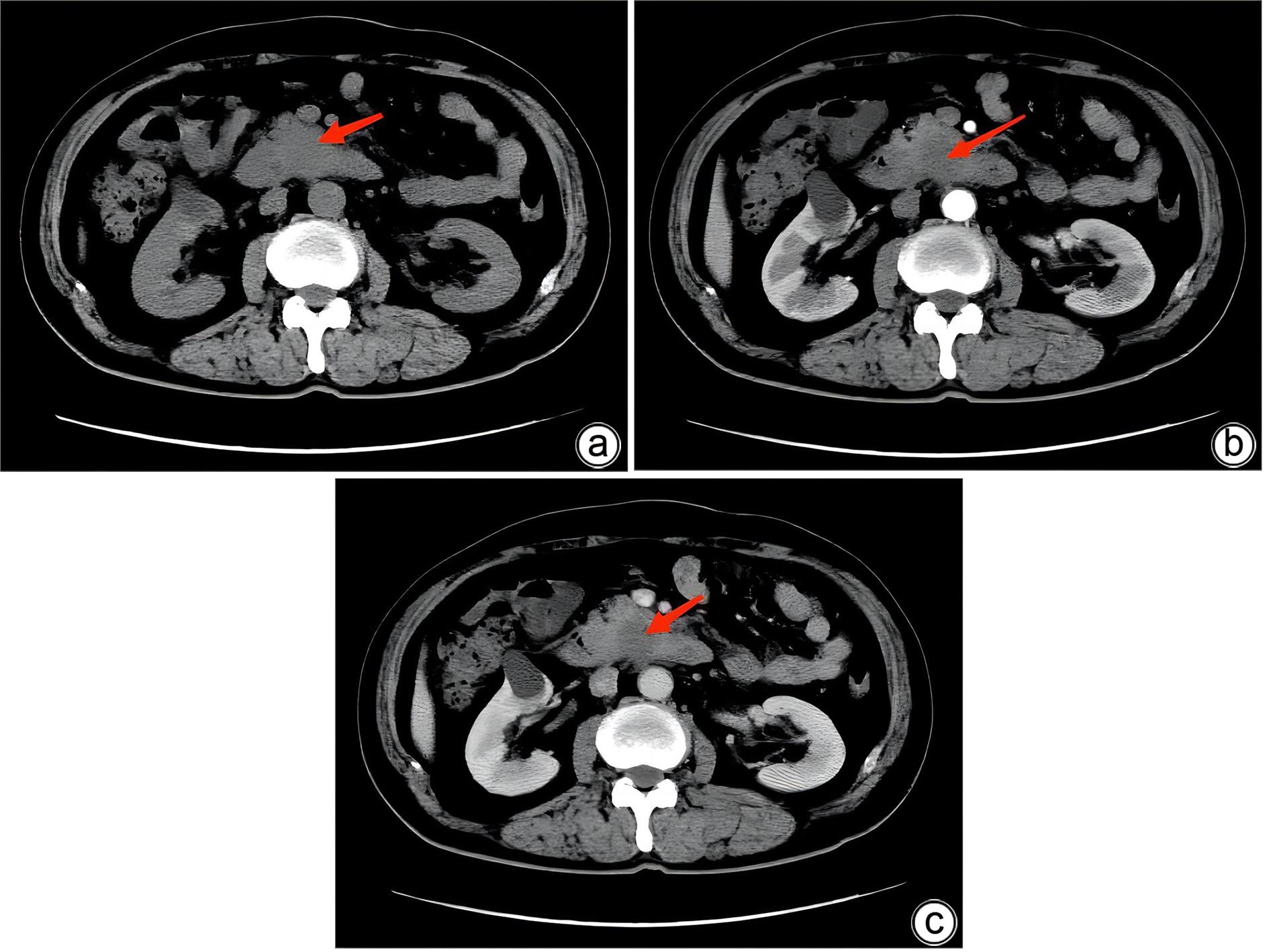
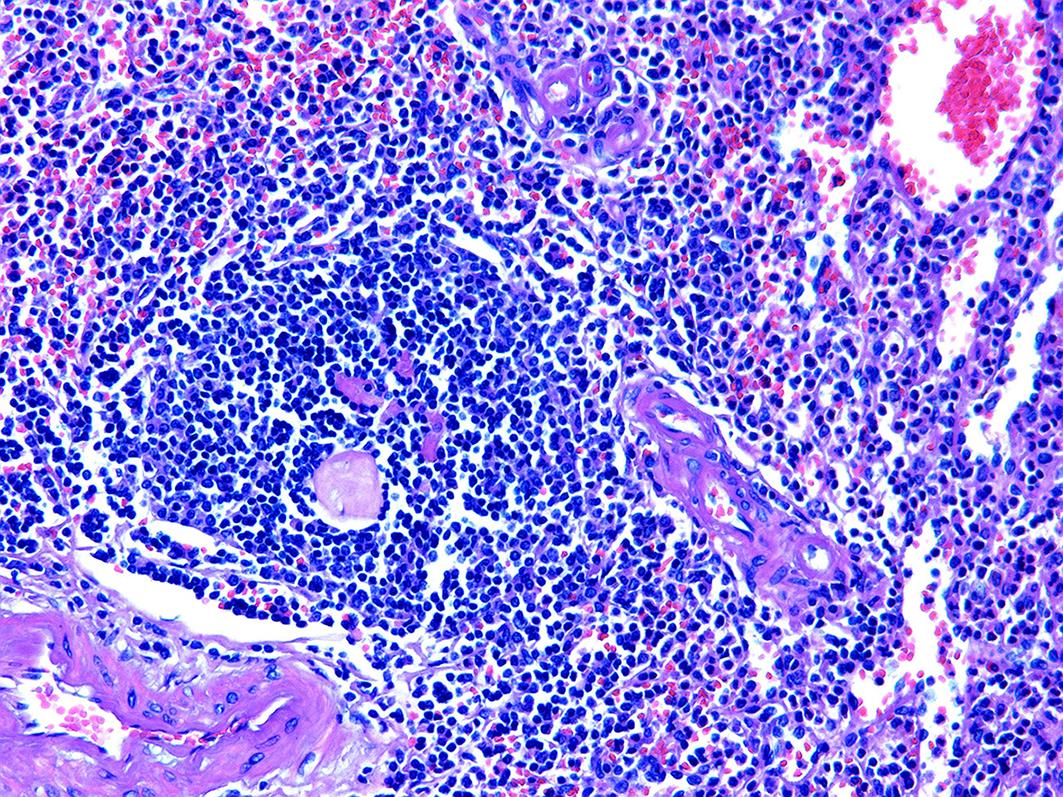
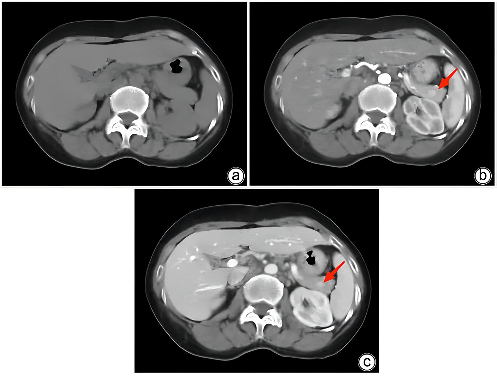
| [1] |
TIPTON SG, SMYRK T, SARR MG, et al. Malignant potential of solid pseudopapillary neoplasm of the pancreas[J]. Br J Surg, 2006, 93( 6): 733- 737. DOI: 10.1002/bjs.5334. |
| [2] |
MOVITZ D. Accessory spleens and experimental splenosis. Principles of growth[J]. Chic Med Sch Q, 1967, 26( 4): 183- 187.
|
| [3] |
HALPERT B, GYORKEY F. Lesions observed in accessory spleens of 311 patients[J]. Am J Clin Pathol, 1959, 32( 2): 165- 168. DOI: 10.1093/ajcp/32.2.165. |
| [4] |
RADOJKOVIC M, RADOJKOVIC D, PREMOVIC N. Intrapancreatic accessory spleen[J]. Med Clínica, 2021, 157( 3): 153- 154. DOI: 10.1016/j.medcli.2020.05.031. |
| [5] |
ZHAO X, ZHOU ZQ, XIANG K, et al. CT diagnosis of intrapancreatic accessory spleen(a report of 2 cases)[J]. Radiol Pract, 2013, 28( 10): 1046- 1048. DOI: 10.13609/j.cnki.1000-0313.2013.10.015. |
| [6] |
KIM SH, LEE JM, HAN JK, et al. Intrapancreatic accessory spleen: Findings on MR Imaging, CT, US and scintigraphy, and the pathologic analysis[J]. Korean J Radiol, 2008, 9( 2): 162- 174. DOI: 10.3348/kjr.2008.9.2.162. |
| [7] |
JANG KM, KIM SH, LEE SJ, et al. Differentiation of an intrapancreatic accessory spleen from a small(<3-cm) solid pancreatic tumor: Value of diffusion-weighted MR imaging[J]. Radiology, 2013, 266( 1): 159- 167. DOI: 10.1148/radiol.12112765. |
| [8] |
|
| [9] |
NI X, HU XM, JIANG H. Clinicopathologic analysis of epidermoid cyst in intrapancreatic accessory spleen: A report of 12 cases[J]. Chin J Pancreatol, 2022, 22( 3): 201- 204. DOI: 10.3760/cma.j.cn115667-20210829-00156. |
| [10] |
OTA T, TEI M, YOSHIOKA A, et al. Intrapancreatic accessory spleen diagnosed by technetium-99m heat-damaged red blood cell SPECT[J]. J Nucl Med, 1997, 38( 3): 494- 495.
|
| [11] |
SCHMID-TANNWALD C, SCHMID-TANNWALD CM, MORELLI JN, et al. Comparison of abdominal MRI with diffusion-weighted imaging to 68Ga-DOTATATE PET/CT in detection of neuroendocrine tumors of the pancreas[J]. Eur J Nucl Med Mol Imaging, 2013, 40( 6): 897- 907. DOI: 10.1007/s00259-013-2371-5. |
| [12] |
MAKINO Y, IMAI Y, FUKUDA K, et al. Sonazoid-enhanced ultrasonography for the diagnosis of an intrapancreatic accessory spleen: A case report[J]. J Clin Ultrasound, 2011, 39( 6): 344- 347. DOI: 10.1002/jcu.20798. |
| [13] |
PENG N, MI JW, ZHAO DQ. Endoscopic ultrasonography in the diagnosis and treatment of pancreatic neuroendocrine tumors[J]. Chin J Ultrason, 2020( 1): 87- 90. DOI: 10.3760/cma.j.issn.1004-4477.2020.01.017. |
| [14] |
XIA XX, LYU GY, QIU XT, et al. Intrapancreatic accessory spleen misdiagnosed as pancreatic neuroendocrine tumor: A case report[J]. J Clin Hepatol, 2022, 38( 2): 436- 438. DOI: 10.3969/j.issn.1001-5256.2022.02.036. |
| [15] |
TATSAS AD, OWENS CL, SIDDIQUI MT, et al. Fine-needle aspiration of intrapancreatic accessory spleen: Cytomorphologic features and differential diagnosis[J]. Cancer Cytopathol, 2012, 120( 4): 261- 268. DOI: 10.1002/cncy.21185. |
| [16] |
BASTIDAS AB, HOLLOMAN D, LANKARANI A, et al. Endoscopic ultrasound-guided needle-based probe confocal laser endomicroscopy(nCLE) of intrapancreatic ectopic spleen[J]. ACG Case Rep J, 2016, 3( 3): 196- 198. DOI: 10.14309/crj.2016.48. |


 DownLoad: