
 PDF下载 ( 3391 KB)

18F-FDG PET/CT、超声造影及联合应用对胰腺良恶性病变的鉴别诊断价值比较
DOI: 10.3969/j.issn.1001-5256.2022.12.017
Value of 18F-FDG PET/CT, contrast-enhanced ultrasound, and their combined use in differential diagnosis of benign and malignant pancreatic lesions: A comparative study
-
摘要:
目的 探讨18F-FDG PET/CT、超声造影及联合应用对胰腺病变良恶性鉴别的诊断价值。 方法 回顾性分析2015年1月—2020年12月于唐山市工人医院行18F-FDG PET/CT和超声造影检查的胰腺病变患者资料,以病理结果为标准,分析18F-FDG P ET/CT、超声造影及两者联合时对胰腺病变良恶性鉴别诊断的灵敏度、特异度、准确度、阳性预测值及阴性预测值。计量资料组间比较采用t检验;计数资料组间比较采用χ2检验。 结果 108例病变中恶性83例、良性25例,18F-FDG PET/CT诊断灵敏度、特异度、准确度、阳性预测值及阴性预测值分别为86.75%、80.00%、85.19%、93.51%及64.52%,超声造影分别为69.88%、76.00%、71.30%、90.63%及43.18%,两者联合应用时分别为90.36%、84.00%、88.89%、94.94%及72.41%。18F-FDG PET/CT与超声造影两者间灵敏度、准确度比较差异均有统计学意义(P值均<0.05)。 结论 18F-FDG PET/CT对胰腺病变良恶性鉴别具有较高的诊断价值,高于超声造影,两者联合应用时可进一步提高诊断价值。 -
关键词:
- 胰腺肿瘤 /
- 正电子发射断层显像计算机体层摄影术 /
- 体层摄影术, X线计算机 /
- 氟脱氧葡萄糖F18
Abstract:Objective To assess the value of 18F-FDG PET/CT, contrast-enhanced ultrasound, and their combination in the differential diagnosis of benign and malignant pancreatic lesions. Methods A retrospective analysis was performed on patients with pancreatic lesions who underwent18F-FDG PET/CT and contrast-enhanced ultrasound who were admitted to Tangshan Gongren Hospital from January 2015 to December 2020. The imaging results were confirmed by pathology examination to evaluate diagnostic sensitivity, specificity, accuracy, positive and negative predictive value. The t-test was used for comparison of continuous data between two groups, and the chi-square test was used for comparison of categorical data between groups. Results There were 83 malignant lesions and 25 benign lesions in 108 patients. The sensitivity, specificity, accuracy, positive and negative predictive value were 86.75%, 80.00%, 85.19%, 93.51% and 64.52% for 18F-FDG PET/CT; and 69.88%, 76.00%, 71.30%, 90.63% and 43.18% for contrast-enhanced ultrasound, respectively. The two methods differed significantly in sensitivity and accuracy (all P < 0.05), but not in specificity, negative and positive predictive value (all P > 0.05). When combined with the contrast-enhanced ultrasound, 18F-FDG PET/CT had an increased sensitivity, specificity, accuracy, positive and negative predictive value of 90.36%, 84.00%, 88.89%, 94.94% and 72.41%, respectively, though this was not statistically significant due to the increased signal of blood supply in the lesions. Conclusion 18F-FDG PET/CT has a better performance than contrast-enhanced ultrasound in the differential diagnosis of benign and malignant pancreatic lesions, and their combination can improve the diagnostic value. -

图 1 PDAC18F-FDG PET/CT图像
注:患者,男,58岁,胰头导管腺癌,18F-FDG PET/CT显示18F-FDG明显不均匀性增高,SUVmax为5.4(箭头)。a,PET;b,CT;c,PET/CT;d,PET MIP。
Figure 1. 18F-FDG PET/CT imaging of pancreatic ductal adenocarcinoma

图 2 AIP18F-FDG PET/CT图像
注:患者,男,36岁,AIP,18F-FDG PET/CT显示胰腺整体弥漫性肿大,18F-FDG摄取呈弥漫性增高,呈“腊肠样”表现,SUVmax为6.1 (箭头)。a,PET;b,CT;c,PET/CT;d,PET MIP。
Figure 2. 18F-FDG PET/CT imaging of autoimmune pancreatitis

图 3 PDAC超声造影动脉期图像
注:患者,男,58岁,胰头导管腺癌(与图 1同一患者),超声造影显示胰头病变动脉期呈低增强(箭头)。a,常规超声;b,超声造影动脉期。
Figure 3. Contrast-enhanced ultrasound imaging of pancreatic ductal adenocarcinoma in arterial phase

图 4 PDAC超声造影静脉期图像
注:患者,男,58岁,胰头导管腺癌(与图 1同一患者),超声造影显示胰头病变静脉期呈低增强(箭头)。a,常规超声;b,超声造影静脉期。
Figure 4. Contrast-enhanced ultrasound imaging of pancreatic ductal adenocarcinoma in venous phase

图 5 AIP超声造影动脉期图像
注:患者,男,36岁,AIP(与图 2同一患者),超声显示胰腺整体弥漫性肿大,动脉期造影呈均匀性等增强表现(箭头)。a,常规超声;b,超声造影动脉期。
Figure 5. Contrast-enhanced ultrasound imaging of autoimmune pancreatitis in arterial phase
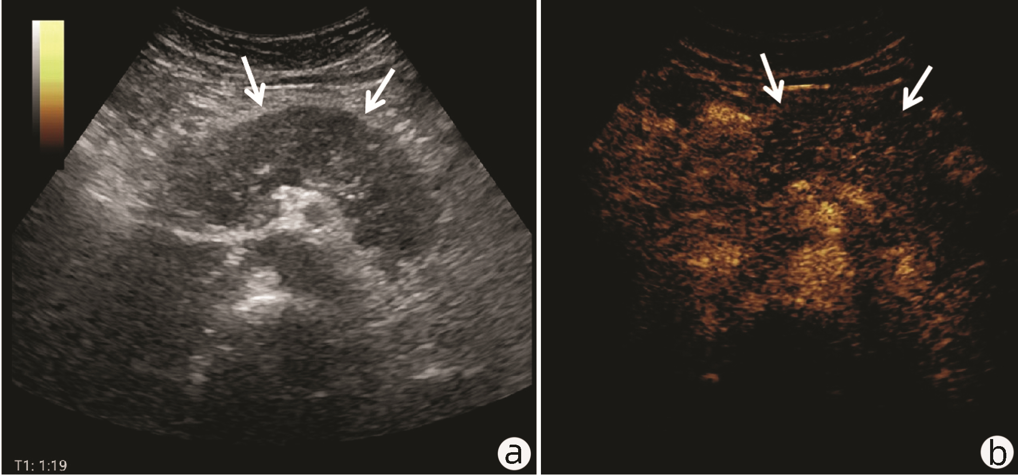
图 6 AIP超声造影静脉期图像
注:患者,男,36岁,AIP(与图 2同一患者),超声显示胰腺整体弥漫性肿大,静脉期造影呈均匀性等增强表现(箭头)。a,常规超声;b,超声造影静脉期。
Figure 6. Contrast-enhanced ultrasound imaging of autoimmune pancreatitis in venous phase
表 1 3种诊断方法分别对胰腺良恶性病变检出的数目比较
Table 1. Comparison of 3 diagnostic results with the number of benign and malignant pancreatic lesions
病理 例数 PET/CT(例) 超声造影(例) PET/CT+超声造影(例) + - + - + - 恶性 83 72 11 58 25 75 8 良性 25 5 20 6 19 4 21  下载: 导出CSV
表 2 3种诊断方法对胰腺良恶性病变诊断指标比较
Table 2. Comparison of 3 diagnostic indexes of benign and malignant pancreatic lesions
组别 灵敏度 特异度 准确度 阳性预测值 阴性预测值 PET/CT 86.75%1) 80.00% 85.19%1) 93.51% 64.52% 超声造影 69.88% 76.00% 71.30% 90.63% 43.18% PET/CT+超声造影 90.36%1) 84.00% 88.89%1) 94.94% 72.41% χ2值 6.952 0.117 6.119 0.403 3.316 P值 0.008 0.733 0.013 0.546 0.069 注:与超声造影比,1)P<0.05。
下载: 导出CSV
-
[1] Chinese Pancreatic Surgery Association, Chinese Society of Surgery, Chinese Medical Association. Guidelines for the diagnosis and treatment of pancreatic cancer in China(2021)[J]. Chin J Dig Surg, 2021, 20(7): 713-729. DOI: 10.3760/cma.j.cn115610-20210618-00289.中华医学会外科学分会胰腺外科学组. 中国胰腺癌诊治指南(2021)[J]. 中华消化外科杂志, 2021, 20(7): 713-729. DOI: 10.3760/cma.j.cn115610-20210618-00289. [2] CAI J, CHEN HD, LU M, et al. Trend analysis on morbidity and mortality of pancreatic cancer in China, 2005-2015[J]. Chin J Epidemiol, 2021, 42(5): 794-800. DOI: 10.3760/cma.j.cn112338-20201115-01328.蔡洁, 陈宏达, 卢明, 等. 2005-2015年中国胰腺癌发病与死亡趋势分析[J]. 中华流行病学杂志, 2021, 42(5): 794-800. DOI: 10.3760/cma.j.cn112338-20201115-01328. [3] YANG H, WANG XK, FAN JH. Present status of epidemiology, risk factors and screening of pancreatic cancer in China[J]. Cancer Res Prev Treat, 2021, 48(10): 909-915. DOI: 10.3971/j.issn.1000-8578.2021.21.0789.杨欢, 王晓坤, 范金虎. 中国胰腺癌流行病学、危险因素及筛查现况[J]. 肿瘤防治研究, 2021, 48(10): 909-915. DOI: 10.3971/j.issn.1000-8578.2021.21.0789. [4] ZHENG LC, LIU GC, OUYANG XL, et al. Diagnosis of 18F-FDG PET/CT imaging in benign and malignant pancreatic lesions[J]. Chin J Med Imaging, 2018, 26(9): 680-684. DOI: 10.3969/j.issn.1005-5185.2018.09.010.郑立春, 刘桂超, 欧阳向柳, 等. ~(18)F-FDG PET/CT显像对胰腺良恶性病变的诊断价值[J]. 中国医学影像学杂志, 2018, 26(9): 680-684. DOI: 10.3969/j.issn.1005-5185.2018.09.010. [5] PU Y, WANG C, ZHAO S, et al. The clinical application of 18F-FDG PET/CT in pancreatic cancer: a narrative review[J]. Transl Cancer Res, 2021, 10(7): 3560-3575. DOI: 10.21037/tcr-21-169. [6] GAO B, OUYANG XL, ZHANG HL, et al. Diagnostic value of contrast-enhanced ultrasound in aged patients with pancreatic tumors[J]. Chin J Ultrasound in Med, 2018, 34(8): 706-709. DOI: 10.3969/j.issn.1002-0101.2018.08.010.高蓓, 欧阳向柳, 张浩良, 等. 超声造影对于老年胰腺肿瘤患者的诊断价值分析[J]. 中国超声医学杂志, 2018, 34(8): 706-709. DOI: 10.3969/j.issn.1002-0101.2018.08.010. [7] LI S, JIANG H, WANG Z, et al. An effective computer aided diagnosis model for pancreas cancer on PET/CT images[J]. Comput Methods Programs Biomed, 2018, 165: 205-214. DOI: 10.1016/j.cmpb.2018.09.001. [8] MYSSAYEV A, MYSSAYEV A, IDEGUCHI R, et al. Usefulness of FDG PET/CT derived parameters in prediction of histopathological finding during the surgery in patients with pancreatic adenocarcinoma[J]. PLoS One, 2019, 14(1): e0210178. DOI: 10.1371/journal.pone.0210178. [9] YAMASHITA YI, OKABE H, HAYASHI H, et al. Usefulness of 18-FDG PET/CT in detecting malignancy in intraductal papillary mucinous neoplasms of the pancreas[J]. Anticancer Res, 2019, 39(5): 2493-2499. DOI: 10.21873/anticanres.13369. [10] REN SN, LI DN, PAN GX, et al. Research progress in diagnosis and prognosis evaluation of pancreatic cancer by~(18)F-FDG PET/CT[J]. Chin J Pancreatol, 2019, 19(4): 307-310. DOI: 10.3760/cma.j.issn.1674-1935.2019.04.018.任胜男, 李丹妮, 潘桂霞, 等. 胰腺癌~(18)F-FDG PET/CT诊断及预后评估的研究进展[J]. 中华胰腺病杂志, 2019, 19(4): 307-310. DOI: 10.3760/cma.j.issn.1674-1935.2019.04.018. [11] SRINIVASAN N, KOH YX, GOH B. Systematic review of the utility of 18-FDG PET in the preoperative evaluation of IPMNs and cystic lesions of the pancreas[J]. Surgery, 2019, 165(5): 929-937. DOI: 10.1016/j.surg.2018.11.006. [12] DUNET V, HALKIC N, SEMPOUX C, et al. Use of PET and MRI imaging features to predict grade and survival of resectable pancreatic ductal adenocarcinoma[J]. Inter J Med Rad, 2021, 44(2): 247. DOI: 10.19300/j.2021.e0211.DUNET V, HALKIC N, SEMPOUX C, 等. 应用PET和MRI影像学特征预测可切除的胰腺导管腺癌肿瘤分级及生存率[J]. 国际医学放射学杂志, 2021, 44(2): 247. DOI: 10.19300/j.2021.e0211. [13] WANG PP, HUO L, LIU Y, et al. Clinical value of 18F-FDG PET/CT imaging in non-functional pancreatic neuroendocrine neoplasms[J]. Chin J Nuclear Med Mol Imaging, 2022, 42(3): 139-143. DOI: 10.3760/cma.j.cn321828-20200721-00288.王佩佩, 霍力, 刘宇, 等. 无功能胰腺神经内分泌肿瘤~(18)F-FDG PET/CT显像的临床应用价值[J]. 中华核医学与分子影像杂志, 2022, 42(3): 139-143. DOI: 10.3760/cma.j.cn321828-20200721-00288. [14] YAMANE T, AIKAWA M, YASUDA M, et al. [18F]FMISO PET/CT as a preoperative prognostic factor in patients with pancreatic cancer[J]. EJNMMI Res, 2019, 9(1): 39. DOI: 10.1186/s13550-019-0507-8. [15] LIERMANN J, SYED M, BEN-JOSEF E, et al. Impact of FAPI-PET/CT on target volume definition in radiation therapy of locally recurrent pancreatic cancer[J]. Cancers (Basel), 2021, 13(4). DOI: 10.3390/cancers13040796. [16] BONACINA M, GHIRARDELLI P, SETTI L, et al. 68Ga-PSMA and 68Ga-DOTATOC PET/CT imaging mismatch of primary pancreatic adenocarcinoma in prostate cancer patient[J]. Eur J Nucl Med Mol Imaging, 2022, 49(2): 781-782. DOI: 10.1007/s00259-021-05523-9. [17] ZHANG Z, JIA G, PAN G, et al. Comparison of the diagnostic efficacy of 68 Ga-FAPI-04 PET/MR and 18F-FDG PET/CT in patients with pancreatic cancer[J]. Eur J Nucl Med Mol Imaging, 2022, 49(8): 2877-2888. DOI: 10.1007/s00259-022-05729-5. [18] JIA W, YIN LL, JI B, et al. Comparison of 18F-FDG PET/CT and enhanced CT to assess the tumor stage, vascular invasion, distant metastasis and surgical indications of pancreatic cancer[J]. Chin J Gen Surg, 2019, 28(3): 360-365. DOI: 10.7659/j.issn.1005-6947.2019.03.017.贾维, 印隆林, 季冰, 等. ~(18)F-FDG PET/CT显像与增强CT评估胰腺癌分期、血管侵犯、远处转移和手术指征的比较[J]. 中国普通外科杂志, 2019, 28(3): 360-365. DOI: 10.7659/j.issn.1005-6947.2019.03.017. [19] LIN ZM, PAN MQ, XU YY, et al. Contrast enhanced ultrasonography vs. contrast enhanced computed tomography for the diagnosis of focal lesions of the pancreas[J]. Chin J Gen Surg, 2018, 33(10): 849-852. DOI: 10.3760/cma.j.issn.1007-631X.2018.10.013.林子梅, 潘敏强, 徐永远, 等. 胰腺实性局灶性病变的超声造影与增强CT对照研究[J]. 中华普通外科杂志, 2018, 33(10): 849-852. DOI: 10.3760/cma.j.issn.1007-631X.2018.10.013. [20] LIN ZM, WEN Q, XU YY, et al. Application of contrast enhanced ultrasound in TN staging of pancreas cancer: comparsion with contrast enhanced computed tomography[J]. China J Ultrasonography, 2018, 27(7): 614-617. DOI: 10.3760/cma.j.issn.1004-4477.2018.07.014.林子梅, 闻卿, 徐永远, 等. 超声造影在胰腺癌T、N分期中的应用价值[J]. 中华超声影像学杂志, 2018, 27(7): 614-617. DOI: 10.3760/cma.j.issn.1004-4477.2018.07.014. [21] WU LQ, LIU XH, LUAN ZY, et al. Meta-analysis on the differential diagnosis of benign and malignant pancreatic masses by contrast-enhanced ultrasound[J]. J Clin Ultrasound Med, 2018, 20(6): 383-387. DOI: 10.3969/j.issn.1008-6978.2018.06.009.吴亮群, 刘雪红, 栾智勇, 等. 超声造影鉴别诊断胰腺良恶性病变的Meta分析[J]. 临床超声医学杂志, 2018, 20(6): 383-387. DOI: 10.3969/j.issn.1008-6978.2018.06.009. [22] LI Y, XU Y, SONG YF, et al. Clinical value of contrast-enhanced ultrasound in differentiating focal pancreatitis from pancreatic cancer: a comparative study with conventional ultrasound[J]. J Chin Med Imaging, 2021, 32(10): 729-732. DOI: 10.12117/jccmi.2021.10.011.李煜, 许芸, 宋一凡. 超声造影增强模式鉴别局灶性胰腺炎与胰腺癌的临床价值: 与常规超声的对照研究[J]. 中国临床医学影像杂志, 2021, 32(10): 729-732. DOI: 10.12117/jccmi.2021.10.011. [23] XU J, CHEN YL, WANG XW. Comparison on Clinical Value of 18F-FDG PET/CT, Enhanced CT, and MRI in Differential Diagnosis of Benign and Malignant Pancreas Cystic Lesions[J]. Chin J CT and MRI, 2022, 20(2): 99-101. DOI: 10.3969/j.issn.1672-5131.2022.02.033.徐杰, 陈艳林, 王雪伟. ~(18)F-FDG PET/CT与增强CT、MRI在诊断鉴别胰腺囊性良恶性病变的临床价值比较[J]. 中国CT和MRI杂志, 2022, 20(2): 99-101. DOI: 10.3969/j.issn.1672-5131.2022.02.033. -

 本文二维码
本文二维码
计量
- 文章访问数: 1547
- HTML全文浏览量: 1036
- PDF下载量: 39
- 被引次数: 0

