
 PDF下载 ( 3726 KB)

利多卡因抑制糖尿病小鼠Kupffer细胞炎症反应及对肝脓肿形成的影响
DOI: 10.3969/j.issn.1001-5256.2022.06.023
Inhibitory effect of lidocaine on Kupffer cell inflammatory response and its effect on liver abscess formation in diabetic mice
-
摘要:
目的 观察利多卡因是否能逆转糖尿病小鼠Kupffer细胞功能障碍,阐明利多卡因通过改善糖尿病小鼠Kupffer细胞吞噬功能并影响肝脓肿形成的机制。 方法 将C57BLKS/J db/db小鼠分为糖尿病对照组和糖尿病+利多卡因组,将C57BLKS/J db/m小鼠分为非糖尿病对照组和非糖尿病+利多卡因组,每组5只,均喂食肺炎克雷伯杆菌悬液。分别取Kupffer细胞体外培养,电镜观察超微结构改变,测定Kupffer细胞的炎症介质分泌水平,细胞间黏附分子1(ICAM-1)表达水平,中性粒细胞趋化功能,吞噬功能,以及肝脓肿的形成。计量资料多组间比较采用Kruskal-Wallis H秩和检验,进一步两两比较采用Mann-Whitney U检验;计数资料组间比较采用χ2检验。 结果 糖尿病小鼠Kupffer细胞中线粒体和粗面内质网的数量减少, 内质网扩张,线粒体肿胀及脂滴增多。与非糖尿病对照组比较,糖尿病对照组Kupffer细胞表达NO[(4.95±0.06) μmol/L vs (1.34±0.13)μmol/L]、IL-6[(740.04±8.58) pg/mL vs (515.77±4.62)pg/mL]、TNFα[(774.23±7.98) pg/mL vs (461.51±1.76)pg/mL]、IFNγ[(842.33±14.79) pg/mL vs (542.47±6.75)pg/mL]、ICAM-1(2.40±0.02 vs 1.33±0.01)水平明显升高(P值均<0.05),中性粒细胞趋化增强(100.80±10.18 vs 13.80±3.70,P<0.05),吞噬能力减弱(9.86±1.82 vs 60.00±3.54,P<0.05),肝脓肿形成无影响(40% vs 0,P>0.05)。与糖尿病对照组比较,糖尿病+利多卡因组Kupffer细胞表达NO[(3.35±0.28) μmol/L vs (4.95±0.06)μmol/L]、IL-6[(688.42±36.34) pg/mL vs (740.04±8.58) pg/mL]、TNFα[(631.15±4.30) pg/mL vs (774.23±7.98) pg/mL]、IFNγ[(704.56±3.64) pg/mL vs (842.33±14.79)pg/mL]、ICAM-1(1.50±0.02 vs 2.40±0.02) 水平明显降低(P值均<0.05),中性粒细胞趋化减弱(33.40±5.60 vs 100.80±10.18,P<0.05),吞噬能力增强(49.20±2.59 vs 9.86±1.82,P<0.05)、肝脓肿形成无影响(0 vs 40%,P>0.05)。 结论 利多卡因可以抑制糖尿病小鼠Kupffer细胞炎症反应并改善其吞噬功能,对Kupffer细胞起到保护作用,但对肝脓肿的形成无影响。 Abstract:Objective To investigate whether lidocaine can reverse Kupffer cell dysfunction in diabetic mice, as well as the mechanism of lidocaine in affecting liver abscess formation by improving the phagocytic function of Kupffer cells. Methods C57BLKS/J db/db mice were divided into diabetes control group and diabetes+lidocaine group, and C57BLKS/J db/m mice were divided into non-diabetes control group and non-diabetes+lidocaine group, with 5 mice in each group. All mice were fed with the suspension of Klebsiella pneumoniae. Kupffer cells were collected from each group and were cultured in vitro; an electron microscope was used to measure the change in ultrastructure, and Kupffer c ells were measured in terms of the levels of inflammatory mediators, the expression level of intercellular adhesion molecule-1 (ICAM-1), the chemotactic function of neutrophils, and phagocytic function; liver abscess formation was also observed. The Kruskal-Wallis H test was used for comparison of continuous data between multiple groups, and the Mann-Whitney U test was used for further comparison between two groups; the chi-square test was used for comparison of categorical data between groups. Results Compared with the non-diabetic mice, the diabetic mice had significant reductions in mitochondria and rough endoplasmic reticulum, endoplasmic reticulum dilation, mitochondrial swelling, and an increase in lipid droplets in Kupffer cells. Compared with the non-diabetes control group, the diabetes control group had significant increases in the levels of nitric oxide (NO) (4.95±0.06 μmol/L vs 1.34±0.13 μmol/L, P < 0.05), interleukin-6 (IL-6) (740.04±8.58 pg/mL vs 515.77±4.62 pg/mL, P < 0.05), tumor necrosis factor-α (TNFα) (774.23±7.98 pg/mL vs 461.51±1.76 pg/mL, P < 0.05), interferon gamma (IFNγ) (842.33±14.79 pg/mL vs 542.47±6.75 pg/mL, P < 0.05), and ICAM-1 (2.40±0.02 vs 1.33±0.01, P < 0.05) in Kupffer cells, a significant increase in neutrophil chemotaxis (100.80±10.18 vs 13.80±3.70, P < 0.05), and a significant reduction in phagocytic capacity (9.86±1.82 vs 60.00±3.54, P < 0.05), with no effect on liver abscess formation (40% vs 0, P > 0.05). Compared with the diabetes control group, the diabetes+lidocaine group had significant reductions in the levels of NO (3.35±0.28 μmol/L vs 4.95±0.06 μmol/L, P < 0.05), IL-6 (688.42±36.34 pg/mL vs 740.04±8.58 pg/mL, P < 0.05), TNFα (631.15±4.30 pg/mL vs 774.23±7.98 pg/mL, P < 0.05), IFNγ (704.56±3.64 pg/mL vs 842.33±14.79 pg/mL, P < 0.05), and ICAM-1 (1.50±0.02 vs 2.40±0.02, P < 0.05) in Kupffer cells, a significant reduction in neutrophil chemotaxis (33.40±5.60 vs 100.80±10.18, P < 0.05), and a significant increase in phagocytic capacity (49.20±2.59 vs 9.86±1.82, P < 0.05), with no effect on liver abscess formation (0 vs 40%, P > 0.05). Conclusion Lidocaine can inhibit Kupffer cell inflammatory response and improve the phagocytic function of Kupffer cells in diabetic mice, thereby exerting a protective effect on Kupffer cells, but it had no effect on liver abscess formation. -
Key words:
- Diabetes Mellitus /
- Liver Abscess /
- Kupffer Cells /
- Lidocaine
-
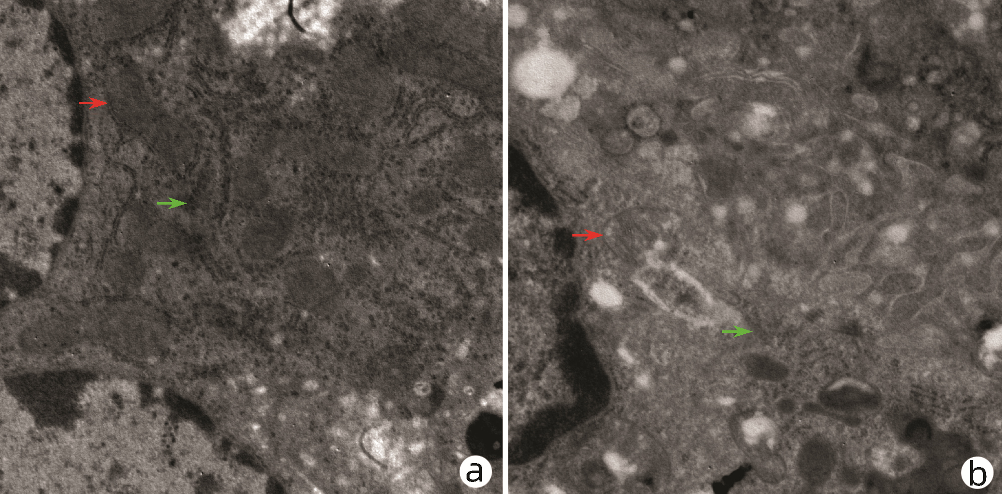
图 1 非糖尿病与糖尿病小鼠Kupffer细胞超微结构观察(透射电镜,×5000)
注:a,糖尿病小鼠; b,非糖尿病小鼠。红色箭头为线粒体; 绿色箭头为粗面内质网。
Figure 1. Ultrastructural observation of mitochondria and endoplasmic reticulum in Kupffer cells in nondiabetic vs diabetic mice (transmission electron microscopy, ×5000)
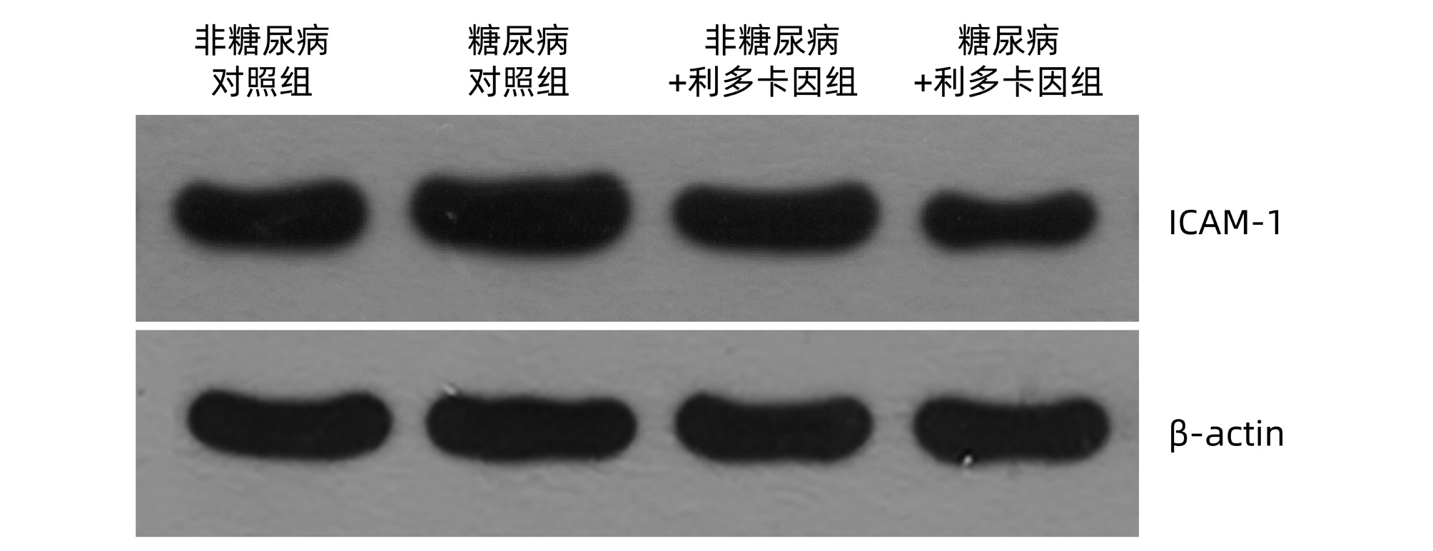
图 2 Western Blot检测各组小鼠Kupffer细胞ICAM-1表达
Figure 2. Expression of ICAM-1 in Kupffer cells detected by Western Blot

图 3 各组小鼠中性粒细胞趋化(0.1%结晶紫染色,×100)
注:a,非糖尿病对照组; b,糖尿病对照组; c,非糖尿病+利多卡因组; d,糖尿病+利多卡因组。
Figure 3. Neutrophil chemotaxis of mice in each group (0.1% crystal violet staining, ×100)

图 4 各组小鼠Kupffer细胞吞噬功能(Giemsa染色,×100)
注:a,非糖尿病对照组; b,糖尿病对照组; c,非糖尿病+利多卡因组; d,糖尿病+利多卡因组。
Figure 4. Phagocytosis of Kupffer cells in each group (Giemsa staining, ×100)
表 1 各组小鼠Kupffer细胞NO、IL-6、TNFα、IFNγ、ICAM-1水平
Table 1. Levels of NO, IL-6, TNFα, IFNγ and ICAM-1 in Kupffer cells of mice in each group
指标 非糖尿病对照组
(n=5)糖尿病对照组
(n=5)非糖尿病+利多
卡因组(n=5)糖尿病+利多
卡因组(n=5)H值 P值 NO(μmol/L) 1.34±0.13 4.95±0.061) 1.47±0.16 3.35±0.282) 16.50 <0.001 IL-6(pg/mL) 515.77±4.62 740.04±8.581) 512.79±2.24 688.42±36.342) 15.79 0.001 TNFα(pg/mL) 461.51±1.76 774.23±7.981) 468.48±7.42 631.15±4.302) 16.71 <0.001 IFNγ(pg/mL) 542.47±6.75 842.33±14.791) 543.49±8.38 704.56±3.642) 16.07 0.001 ICAM-1 1.33±0.01 2.40 ±0.021) 1.31±0.03 1.50±0.022) 16.52 0.001 注:与非糖尿病对照组相比,1)P<0.05;与糖尿病对照组相比,2)P<0.05。  下载: 导出CSV
表 2 各组小鼠中性粒细胞招募、吞噬功能水平
Table 2. Neutrophil recruitment and phagocytic function levels in each group
项目 非糖尿病对照组
(n=5)糖尿病对照组
(n=5)非糖尿病+利多
卡因组(n=5)糖尿病+利多
卡因组(n=5)H值 P值 中性粒细胞趋化 13.80±3.70 100.80±10.181) 14.40±4.67 33.40±5.602) 16.12 0.001 Kupffer细胞吞噬功能 60.00±3.54 9.86 ±1.821) 62.60±1.95 49.20±2.592) 16.87 0.001 注:与非糖尿病对照组相比,1)P<0.05;与糖尿病对照组相比,2)P<0.05。
下载: 导出CSV
-
[1] NCD Risk Factor Collaboration (NCD-RisC). Worldwide trends in diabetes since 1980: a pooled analysis of 751 population- based studies with 4.4 million participants[J]. Lancet, 2016, 387(10027): 1513-1530. DOI: 10.1016/S0140-6736(16)00618-8. [2] FENG J. Retrospective analysis of clinical features and risk factors of patients with diabetic bacterial hepatic abscess complicated with hepatobiliary and pancreatic diseases[J]. J Hepatobiliary Surg, 2020, 28(6): 423-426. DOI: 10.3969/j.issn.1006-4761.2020.06.008.冯静. 回顾性分析糖尿病细菌性肝脓肿患者合并肝胆胰疾病的临床特点及危险因素[J]. 肝胆外科杂志, 2020, 28(6): 423-426. DOI: 10.3969/j.issn.1006-4761.2020.06.008. [3] ATREJA A, KALRA S. Infections in diabetes[J]. J Pak Med Assoc, 2015, 65(9): 1028-1030. [4] PAN JZ, WANG XM, HUANG YQ, et al. Clinical analysis of 424 cases of diabetes mellitus complicated with bacterial liver abscess[J]. Chin J Clin Infect Dis, 2021, 14(3): 199-202. DOI: 10.3760/cma.j.issn.1674-2397.2021.03.007.潘教治, 王仙敏, 黄友全, 等. 糖尿病合并细菌性肝脓肿424例临床分析[J]. 中华临床感染病杂志, 2021, 14(3): 199-202. DOI: 10.3760/cma.j.issn.1674-2397.2021.03.007. [5] WOHLLEBER D, KNOLLE PA. The role of liver sinusoidal cells in local hepatic immune surveillance[J]. Clin Transl Immunology, 2016, 5(12): e117. DOI: 10.1038/cti.2016.74. [6] DONG B, ZHOU Y, WANG W, et al. Vitamin D receptor activation in liver macrophages ameliorates hepatic inflammation, steatosis, and insulin resistance in mice[J]. Hepatology, 2020, 71(5): 1559-1574. DOI: 10.1002/hep.30937. [7] HOTAMISLIGIL GS. Inflammation, metaflammation and immunometabolic disorders[J]. Nature, 2017, 542(7640): 177-185. DOI: 10.1038/nature21363. [8] BILZER M, ROGGEL F, GERBES AL. Role of Kupffer cells in host defense and liver disease[J]. Liver Int, 2006, 26(10): 1175-1186. DOI: 10.1111/j.1478-3231.2006.01342.x. [9] DASU MR, DEVARAJ S, ZHAO L, et al. High glucose induces toll-like receptor expression in human monocytes: Mechanism of activation[J]. Diabetes, 2008, 57(11): 3090-3098. DOI: 10.2337/db08-0564. [10] KANETO H, KATAKAMI N, MATSUHISA M, et al. Role of reactive oxygen species in the progression of type 2 diabetes and atherosclerosis[J]. Mediators Inflamm, 2010, 2010: 453892. DOI: 10.1155/2010/453892. [11] TABET F, LAMBERT G, CUESTA TORRES LF, et al. Lipid-free apolipoprotein A-I and discoidal reconstituted high-density lipoproteins differentially inhibit glucose-induced oxidative stress in human macrophages[J]. Arterioscler Thromb Vasc Biol, 2011, 31(5): 1192-1200. DOI: 10.1161/ATVBAHA.110.222000. [12] YUAN T, LI Z, LI X, et al. Lidocaine attenuates lipopolysaccharide-induced inflammatory responses in microglia[J]. J Surg Res, 2014, 192(1): 150-162. DOI: 10.1016/j.jss.2014.05.023. [13] International Diabetes Federation: A global emergency. IDF diabetes atlas seventh edition[C]. Belgium: International Diabetes Federation, 2015: 11-13. [14] PEARSON-STUTTARD J, BLUNDELL S, HARRIS T, et al. Diabetes and infection: Assessing the association with glycaemic control in population-based studies[J]. Lancet Diabetes Endocrinol, 2016, 4(2): 148-158. DOI: 10.1016/S2213-8587(15)00379-4. [15] BALMER ML, SLACK E, de GOTTARDI A, et al. The liver may act as a firewall mediating mutualism between the host and its gut commensal microbiota[J]. Sci Transl Med, 2014, 6(237): 237ra66. DOI: 10.1126/scitranslmed.3008618. [16] CHEN YC, LIN CH, CHANG SN, et al. Epidemiology and clinical outcome of pyogenic liver abscess: An analysis from the National Health Insurance Research Database of Taiwan, 2000-2011[J]. J Microbiol Immunol Infect, 2016, 49(5): 646-653. DOI: 10.1016/j.jmii.2014.08.028. [17] FOO NP, CHEN KT, LIN HJ, et al. Characteristics of pyogenic liver abscess patients with and without diabetes mellitus[J]. Am J Gastroenterol, 2010, 105(2): 328-335. DOI: 10.1038/ajg.2009.586. [18] SOHN SH, KIM KH, PARK JH, et al. Predictors of mortality in Korean patients with pyogenic liver abscess: A single center, retrospective study[J]. Korean J Gastroenterol, 2016, 67(5): 238-244. DOI: 10.4166/kjg.2016.67.5.238. [19] GREGORY SH, COUSENS LP, van ROOIJEN N, et al. Complementary adhesion molecules promote neutrophil-Kupffer cell interaction and the elimination of bacteria taken up by the liver[J]. J Immunol, 2002, 168(1): 308-315. DOI: 10.4049/jimmunol.168.1.308. [20] OZCAN U, CAO Q, YILMAZ E, et al. Endoplasmic reticulum stress links obesity, insulin action, and type 2 diabetes[J]. Science, 2004, 306(5695): 457-461. DOI: 10.1126/science.1103160. [21] LOHMANN-MATTHES ML, STEINMVLLER C, FRANKE-ULLMANN G. Pulmonary macrophages[J]. Eur Respir J, 1994, 7(9): 1678-1689. [22] GALASSETTI P. Inflammation and oxidative stress in obesity, metabolic syndrome, and diabetes[J]. Exp Diabetes Res, 2012, 2012: 943706. DOI: 10.1155/2012/943706. [23] HOTAMISLIGIL GS. Inflammation and metabolic disorders[J]. Nature, 2006, 444(7121): 860-867. DOI: 10.1038/nature05485. [24] ESSER N, LEGRAND-POELS S, PIETTE J, et al. Inflammation as a link between obesity, metabolic syndrome and type 2 diabetes[J]. Diabetes Res Clin Pract, 2014, 105(2): 141-150. DOI: 10.1016/j.diabres.2014.04.006. [25] DUSTIN ML, ROTHLEIN R, BHAN AK, et al. Induction by IL 1 and interferon-γ: Tissue distribution, biochemistry, and function of a natural adherence molecule (ICAM-1)[J]. J Immunol, 1986. 137: 245-254. [26] BAKER RG, HAYDEN MS, GHOSH S. NF-κB, inflammation, and metabolic disease[J]. Cell Metab, 2011, 13(1): 11-22. DOI: 10.1016/j.cmet.2010.12.008. [27] KAUPPINEN A, SUURONEN T, OJALA J, et al. Antagonistic crosstalk between NF-κB and SIRT1 in the regulation of inflammation and metabolic disorders[J]. Cell Signal, 2013, 25(10): 1939-1948. DOI: 10.1016/j.cellsig.2013.06.007. [28] PIEGELER T, VOTTA-VELIS EG, BAKHSHI FR, et al. Endothelial barrier protection by local anesthetics: Ropivacaine and lidocaine block tumor necrosis factor-α-induced endothelial cell Src activation[J]. Anesthesiology, 2014, 120(6): 1414-1428. DOI: 10.1097/ALN.0000000000000174. [29] PIEGELER T, VOTTA-VELIS EG, LIU G, et al. Antimetastatic potential of amide- linked local anesthetics: Inhibition of lung adenocarcinoma cell migration and inflammatory Src signaling independent of sodium channel blockade[J]. Anesthesiology, 2012, 117(3): 548-559. DOI: 10.1097/ALN.0b013e3182661977. -

 本文二维码
本文二维码
计量
- 文章访问数: 393
- HTML全文浏览量: 209
- PDF下载量: 31
- 被引次数: 0

