
 PDF下载 ( 6477 KB)

大鼠感染泡球蚴后外周血及脾脏中滤泡辅助T淋巴细胞的变化
DOI: 10.3969/j.issn.1001-5256.2021.07.035
利益冲突声明:本研究不存在研究者、伦理委员会成员、受试者监护人以及与公开研究成果有关的利益冲突。
作者贡献声明:马艳艳、樊海宁负责课题设计,拟定写作思路;张耀刚、李建华负责资料分析,撰写论文;侯静、孙莉、田美媛、江源、张涛、黄登亮参与收集数据,修改论文;马艳艳、樊海宁指导撰写文章并最后定稿。
Change in follicular helper T cells in the peripheral blood and spleen of rats after Echinococcus multilocularis infection
-
摘要:
目的 分析泡球蚴感染宿主后外周血和脾脏中滤泡辅助T淋巴细胞(Tfh)水平与包虫病进展的关系。 方法 20只SD大鼠随机分为正常对照组和模型组,每组10只,模型组开腹直视下在右肝接种约2000个原头节,对照组不做任何处理,3个月后麻醉处死SD大鼠,取外周血和脾脏细胞,以CD4+CXCR5+PD1+为Tfh细胞的标记,使用流式细胞术检测外周血和脾脏中Tfh细胞的水平。使用t检验比较两组之间Tfh细胞的差异。 结果 大鼠感染泡球蚴3个月后,肝脏可见明显病灶,HE染色显示病灶中可见原头节。泡球蚴感染模型组外周血中CD4+CXCR5+PD1+Tfh细胞在CD4+细胞中的平均占比为25.63%±3.47%,正常对照组为11.12%±2.94%,2组比较差异有统计学意义(t=10.230,P<0.001),模型组CD4+CXCR5+PD1+Tfh细胞在所有细胞中所占的比例较正常对照组下降显著(0.08%±0.02% vs 0.18%±0.05%, t=5.520,P<0.001);在所有细胞中,模型组脾脏中CD4+CXCR5+PD1+Tfh细胞所占的比例(3.00%±0.42%)显著低于正常对照组(5.30%±1.40%)(t=4.769,P<0.001)。 结论 外周血中Tfh细胞与包虫病进展密切相关,有望成为反应泡球蚴感染的指标。 -
关键词:
- 棘球蚴病, 肝 /
- T淋巴细胞 /
- 大鼠, Sprague-Dawley
Abstract:Objective To investigate the level of follicular helper T (Tfh) cells in the peripheral blood and spleen of the host after Echinococcus multilocularis infection and its association with the progression of echinococcosis. Methods A total of 20 Sprague-Dawley rats were randomly divided into normal control group and model group, with 10 rats in each group. The rats in the model group were inoculated with about 2000 protoscoleces in the right liver under direct-view laparotomy, and those in the control group were not given any treatment. The rats were anesthetized and sacrificed after 3 months to collect peripheral blood and spleen cells, and with CD4+CXCR5+PD1+ as the marker of Tfh cells, flow cytometry was used to measure the level of Tfh cells in peripheral blood and spleen. The t-test was used for comparison of Tfh cells between the two groups. Results After 3 months of Echinococcus multilocularis infection, marked lesions were observed in the liver, and HE staining showed the presence of protoscoleces in the lesions. The proportion of CD4+CXCR5+PD1+Tfh cells in CD4+ cells in peripheral blood was 25.63%±3.47% in the model group and 11.12%±2.94% in the normal control group (t=10.230, P < 0.001), a nd the model group had a significantly lower proportion of CD4+CXCR5+PD1+Tfh cells in all cells than the normal control group (0.08%±0.02% vs 0.18%±0.05%, t=5.520, P < 0.001). For the model group, the proportion of CD4+CXCR5+PD1+Tfh cells in all cells in the spleen decreased to 3.00%±0.42%, which was significantly lower than the proportion of 5.30%±1.40% in the normal control group (t=4.769, P < 0.001). Conclusion Tfh cells in peripheral blood are closely associated with the progression of echinococcosis and are expected to become an indicator of Echinococcus multilocularis infection. -
Key words:
- Echinococcosis, Hepatic /
- T-Lymphocytes /
- Rats, Sprague-Dawley
-
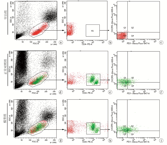
图 2 AE模型鼠外周血中Tfh细胞的流式细胞检测结果
注:空白组,未标记荧光抗体用于流式细胞术圈门设定界限的组;a、d、g, 外周血中淋巴细胞;b、e、h, CD4+细胞;c、f、i,CD4+中CXCR5和PD1阳性细胞,其中Q2门表示CD4+CXCR5+PD1+Tfh细胞。
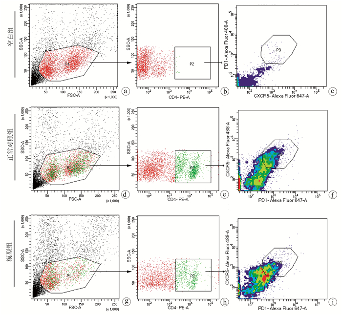
图 3 AE模型鼠脾脏中Tfh细胞的流式细胞检测结果
注:a、d、g, 目的细胞群;b、e、h, CD4+细胞;c、f、i, CD4+中CXCR5和PD1阳性细胞, 其中Q2门表示CD4+CXCR5+PD1+Tfh细胞。
表 1 AE模型鼠外周血中Tfh细胞表达情况
组别 动物数(只) Tfh细胞在CD4+细胞中百分比(%) Tfh细胞在所有细胞中百分比(%) 正常对照组 10 11.12±2.94 0.18±0.05 模型组 10 25.63±3.47 0.08±0.02 t值 10.230 5.520 P值 <0.001 <0.001  下载: 导出CSV
表 2 AE模型鼠脾脏中Tfh细胞表达情况
组别 动物数(只) Tfh细胞在CD4+细胞中百分比(%) Tfh细胞在所有细胞中百分比(%) 正常对照组 10 35.82±4.13 5.30±1.40 模型组 10 33.50±6.40 3.00±0.42 t值 0.974 4.769 P值 0.343 <0.001
下载: 导出CSV
-
[1] CASULLI A, BARTH T, TAMAROZZI F. Echinococcus multilocularis[J]. Trends Parasitol, 2019, 35(9): 738-739. DOI: 10.1016/j.pt.2019.05.005. [2] WEN H, VUITTON L, TUXUN T, et al. Echinococcosis: Advances in the 21st Century[J]. Clin Microbiol Rev, 2019, 32(2): e00075-18. DOI: 10.1128/CMR.00075-18. [3] CAI H, GUAN Y, MA X, et al. Epidemiology of echinococcosis among schoolchildren in golog tibetan autonomous prefecture, qinghai, China[J]. Am J Trop Med Hyg, 2017, 96(3): 674-679. DOI: 10.4269/ajtmh.16-0479. [4] WANG J, GOTTSTEIN B. Immunoregulation in larval Echinococcus multilocularis infection[J]. Parasite Immunol, 2016, 38(3): 182-192. DOI: 10.1111/pim.12292. [5] GLATMAN ZARETSKY A, TAYLOR JJ, KING IL, et al. T follicular helper cells differentiate from Th2 cells in response to helminth antigens[J]. J Exp Med, 2009, 206(5): 991-999. DOI: 10.1084/jem.20090303. [6] HE L, GU W, WANG M, et al. Extracellular matrix protein 1 promotes follicular helper T cell differentiation and antibody production[J]. Proc Natl Acad Sci U S A, 2018, 115(34): 8621-8626. DOI: 10.1073/pnas.1801196115. [7] CROTTY S. T follicular helper cell biology: A decade of discovery and diseases[J]. Immunity, 2019, 50(5): 1132-1148. DOI: 10.1016/j.immuni.2019.04.011. [8] CROTTY S. T follicular helper cell differentiation, function, and roles in disease[J]. Immunity, 2014, 41(4): 529-542. DOI: 10.1016/j.immuni.2014.10.004. [9] SONG W, CRAFT J. T follicular helper cell heterogeneity: Time, space, and function[J]. Immunol Rev, 2019, 288(1): 85-96. DOI: 10.1111/imr.12740. [10] VINUESA CG, LINTERMAN MA, YU D, et al. Follicular helper T cells[J]. Annu Rev Immunol, 2016, 34: 335-368. DOI: 10.1146/annurev-immunol-041015-055605. [11] HELMOLD HAIT S, VARGAS-INCHAUSTEGUI DA, MUSICH T, et al. Early T follicular helper cell responses and germinal center reactions are associated with viremia control in immunized rhesus macaques[J]. J Virol, 2019, 93(4). DOI: 10.1128/JVI.01687-18. [12] XU W, ZHAO X, WANG X, et al. The Transcription factor tox2 drives T follicular helper cell development via regulating chromatin accessibility[J]. Immunity, 2019, 51(5): 826-839.e5. DOI: 10.1016/j.immuni.2019.10.006. [13] WANG B, LI H, SA RL, et al. The expression of ICOS in Tfh cells and the effect of ICOS blocker on the expression of IL-21 in Tfh cells with liver fibrosis[J]. Int J Immunol, 2020, 43(5): 483-487. DOI: 10.3760/cma.j.issn.1673-4394.2020.05.001.王波, 李慧, 萨茹拉, 等. 四氯化碳诱导肝纤维化小鼠Tfh中ICOS的表达及ICOS阻断剂对IL-21表达的影响[J]. 国际免疫学杂志, 2020, 43(5): 483-487. DOI: 10.3760/cma.j.issn.1673-4394.2020.05.001. [14] LINDQVIST M, van LUNZEN J, SOGHOIAN DZ, et al. Expansion of HIV-specific T follicular helper cells in chronic HIV infection[J]. J Clin Invest, 2012, 122(9): 3271-3280. DOI: 10.1172/JCI64314. [15] VELU V, MYLVAGANAM G, IBEGBU C, et al. Tfh1 cells in germinal centers during chronic HIV/SIV infection[J]. Front Immunol, 2018, 9: 1272. DOI: 10.3389/fimmu.2018.01272. [16] GLATMAN ZA, TAYLOR JJ, KING IL, et al. T follicular helper cells differentiate from Th2 cells in response to helminth antigens[J]. J Exp Med, 2009, 206(5): 991-999. DOI: 10.1126/sciimmunol.aan8884. [17] KLEIN F, MOUQUET H, DOSENOVIC P, et al. Antibodies in HIV-1 vaccine development and therapy[J]. Science, 2013, 341(6151): 1199-1204. DOI: 10.1126/science.1241144. [18] HANSEN DS, OBENG-ADJEI N, LY A, et al. Emerging concepts in T follicular helper cell responses to malaria[J]. Int J Parasitol, 2017, 47(2-3): 105-110. DOI: 10.1016/j.ijpara.2016.09.004. [19] DÍAZ A, CASARAVILLA C, ALLEN JE, et al. Understanding the laminated layer of larval Echinococcus Ⅱ: Immunology[J]. Trends Parasitol, 2011, 27(6): 264-273. DOI: 10.1016/j.pt.2011.01.008. [20] HOU YJ, ZHANG LQ, FAN HN. Research advances in circulating free DNA in liver cancer and liver-related parasitic diseases[J]. J Clin Hepatol, 2020, 36(2): 430-432. DOI: 10.3969/j.issn.1001-5256.2020.02.043.后亚军, 张灵强, 樊海宁. 循环游离DNA在肝癌和肝相关性寄生虫病中的应用进展[J]. 临床肝胆病杂志, 2020, 36(2): 430-432. DOI: 10.3969/j.issn.1001-5256.2020.02.043. [21] WANG ZX, GOU P, YU WH, et al. Measurement and bioinformatics analysis of exosomes microRNAs in bile of hepatic alveolar echinococcosis patients with biliary tract invasion[J]. J Clin Hepatol, 2020, 36(9): 2045-2049. DOI: 10.3969/j.issn.1001-5256.2020.09.027.王志鑫, 苟平, 于文昊, 等. 肝泡型包虫病侵及胆道患者胆汁外泌体microRNA的检测及生物信息学分析[J]. 临床肝胆病杂志, 2020, 36(9): 2045-2049. DOI: 10.3969/j.issn.1001-5256.2020.09.027. [22] DING JB, LI YJ, ZHANG FB. Research progress of hydatidosis immunity and vaccine[J]. J Xinjiang Med Univ, 2019, 42(1): 24-28. DOI: 10.3969/j.issn.1009-5551.2019.01.005.丁剑冰, 李玉娇, 张峰波. 包虫病免疫及疫苗的研究进展[J]. 新疆医科大学学报, 2019, 42(1): 24-28. DOI: 10.3969/j.issn.1009-5551.2019.01.005. -

 本文二维码
本文二维码

图(3) / 表(2)
计量
- 文章访问数: 914
- HTML全文浏览量: 142
- PDF下载量: 27
- 被引次数: 0

