
 PDF下载 ( 2308 KB)

二维剪切波弹性成像测量脾硬度联合血小板/脾直径对乙型肝炎肝硬化患者中重度食管胃静脉曲张的评估价值
DOI: 10.3969/j.issn.1001-5256.2021.07.019
利益冲突声明:本研究不存在研究者、伦理委员会成员、受试者监护人以及与公开研究成果有关的利益冲突。
作者贡献声明:余敏睿负责整理分析数据,制订写作思路,撰写论文;杨杰、王进勇参与数据收集及分析;周波参与统计分析;姜镔参与数据收集,实施研究;邓宝成负责研究设计,指导撰写论文并最后定稿。
Value of spleen stiffness measured by two-dimensional shear wave elastography combined with platelet count/spleen diameter ratio in evaluating moderate-to-severe gastroesophageal varices in patients with hepatitis B cirrhosis
-
摘要:
目的 建立乙型肝炎肝硬化患者中重度食管胃静脉曲张(GEV)的无创性诊断模型。 方法 选取2017年10月—2019年12月于中国医科大学附属第一医院就诊的乙型肝炎肝硬化患者。以胃镜检查结果为金标准,分为无/轻度GEV组和中重度GEV组。不符合正态分布的计量资料两组间比较采用Mann-Whitney U检验;计数资料两组间比较采用χ2检验。采用logistic回归分析乙型肝炎肝硬化患者中重度食管胃静脉发生与二维剪切波弹性成像(2D-SWE)测量脾硬度、PLT/脾直径之间的关系,以后退法作为自变量筛选方法,建立回归方程即诊断模型并进行检验。绘制受试者工作特征曲线(ROC曲线)判断肝和脾硬度、PLT/脾直径以及诊断模型等无创性检查指标的诊断价值,得出最佳截断值,DeLong检验比较几种无创性检查指标的ROC曲线之间是否具有统计学意义。 结果 共纳入168例乙型肝炎肝硬化患者,其中有67例诊断为中重度GEV。肝硬化无/轻度和中重度GEV组PLT、ALT、Alb、WBC以及INR比较差异均有统计学意义(Z值分别为-6.508、-2.132、-2.470、-4.510、-5.298,P值均<0.05)。2D-SWE测量的脾硬度、脾脏直径以及PLT/脾脏直径在两组间差异均有统计学意义(Z值分别为-7.264、-5.924、-7.028,P值均<0.05)。PLT/脾直径ROC曲线下面积(AUC)为0.821(95%CI:0.754~0.875),截断值为≤6.7,敏感度为83.58%,特异度为74.26%。脾硬度AUC为0.831(95%CI:0.766~0.885),脾硬度截断值≥34.2 kPa,敏感度为85.07%,特异度为73.27%。肝硬度AUC为0.557(95%CI:0.479~0.634),肝硬度的截断值≥10.8 kPa,特异度为79.10%,敏感度为40.59%。肝硬度与脾硬度、PLT/脾直径对GEV诊断价值的AUC比较,差异均有统计学意义(Z值分别为4.878、5.536,P值均<0.001)。建立判断乙型肝炎肝硬化患者中重度GEV模型:Y=-0.682+0.068×脾硬度-0.225(PLT/脾直径)。诊断模型AUC为0.860(95%CI:0.799~0.909),敏感度为79.10%,特异度为81.19%,准确度为79.1%。 结论 据2D-SWE测量的脾硬度联合PLT/脾直径建立的无创性诊断模型可用于辅助判断乙型肝炎肝硬化患者中重度GEV,准确度较单独使用肝硬度或脾硬度判断中重度GEV高。 Abstract:Objective To establish a noninvasive diagnostic model for moderate-to-severe gastroesophageal varices (GEV) in patients with hepatitis B cirrhosis. Methods The patients with hepatitis B cirrhosis who attended The First Affiliated Hospital of China Medical University from October 2017 to December 2019 were enrolled, and with the results of gastroscopy as the gold standard, the patients were divided into none-to-mild GEV group and moderate-to-severe GEV group. The Mann-Whitney U test was used for comparison of non-normally distributed continuous data between groups, and the chi-square test was used for comparison of categorical data. A logistic regression analysis was used to investigate the association of moderate-to-severe GEV with spleen stiffness measured by two-dimensional shear wave elastography (2D-SWE) and platelet count (PLT)/spleen diameter ratio in patients with hepatitis B cirrhosis, and with the backward method for independent variable screening, a regression equation, i.e., a diagnostic model, was established and validated. The receiver operating characteristic (ROC) curve was plotted to investigate the diagnostic value of noninvasive examination indices including liver and spleen stiffness, PLT/spleen diameter ratio, and the above diagnostic model and obtain their cut-off values, the DeLong test was used to compare whether there is a statistical significance between the ROC curves of the above noninvasive indices. Results A total of 168 patients with hepatitis B cirrhosis were enrolled, among whom 67 were diagnosed with moderate-to-severe GEV. There were significant differences in PLT, alanine aminotransferase, albumin, white blood cell count, and international normalized ratio between the none-to-mild GEV group and the moderate-to-severe GEV group (Z=-6.508, -2.132, -2.470, -4.510, and -5.298, all P < 0.05). There were also significant differences in spleen stiffness measured by 2D-SWE, spleen diameter, and PLT/spleen diameter ratio between the two groups (Z=-7.264, -5.924, and -7.028, all P < 0.05). The PLT/spleen diameter ratio had an area under the ROC curve (AUC) of 0.821 (95% confidence interval [CI]: 0.754-0.875) at the cut-off value of ≤6.7, with a sensitivity of 83.58% and a specificity of 74.26%; spleen stiffness had an AUC of 0.831 (95%CI: 0.766-0.885) at the cut-off value of ≥34.2 kPa, with a sensitivity of 85.07% and a specificity of 73.27%; liver stiffness had an AUC of 0.557 (95%CI: 0.479-0.634) at the cut-off value of ≥10.8 kPa, with a specificity of 79.10% and a sensitivity of 40.59%. There was a significant difference in AUC between liver stiffness and spleen stiffness, as well as between liver stiffness and PLT/spleen diameter ratio (Z=4.878 and 5.536, P < 0.001). The model of Y=-0.682+0.068×spleen stiffness-0.225 (PLT/spleen diameter ratio) was established for predicting moderate-to-severe GEV in patients with hepatitis B cirrhosis, which had an AUC of 0.860 (95%CI: 0.799-0.909), a sensitivity of 79.10%, a specificity of 81.19%, and an accuracy of 79.1%. Conclusion The noninvasive diagnostic model based on spleen stiffness measured by 2D-SWE and PLT/spleen diameter ratio can be used to assist the judgment of moderate-to-severe GEV in patients with hepatitis B cirrhosis, with a higher accuracy than liver stiffness or spleen stiffness alone. -
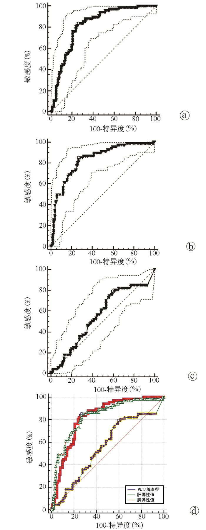
图 1 肝硬度、脾硬度及PLT/脾直径对GEV诊断价值的ROC曲线
注:a,PLT/脾直径的ROC曲线;b,脾硬度值的ROC曲线;c,肝硬度的ROC曲线;d,PLT/脾直径、脾硬度、肝硬度三者的ROC曲线。
表 1 患者基本特征
项目 结果 男/女(例) 118/50 年龄(岁) 53.42(46.17~61.06) 实验室检查 WBC(×109/L) 3.99(2.56~5.46) AST(U/L) 37.43(25.33~60.50) ALT(U/L) 30.71(20.14~58.50) Alb(g/L) 32.40(28.16~39.00) TBil(μmol/L) 21.65(13.00~39.95) DBil(μmol/L) 7.90(4.66~18.55) PLT(×109/L) 87.00(60.00~134.50) INR 1.19(1.05~1.41) 肌酐(μmol/L) 61.00(48.00~72.14) GEV(例) 无/轻 101 中重 67 弹性成像结果 门静脉直径(cm) 1.47(1.29~1.65) 肝硬度(kPa) 15.20(9.05~20.35) 脾硬度(kPa) 34.35(22.80~40.20) 脾直径(cm) 12.86(10.79~14.94)  下载: 导出CSV
表 2 无/轻度GEV与中重度GEV肝硬化患者临床特征比较
项目 无/轻度GEV(n=101) 中重度GEV(n=67) 统计值 P值 男/女(例) 72/29 46/21 χ2=0.133 0.716 年龄(岁) 53.00(44.50~61.00) 54.00(47.00~61.00) Z=-0.905 0.365 实验室指标 WBC(×109/L) 4.82(3.11~6.02) 3.19(2.02~4.04) Z=-4.510 <0.001 AST(U/L) 38.00(25.00~77.50) 33.00(25.00~55.00) Z=-1.115 0.265 ALT(U/L) 37.00(21.00~81.00) 29.00(18.00~42.00) Z=-2.132 0.033 Alb(g/L) 34.90(28.75~40.50) 30.30(27.60~35.40) Z=-2.470 0.014 TBil(μmol/L) 19.70(11.20~45.20) 23.00(15.20~35.10) Z=-1.095 0.274 DBil(μmol/L) 7.60(3.70~20.30) 8.70(5.70~18.20) Z=-1.545 0.122 PLT(109/L) 116.00(79.00~169.00) 64.00(51.00~83.00) Z=-6.508 <0.001 INR 1.07(1.00~1.29) 1.30(1.17~1.61) Z=-5.298 <0.001 肌酐(μmol/L) 61.00(49.00~74.00) 61.00(44.00~69.00) Z=-0.987 0.324 门静脉主干(cm) 1.45(1.20~1.62) 1.50(1.39~1.66) Z=-1.367 0.172 肝硬度(kPa) 14.00(8.45~20.20) 16.9(11.60~21.00) Z=-1.261 0.207 脾硬度(kPa) 26.60(20.55~35.70) 39.90(35.70~43.30) Z=-7.264 <0.001 脾直径(cm) 11.60(10.00~13.94) 14.45(12.80~16.30) Z=-5.942 <0.001 PLT/脾直径 10.15(6.54~16.98) 4.78(2.99~6.19) Z=-7.028 <0.001
下载: 导出CSV
表 3 肝硬度、脾硬度及PLT/脾直径对GEV诊断价值的ROC曲线比较
组别 AUC差异值 95%CI Z值 P值 PLT/脾直径vs脾硬度 0.011 -0.066~0.088 0.274 0.784 PLT/脾直径vs肝硬度 0.263 0.157~0.369 4.878 <0.001 脾硬度vs肝硬度 0.274 0.177~0.371 5.536 <0.001
下载: 导出CSV
表 4 乙型肝炎肝硬化患者中重度GEV的诊断模型
变量 B值 SE Wald P值 OR 95%CI 脾硬度 0.068 0.023 8.578 0.003 1.070 1.023~1.119 PLT/脾直径 -0.225 0.061 17.282 0.000 0.775 0.687~0.874 常量 -0.682 1.008 0.458 0.499 0.506
下载: 导出CSV
表 5 诊断乙型肝炎肝硬化患者中重度GEV不同参数的正确百分比
参数 实测GEV 例数 预测GEV(例) 正确率(%) 无/轻度 中重度 PLT/脾直径 无/轻度 101 80 21 79.2 中重度 67 17 50 74.6 脾硬度 无/轻度 101 84 17 83.2 中重度 67 22 45 67.2 诊断模型 无/轻度 101 701) 21 76.9 中重度 67 12 55 82.1 注:PLT/脾直径、脾硬度和诊断模型的总体正确率分别为77.4%、76.8%和79.1%。1)10例患者因数据缺失,未使用诊断模型。
下载: 导出CSV
-
[1] Chinese Society of Spleen and Portal Hypertension Surgery, Chinese Society of Surgery, Chinese Medical Association. Expert consensus on diagnosis and treatment of esophagogastric variceal bleeding in cirrhotic portal hypertension (2019 edition)[J]. Chin J Surg, 2019, 57(12): 885-892. DOI: 10.3760/cma.j.issn.0529-5815.2019.12.002.中华医学会外科学分会脾及门静脉高压外科学组. 肝硬化门静脉高压症食管、胃底静脉曲张破裂出血诊治专家共识(2019版)[J]. 中华外科杂志, 2019, 57(12): 885-892. DOI: 10.3760/cma.j.issn.0529-5815.2019.12.002. [2] de FRANCHIS R, Baveno VI Faculty. Expanding consensus in portal hypertension: Report of the Baveno VI Consensus Workshop: Stratifying risk and individualizing care for portal hypertension[J]. J Hepatol, 2015, 63(3): 743-752. DOI: 10.1016/j.jhep.2015.05.022. [3] Chinese Society of Hepatology, Chinese Medical Association; Chinese Society of Gastroenterology, Chinese Medical Association; Chinese Society of Endoscopy, Chinese Medical Association. Guidelines for the diagnosis and treatment of esophageal and gastric variceal bleeding in cirrhotic portal hypertension[J]. J Clin Hepatol, 2016, 32(2): 203-219. DOI: 10.3969/j.issn.1001-5256.2016.02.002.中华医学会肝病学分会, 中华医学会消化病学分会, 中华医学会内镜学分会. 肝硬化门静脉高压食管胃静脉曲张出血的防治指南[J]. 临床肝胆病杂志, 2016, 32(2): 203-219. DOI: 10.3969/j.issn.1001-5256.2016.02.002. [4] BUECHTER M, KAHRAMAN A, MANKA P, et al. Spleen and liver stiffness is positively correlated with the risk of esophageal variceal bleeding[J]. Digestion, 2016, 94(3): 138-144. DOI: 10.1159/000450704. [5] PU K, SHI JH, WANG X, et al. Diagnostic accuracy of transient elastography (FibroScan) in detection of esophageal varices in patients with cirrhosis: A meta-analysis[J]. World J Gastroenterol, 2017, 23(2): 345-356. DOI: 10.3748/wjg.v23.i2.345. [6] ZHU YL, DING H, FU TT, et al. Portal hypertension in hepatitis B-related cirrhosis: Diagnostic accuracy of liver and spleen stiffness by 2-D shear-wave elastography[J]. Hepatol Res, 2019, 49(5): 540-549. DOI: 10.1111/hepr.13306. [7] ZHANG XY, TANG SS. Real-time shear wave elastography in quantitative measurement of tissue elasticity on normal spleens[J]. Chin J Med Imaging Technol, 2016, 32(10): 1523-1526. DOI: 10.13929/j.1003-3289.2016.10.013.张潇月, 唐少珊. 实时剪切波弹性成像定量评价正常脾脏组织弹性[J]. 中国医学影像技术, 2016, 32(10): 1523-1526. DOI: 10.13929/j.1003-3289.2016.10.013. [8] Panel of Elastography Assessment of Liver Fibrosis, Study Group of Interventional Ultrasound, Society of Ultrasound in Medicine of Chinese Medical Association. Guidelines for clinical application of two-dimensional shear wave elastography in assessment of liver fibrosis in chronic hepatitis B[J]. J Clin Hepatol, 2018, 34(2): 255-261. DOI: 10.3969/j.issn.1001-5256.2018.02.008.中华医学会超声医学分会介入超声学组弹性成像评估肝纤维化专家组. 二维剪切波弹性成像评估慢性乙型肝炎肝纤维化临床应用指南[J]. 临床肝胆病杂志, 2018, 34(2): 255-261. DOI: 10.3969/j.issn.1001-5256.2018.02.008. [9] FERRAIOLI G, TINELLI C, DAL BELLO B, et al. Accuracy of real-time shear wave elastography for assessing liver fibrosis in chronic hepatitis C: A pilot study[J]. Hepatology, 2012, 56(6): 2125-2133. DOI: 10.1002/hep.25936. [10] GIANNINI E, BOTTA F, BORRO P, et al. Platelet count/spleen diameter ratio: Proposal and validation of a non-invasive parameter to predict the presence of oesophageal varices in patients with liver cirrhosis[J]. Gut, 2003, 52(8): 1200-1205. DOI: 10.1136/gut.52.8.1200. [11] Committee of esophageal varicosity, Society of Digestive Endoscopy of Chinese Medical Association. Tentative guidelines for endoscopic diagnosis and treatment of varicosity and variceal bleeding in digestive tract (2009)[J]. Chin J Dig Endosc, 2010, 27(1): 1-4. DOI: 10.3760/cma.j.issn.1007-5232.2010.01.001.中华医学会消化内镜学分会食管胃静脉曲张学组. 消化道静脉曲张及出血的内镜诊断和治疗规范试行方案(2009年)[J]. 中华消化内镜杂志, 2010, 27(1): 1-4. DOI: 10.3760/cma.j.issn.1007-5232.2010.01.001. [12] Chinese Society of Infectious Diseases, Chinese Medical Association; Chinese Society of Hepatology, Chinese Medical Association. Guidelines for the prevention and treatment of chronic hepatitis B (version 2019)[J]. J Clin Hepatol, 2019, 35(12): 2648-2669. DOI: 10.3969/j.issn.1001-5256.2019.12.007.中华医学会感染病学分会, 中华医学会肝病学分会. 慢性乙型肝炎防治指南(2019年版)[J]. 临床肝胆病杂志, 2019, 35(12): 2648-2669. DOI: 10.3969/j.issn.1001-5256.2019.12.007. [13] DELONG ER, DELONG DM, CLARKE-PEARSON DL. Comparing the areas under two or more correlated receiver operating characteristic curves: A nonparametric approach[J]. Biometrics, 1988, 44(3): 837-845. https://pubmed.ncbi.nlm.nih.gov/3203132/ [14] WANG J, ZHENG GZ. Factors related to esophageal variceal bleeding in patients with liver cirrhosis[J/CD]. Chin J Liver Dis(Electronic Edition), 2019, 11(1): 42-46. DOI: 10.3969/j.issn.1674-7380.2019.01.008.王娟, 郑鸽之. 肝硬化食管静脉曲张破裂出血相关因素分析[J/CD]. 中国肝脏病杂志(电子版), 2019, 11(1): 42-46. DOI: 10.3969/j.issn.1674-7380.2019.01.008. [15] ZHAO ZZ, LUO BM. The Principle and techniques of ultrasonic elastography[J]. China Med Devices Information, 2008, 14(4): 6-8. DOI: 10.3969/j.issn.1006-6586.2008.04.003.赵子卓, 罗葆明. 超声弹性成像基本原理及技术[J]. 中国医疗器械信息, 2008, 14(4): 6-8. DOI: 10.3969/j.issn.1006-6586.2008.04.003. [16] MERCHANTE N, RIVERO-JUÁREZ A, TÉLLEZ F, et al. Liver stiffness predicts variceal bleeding in HIV/HCV-coinfected patients with compensated cirrhosis[J]. AIDS, 2017, 31(4): 493-500. DOI: 10.1097/QAD.0000000000001358. [17] COLECCHIA A, MONTRONE L, SCAIOLI E, et al. Measurement of spleen stiffness to evaluate portal hypertension and the presence of esophageal varices in patients with HCV-related cirrhosis[J]. Gastroenterology, 2012, 143(3): 646-654. DOI: 10.1053/j.gastro.2012.05.035. [18] GIBⅡNO G, GARCOVICH M, AINORA ME, et al. Spleen ultrasound elastography: State of the art and future directions-a systematic review[J]. Eur Rev Med Pharmacol Sci, 2019, 23(10): 4368-4381. DOI: 10.26355/eurrev_201905_17944. [19] TSENG Y, LI F, WANG J, et al. Spleen and liver stiffness for noninvasive assessment of portal hypertension in cirrhotic patients with large esophageal varices[J]. J Clin Ultrasound, 2018, 46(7): 442-449. DOI: 10.1002/jcu.22635. [20] MANATSATHIT W, SAMANT H, KAPUR S, et al. Accuracy of liver stiffness, spleen stiffness, and LS-spleen diameter to platelet ratio score in detection of esophageal varices: Systemic review and meta-analysis[J]. J Gastroenterol Hepatol, 2018, 33(10): 1696-1706. DOI: 10.1111/jgh.14271. [21] CHEN YP, HUANG LW, LIN XY, et al. Alanine aminotransferase influencing performances of routine available tests detecting hepatitis B-related cirrhosis[J]. J Viral Hepat, 2020, 27(8): 826-836. DOI: 10.1111/jvh.13293. [22] PATEL DM, NAIR S, PATEL PD. Clinical assessment of cases presented with esophageal varices and portal hypertension in a tertiary healthcare institute: An observational study[J]. Int J Contemporary Med Res, 2019, 6(5): e22-e44. DOI: 10.21276/ijcmr.2019.6.5.59. [23] XU XZ. The value of non-invasive test to predicting risk of esophageal varices in patients with hepatitis B cirrhosis[D]. Changchun: Jilin University China, 2018.许馨之. 无创性手段预测乙肝肝硬化食管静脉曲张发生风险的临床价值[D]. 长春: 吉林大学, 2018. [24] ELALFY H, ELSHERBINY W, ABDEL RAHMAN A, et al. Diagnostic non-invasive model of large risky esophageal varices in cirrhotic hepatitis C virus patients[J]. World J Hepatol, 2016, 8(24): 1028-1037. DOI: 10.4254/wjh.v8.i24.1028. [25] CHEN XL, CHEN TW, ZHANG XM, et al. Platelet count combined with right liver volume and spleen volume measured by magnetic resonance imaging for identifying cirrhosis and esophageal varices[J]. World J Gastroenterol, 2015, 21(35): 10184-10191. DOI: 10.3748/wjg.v21.i35.10184. -


 本文二维码
本文二维码
图(2) / 表(5)
计量
- 文章访问数: 580
- HTML全文浏览量: 104
- PDF下载量: 36
- 被引次数: 0

