
 PDF下载 ( 3026 KB)

巨噬细胞对小鼠诱导多能干细胞向肝祖细胞分化的影响
DOI: 10.3969/j.issn.1001-5256.2021.04.025
利益冲突声明:本研究不存在研究者、伦理委员会成员、受试者监护人以及与公开研究成果有关的利益冲突。
作者贡献声明:宫甜甜、雷蕾、单智焱负责课题设计,资料分析,撰写论文;黄少刚、申景岭参与收集数据,修改论文;孙瑞珍、李秋明负责拟定写作思路,指导撰写文章并最后定稿。
Effect of macrophages on the differentiation of mouse induced pluripotent stem cells into hepatic progenitor cells
-
摘要:
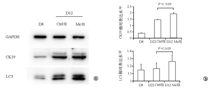
目的 探讨巨噬细胞(Mc)对小鼠诱导多能干细胞(iPSCs)向肝祖细胞(HPCs)分化的影响。 方法 C57BL/6N小鼠24只,采取腹腔冲洗法获得巨噬细胞,收集上清获得巨噬细胞条件培养基(Mc-CDM)。通过激活素A、骨形态发生蛋白4和成纤维细胞生长因子等诱导小鼠iPSCs向HPCs分化。将HPCs的诱导分为两组,一组使用正常培养基,为对照组(Ctrl组); 另一组在诱导D5使用Mc-CDM培养基,为实验组(Mc组)。通过形态学、免疫荧光、Western Blot检测方法,比较正常Ctrl组与Mc组HPCs的形态及相关蛋白表达的差异。计量资料两组间比较采用t检验。 结果 在体外建立了iPSCs来源的HPCs;HPCs具有向肝细胞分化的潜能。免疫荧光结果显示:与第12天的Ctrl组相比,第12天的Mc组的肝祖细胞特异性蛋白CK19的表达显著增高(0.901±0.072 vs 0.686±0.097,t=-3.093,P < 0.05);Western Blot结果显示:与第12天Ctrl组相比,第12天Mc组肝祖细胞相关蛋白CK19的表达显著增高(1.922±0.103 vs 1.448±0.012,t=-7.881,P < 0.05);同时,第12天Mc组自噬相关蛋白LC3的表达亦显著增高(1.392±0.042 vs 1.101±0.048,t=-5.978,P < 0.05)。 结论 巨噬细胞可促进小鼠iPSCs向HPCs分化,其机制可能与HPCs细胞自噬水平增加有关。 -
关键词:
- 多能干细胞 /
- 肝祖细胞 /
- 巨噬细胞 /
- 细胞分化 /
- 小鼠, 近交C57BL
Abstract:Objective To investigate the effect of macrophages (MCs) on the differentiation of mouse induced pluripotent stem cells (iPSCs) into hepatic progenitor cells (HPCs). Methods A total of 24 C57BL/6N mice were used to obtain MCs by peritoneal irrigation, and the supernatant was collected to obtain the conditioned medium of MCs (MC-CDM). Activin A, bone morphogenetic protein 4, and fibroblast growth factor were used to induce the differentiation of mouse iPSCs into HPCs. The differentiation of HPCs were randomly divided into control group (normal medium) and experimental group (MC group; use of MC-CDM medium on day 5 of induction). Morphology, immunofluorescence assay, and Western blot were used to compare the morphology of HPCs and the expression of related proteins between the control group and the MC group. The t-test was used for comparison of continuous data between two groups. Results HPCs derived from iPSCs were established in vitro, and HPCs had the potential to differentiate into hepatocytes. Immunofluorescence assay showed that compared with the D12 control group, the D12 MC group had a significant increase in the protein expression of the HPC-specific protein CK19 (0.901±0.072 vs 0.686±0.097, t=-3.093, P < 0.05). Western blot showed that compared with the D12 control group, the D12 MC group had a significant increase in the protein expression of the HPC-related protein CK19 (1.922±0.103 vs 1.448±0.012, t =-7.881, P < 0.05), as well as a significant increase in the protein expression of the autophagy-related protein LC3 (1.392±0.042 vs 1.101±0.048, t =-5.978, P < 0.05). Conclusion MCs can promote the differentiation of mouse iPSCs into HPCs, possibly by increasing the autophagy level of HPCs. -
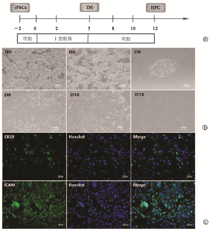
图 1 iPSCs-HPCs的建立
注:a,HPCs诱导流程图;b,细胞各个诱导阶段的形态图(×10);c,免疫荧光染色检测诱导D12细胞中CK19及ICAM的表达(×20)。
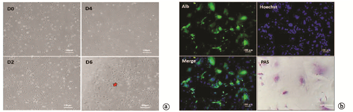
图 2 iPSCs-HPCs可分化为有功能的肝细胞
注:a,肝细胞分化各个阶段的形态图(×10);b,免疫荧光染色检测D6细胞Alb的表达情况(×20),PAS染色鉴定D6细胞功能(×40)。
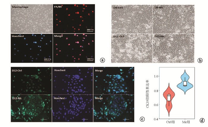
图 3 巨噬细胞鉴定及Ctrl组与Mc组iPSCs-HPCs的CK19表达情况
注:a,巨噬细胞的形态及巨噬细胞标志F4/80的表达(×10);b,Ctrl组与Mc组iPSCs-HPCs诱导形态图(×10);c,Ctrl组与Mc组CK19的表达情况(×20);d, Ctrl组与Mc组CK19阳性表达统计图。
-
[1] KATOONIZADEH A, POUSTCHI H. Adult hepatic progenitor cell niche: How it affects the progenitor cell fate[J]. Middle East J Dig Dis, 2014, 6(2): 57-64. http://pubmedcentralcanada.ca/pmcc/articles/PMC4034666/ [2] LIU LP, YANNAM GR, NISHIKAWAT, et al. The microenvironment in hepatocyte regeneration and function in rats with advanced cirrhosis[J]. Hepatology, 2012, 55: 1529-1539. DOI: 10.1002/hep.24815 [3] SALATI S, LISIGNOLI G, MANFERDINI C, et al. Co-culture of hematopoietic stem/progenitor cells with human osteblasts favours mono/macrophage differentiation at the expense of the erythroid lineage[J]. PLoS One, 2013, 8(1): e53496. DOI: 10.1371/journal.pone.0053496 [4] ZHAO SJ, KONG FQ, JIE J, et al. Macrophage MSR1 promotes BMSC osteogenic differentiation and M2-like polarization by activating PI3K/AKT/GSK3β/β-catenin pathway[J]. Theranostics, 2020, 10(1): 17-35. DOI: 10.7150/thno.36930 [5] ZHOU QJ, XIANG LX, SHAO JZ, et al. In vitro differentiation of hepatic progenitor cells from mouse embryonic stem cells induced by sodium butyrate[J]. J Cell Biochem, 2007, 100(1): 29-42. DOI: 10.1002/jcb.20970 [6] YANAGIDA A, NAKAUCHI H, KAMIYA A. Generation and in vitro expansion of hepatic progenitor cells from human iPS cells[J]. Methods Mol Biol, 2016, 1357: 295-310. [7] BELLANTI F, PANNONE G, TARTAGLIA N. Redox control of the immune response in the hepatic progenitor cell niche[J]. Front Cell Dev Biol, 2020, 8: 295. DOI: 10.3389/fcell.2020.00295 [8] CHEN Y, WANG B, ZHOU H, et al. Autophagy is required for the maintenance of liver progenitor cell functionality[J]. Cell Physiol Biochem, 2015, 36(3): 1163-1174. DOI: 10.1159/000430287 [9] CHANG NC. Autophagy and stem cells: Self-eating for self-renewal[J]. Front Cell Dev Biol, 2020, 8: 138. DOI: 10.3389/fcell.2020.00138 [10] CHEN XD, TAN JL, FENG Y, et al. Autophagy in fate determination of mesenchymal stem cells and bone remodeling[J]. World J Stem Cells, 2020, 12(8): 776-786. DOI: 10.4252/wjsc.v12.i8.776 [11] TOMCZYK S, SUKNOVIC N, SCHENKELAARS Q, et al. Deficient autophagy in epithelial stem cells drives aging in the freshwater cnidarian Hydra[J]. Development, 2020, 147(2): dev177840. DOI: 10.1242/dev.177840 -

 下载:
下载:
 本文二维码
本文二维码
图(4)
计量
- 文章访问数: 378
- HTML全文浏览量: 158
- PDF下载量: 23
- 被引次数: 0

